. 2020 Sep:36:101627.
doi: 10.1016/j.redox.2020.101627. Epub 2020 Jun 30.

Regulation of Nrf2/Keap1 signalling in human skeletal muscle during exercise to exhaustion in normoxia, severe acute hypoxia and post-exercise ischaemia: Influence of metabolite accumulation and oxygenation

Affiliations

Regulation of Nrf2/Keap1 signalling in human skeletal muscle during exercise to exhaustion in normoxia, severe acute hypoxia and post-exercise ischaemia: Influence of metabolite accumulation and oxygenation

Angel Gallego-Selles et al. Redox Biol. 2020 Sep.

Abstract

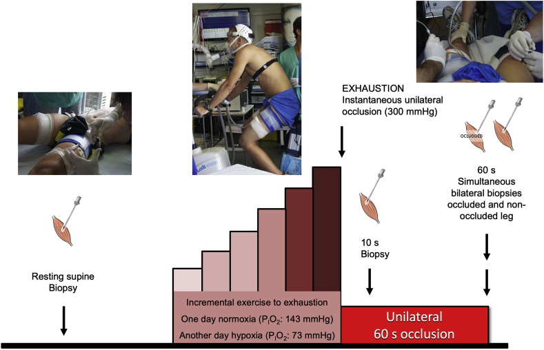
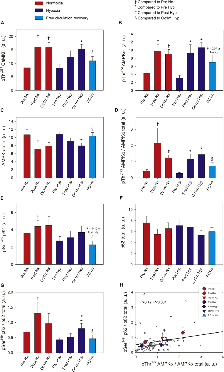
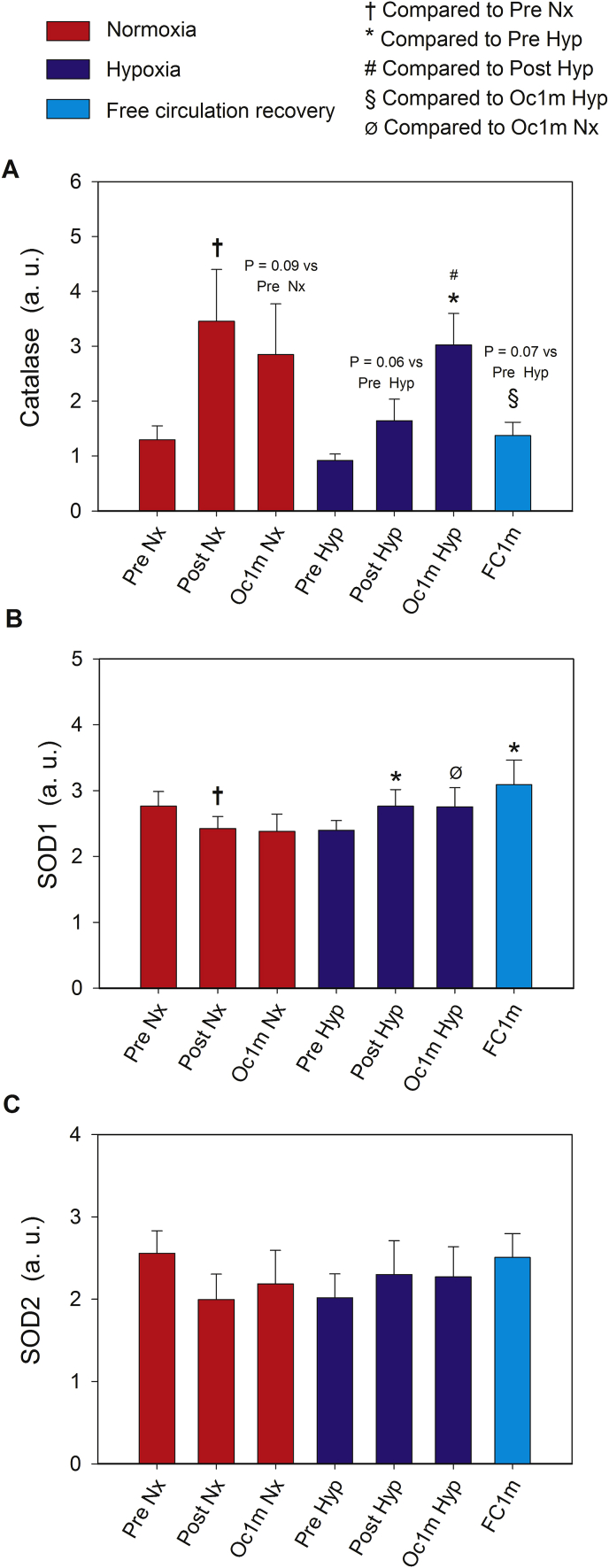
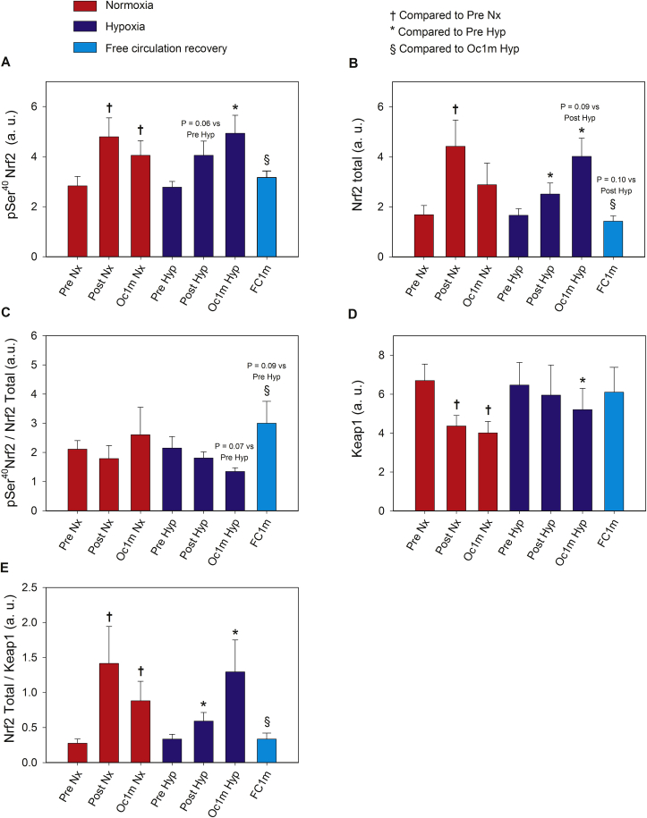
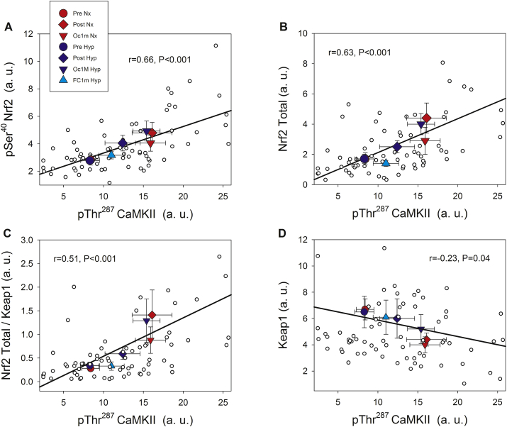
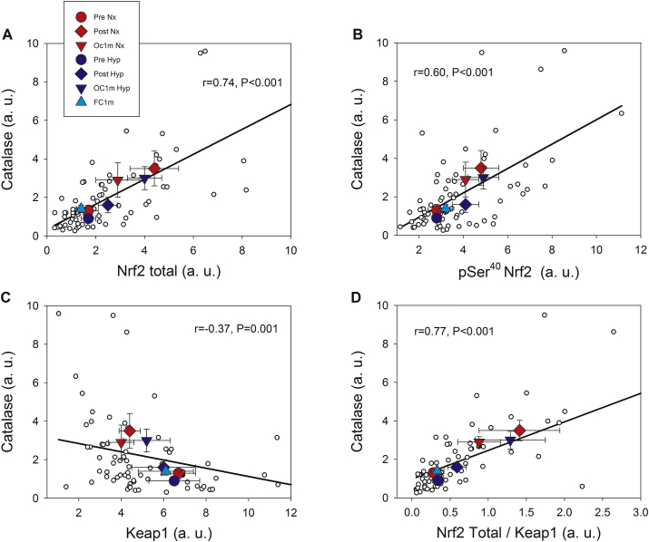
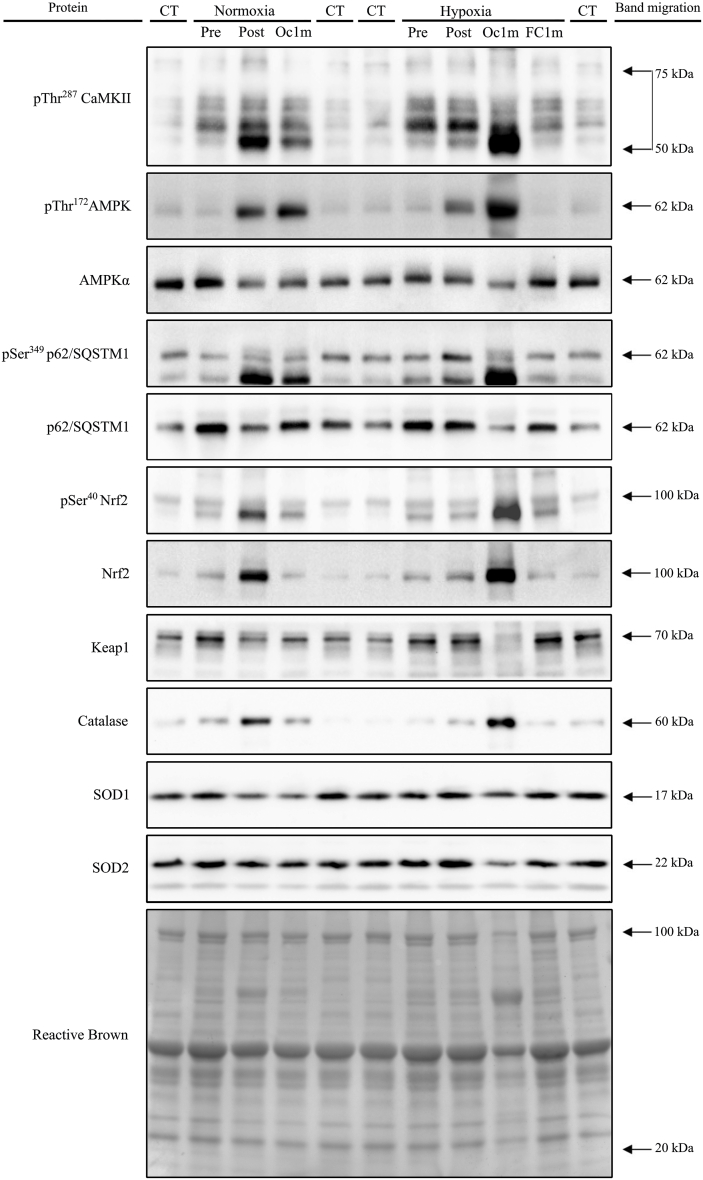
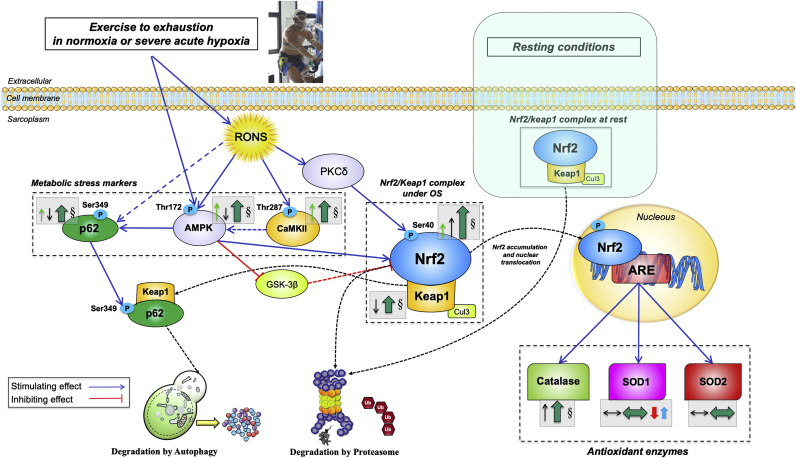
The Nrf2 transcription factor is induced by reactive oxygen and nitrogen species and is necessary for the adaptive response to exercise in mice. It remains unknown whether Nrf2 signalling is activated by exercise in human skeletal muscle. Here we show that Nrf2 signalling is activated by exercise to exhaustion with similar responses in normoxia (PIO2: 143 mmHg) and severe acute hypoxia (PIO2: 73 mmHg). CaMKII and AMPKα phosphorylation were similarly induced in both conditions. Enhanced Nrf2 signalling was achieved by raising Nrf2 total protein and Ser40 Nrf2 phosphorylation, accompanied by a reduction of Keap1. Keap1 protein degradation is facilitated by the phosphorylation of p62/SQSTM1 at Ser349 by AMPK, which targets Keap1 for autophagic degradation. Consequently, the Nrf2-to-Keap1 ratio was markedly elevated and closely associated with a 2-3-fold increase in Catalase protein. No relationship was observed between Nrf2 signalling and SOD1 and SOD2 protein levels. Application of ischaemia immediately at the end of exercise maintained these changes, which were reverted within 1 min of recovery with free circulation. While SOD2 did not change significantly during either exercise or ischaemia, SOD1 protein expression was marginally downregulated and upregulated during exercise in normoxia and hypoxia, respectively. We conclude that Nrf2/Keap1/Catalase pathway is rapidly regulated during exercise and recovery in human skeletal muscle. Catalase emerges as an essential antioxidant enzyme acutely upregulated during exercise and ischaemia. Post-exercise ischaemia maintains Nrf2 signalling at the level reached at exhaustion and can be used to avoid early post-exercise recovery, which is O2-dependent.

Keywords: AMPK; CaMKII; Fatigue; High-intensity exercise; Ischaemia; Performance.


Conflict of interest statement

The authors declare that they have no known competing financial interests or personal relationships that could have appeared to influence the work reported in this paper.

Figures

Fig. 1
Fig. 1
Schematic representation of the experimental protocol. Subjects performed an incremental exercise to exhaustion either in normoxia (Nx; FIO2 = 0.21, PIO2: 143 mmHg) or severe acute normobaric hypoxia (Hyp; FIO2 = 0.104, PIO2: 73 mmHg) in random order. Before warm-up, a resting biopsy was obtained, followed by the exercise test. Immediately at exhaustion, the circulation of one leg was completely occluded by the instantaneous inflation of a cuff at 300 mmHg, which was maintained for 60 s. Skeletal muscle biopsies were taken from the cuffed leg at 10 s and 60 s of occlusion in both trials (Nx and Hyp). In the test performed in hypoxia, another biopsy was obtained 60 s after the end of the incremental exercise from the leg recovering with free circulation, while the subjects recovered breathing room air (i.e., Nx). The application of ischaemia impeded the recovery of muscle metabolites, exhausted the O2 stores and resulted in further accumulation of lactate and H+, Pi, and Cr.
Fig. 2
Fig. 2
Skeletal muscle intracellular CaMKII, AMPKα and p62 signalling in response to incremental exercise to exhaustion in normoxia and severe hypoxia and the application of immediate ischaemic or non-ischaemic recovery. Protein expression levels of pThr287 CaMKII (A), pThr172 AMPKα (B), AMPKα total (C), pThr172 AMPKα/AMPKα total ratio (D), pSer349 p62 (E), p62 total (F), pSer349 p62/p62 total ratio (G), and association between the ratio of pThr172 AMPKα/AMPKα total and the ratio of pSer349 p62/p62 total (H). Nx; test performed in normoxia (FIO2 = 0.21, PIO2: 143 mmHg), Hyp; test performed in severe acute normobaric hypoxia (FIO2 = 0.104, PIO2: 73 mmHg); Pre, before exercise; Post, 10 s after the end of exercise with ischaemic recovery; Oc1m, 60 s after the end of exercise with ischaemic recovery; FC1m, 60 s after the end of exercise in the leg recovering without occlusion (free circulation); p62/SQSTM1 (shortened to p62). n = 11 in all conditions except for Oc1m Nx (n = 9), Post Hyp (n = 10), and Oc1m Hyp (n = 10). In (H), large symbols represent the mean of the subjects studied in each condition. The correlation coefficient and regression line have been calculated using the individual values (small white circles, n = 73). A detailed description of the experimental phases is explained in Fig. 1. The statistical analysis was performed with logarithmically transformed data for the pThr172 AMPKα/AMPKα total ratio and p62 total. The values shown are means ± standard errors and expressed in arbitrary units (a.u.). †p < 0.05 vs Pre Nx; *p < 0.05 vs Pre Hyp; #p < 0.05 vs Post Hyp; §p < 0.05 vs Oc1m Hyp.
Fig. 3
Fig. 3
Skeletal muscle intracellular Catalase, SOD1 and SOD2 signalling in response to incremental exercise to exhaustion in normoxia and severe hypoxia and the application of immediate ischaemic or non-ischaemic recovery. Protein expression levels of Catalase (A), SOD1 (B), and SOD2 (C). Nx; test performed in normoxia (FIO2 = 0.21, PIO2: 143 mmHg), Hyp; test performed in severe acute normobaric hypoxia (FIO2 = 0.104, PIO2: 73 mmHg); Pre, before exercise; Post, 10 s after the end of exercise with ischaemic recovery; Oc1m, 60 s after the end of exercise with ischaemic recovery; FC1m, 60 s after the end of exercise in the leg recovering without occlusion (free circulation). A detailed description of the experimental phases is explained in Fig. 1. The statistical analysis was performed with logarithmically transformed data for Catalase and SOD2. The values shown are means ± standard errors and expressed in arbitrary units (a.u.). n = 11 in all conditions except for Oc1m Nx (n = 9), Post Hyp (n = 10), and Oc1m Hyp (n = 10). †p < 0.05 vs Pre Nx; *p < 0.05 vs Pre Hyp; #p < 0.05 vs Post Hyp; §p < 0.05 vs Oc1m Hyp; Ø p < 0.05 vs Oc1m Nx.
Fig. 4
Fig. 4
Skeletal muscle intracellular Nrf2 and Keap1 signalling in response to incremental exercise to exhaustion in normoxia and severe hypoxia and the application of immediate ischaemic or non-ischaemic recovery. Protein expression levels of pSer40 Nrf2 (A), Nrf2 total (B), pSer40 Nrf2/Nrf2 total ratio (C), Keap1 (D), Nrf2 Total/Keap1 ratio (E). Nx; test performed in normoxia (FIO2 = 0.21, PIO2: 143 mmHg), Hyp; test performed in severe acute normobaric hypoxia (FIO2 = 0.104, PIO2: 73 mmHg); Pre, before exercise; Post, 10 s after the end of exercise with ischaemic recovery; Oc1m, 60 s after the end of exercise with ischaemic recovery; FC1m, 60 s after the end of exercise in the leg recovering without occlusion (free circulation). A detailed description of the experimental phases is explained in Fig. 1. The statistical analysis was performed with logarithmically transformed data for all proteins except for Keap1. The values shown are means ± standard errors and expressed in arbitrary units (a.u.). n = 11 in all conditions except for Oc1m Nx (n = 9), Post Hyp (n = 10), and Oc1m Hyp (n = 10). †p < 0.05 vs Pre Nx; *p < 0.05 vs Pre Hyp; #p < 0.05 vs Post Hyp; §p < 0.05 vs Oc1m Hyp.
Fig. 5
Fig. 5
Associations between the protein expression levels of phosphorylated CaMKII, Nrf2 and Keap1 across experimental phases. pThr287 CaMKII and pSer40 Nrf2 (A) pThr287 CaMKII and Nrf2 total (B), pThr287 CaMKII and Nrf2/Keap1 ratio (C), pThr287 CaMKII and Keap1 (D). A description of the experimental phases is explained in Fig. 1. Nx; test performed in normoxia (FIO2 = 0.21, PIO2: 143 mmHg), Hyp; test performed in severe acute normobaric hypoxia (FIO2 = 0.104, PIO2: 73 mmHg); Pre, before exercise; Post, 10 s after the end of exercise with ischaemic recovery; Oc1m, 60 s after the end of exercise with ischaemic recovery; FC1m, 60 s after the end of exercise in the leg recovering without occlusion (free circulation). n = 11 in all conditions except for Oc1m Nx (n = 9), Post Hyp (n = 10), and Oc1m Hyp (n = 10). Large symbols: each point is representing the mean of the subjects studied in each condition. Correlation coefficients and regression lines have been calculated using the individual values (small white circles, n = 73). The values shown are means ± standard errors and expressed in arbitrary units (a.u.). Statistical significance was set at p < 0.05.
Fig. 6
Fig. 6
Associations between the protein expression levels of Catalase, Nrf2 and Keap1 across experimental phases. Nrf2 total and Catalase (A), pSer40 Nrf2 and Catalase (B), Keap1 and Catalase (C) and Nrf2/Keap1 ratio and Catalase (D). A description of the experimental phases is explained in Fig. 1. Nx; test performed in normoxia (FIO2 = 0.21, PIO2: 143 mmHg), Hyp; test performed in severe acute normobaric hypoxia (FIO2 = 0.104, PIO2: 73 mmHg); Pre, before exercise; Post, 10 s after the end of exercise with ischaemic recovery; Oc1m, 60 s after the end of exercise with ischaemic recovery; FC1m, 60 s after the end of exercise without ischaemic recovery (free circulation). n = 11 in all conditions except for Oc1m Nx (n = 9), Post Hyp (n = 10), and Oc1m Hyp (n = 10). Large symbols: each point is representing the mean of the subjects studied in each condition. Correlation coefficients and regression lines have been calculated using the individual values (small white circles, n = 73). The values shown are means ± standard errors and expressed in arbitrary units (a.u.). Statistical significance was set at p < 0.05.
Fig. 7
Fig. 7
Representative immunoblot images of all proteins and their regulatory phosphorylations and the total amount of protein loaded (Reactive Brown staining) from a single participant. From top to bottom: pThr287 CaMKII, pThr172 AMPKα, AMPKα total, pSer349 p62/SQSTM1, p62/SQSTM1 total, pSer40 Nrf2, Nrf2, Keap1, Catalase, SOD1, SOD2 and Reactive Brown (as protein loading control). Nx; test performed in normoxia (FIO2 = 0.21, PIO2: 143 mmHg), Hyp; test performed in severe acute normobaric hypoxia (FIO2 = 0.104, PIO2: 73 mmHg); Pre, before exercise; Post, 10 s after the end of exercise with ischaemic recovery; Oc1m, 60 s after the end of exercise with ischaemic recovery; FC1m, 60 s after the end of exercise without ischaemic recovery (free circulation); CT, control sample. Arrows indicate estimated molecular weights.
Fig. 8
Fig. 8
Schematic model of the regulation of Nrf2 and Keap1 signalling in human skeletal muscle immediately after incremental exercise to exhaustion in normoxia and severe hypoxia. Under basal conditions, Keap1 continuously targets Nrf2 for ubiquitination and degradation, allowing for minimal levels of Nrf2. The production of RONS during exhaustive exercise stimulates the activation of AMPK and CaMKII. Concomitantly, CaMKII acts indirectly as an upstream AMPK activator (by an unknown mechanism). AMPK promotes the increase of Nrf2 levels by two main mechanisms. Firstly, by phosphorylating p62 at Ser349, which stimulates the p62-mediated degradation of Keap1 via autophagy; secondly, by phosphorylating and blocking GSK3-β, which activates β-TrCP (an E3 ubiquitin-protein ligase) which tags Nrf2 for proteasomal degradation (not measured here). RONS may also activate PKCδ which phosphorylates Nrf2 at its Serine 40 promoting its nuclear translocation and genes transactivation. The lowered levels of Keap1 and the reduced amount of p62 observed here are suggestive of co-degradation following exercise. Overall, the augmented Nrf2 total and phosphorylated protein expression together with the rise in the Nrf2-to-Keap1 ratio elicited by exhausting exercise should be sufficient to enhance the Nrf2-mediated antioxidant response. A central role of Catalase is manifested by a remarkable increase in its protein content following exercise, which was exacerbated during exercise in severe acute hypoxia, likely as a response to increased H2O2 production, by superoxide dismutases. This process is facilitated in hypoxia due to the upregulation of SOD1. No acute changes in SOD2 protein expression were observed. Most changes evoked by the exhaustive exercise bout were almost entirely reverted to baseline in less than 60 s by an O2-dependent mechanism. Activating/inhibiting actions are represented by blue/red connecting lines (dashed if the effect is indirect). Changes on cellular locations are presented with black dashed lines. The arrows and symbols depicted inside dashed grey boxes and located beside the specific markers illustrate the overall protein expression changes in this investigation, as follows: Thin arrows in green (phosphorylated form) and black (total form) depict the overall direction of the outcomes (increase/decrease) for the particular muscle protein; thick arrows in darker green represent the overall effect on stimulation/inhibition of Nrf2 signalling; the symbol § indicates a significant difference between the biopsies taken 60 s after the end of the exercise, i.e., between the legs recovering with and without ischaemia. A differential modulation due to FIO2 is illustrated by the presence of arrows in red (normoxia) and blue (severe hypoxia). The size of each arrow is commensurate with the magnitude of the change. Abbreviations not defined in the text: ARE, antioxidant response element; OS: oxidative stress. (For interpretation of the references to colour in this figure legend, the reader is referred to the Web version of this article.)

References

    1. Powers S.K., Ji L.L., Kavazis A.N., Jackson M.J. Reactive oxygen species: impact on skeletal muscle. Comp. Physiol. 2011;1(2):941–969. - PMC - PubMed
    1. Calbet J.A.L., Martin-Rodriguez S., Martin-Rincon M., Morales-Alamo D. An integrative approach to the regulation of mitochondrial respiration during exercise: focus on high-intensity exercise. Redox Biol. 2020:101478. - PMC - PubMed
    1. Morales-Alamo D., Calbet J.A. Free radicals and sprint exercise in humans. Free Radic. Res. 2014;48(1):30–42. - PubMed
    1. Brooks S.V., Vasilaki A., Larkin L.M., McArdle A., Jackson M.J. Repeated bouts of aerobic exercise lead to reductions in skeletal muscle free radical generation and nuclear factor kappaB activation. J. Physiol. 2008;586(16):3979–3990. - PMC - PubMed
    1. Morales-Alamo D., Ponce-Gonzalez J.G., Guadalupe-Grau A., Rodriguez-Garcia L., Santana A., Cusso R. Critical role for free radicals on sprint exercise-induced CaMKII and AMPKalpha phosphorylation in human skeletal muscle. J. Appl. Physiol. 2013;114(5):566–577. - PubMed

Publication types

Substances

LinkOut - more resources