Review
. 2020 Sep:36:101664.
doi: 10.1016/j.redox.2020.101664. Epub 2020 Aug 1.

Nrf2/Wnt resilience orchestrates rejuvenation of glia-neuron dialogue in Parkinson's disease

Affiliations
Review

Nrf2/Wnt resilience orchestrates rejuvenation of glia-neuron dialogue in Parkinson's disease

Bianca Marchetti. Redox Biol. 2020 Sep.

Abstract

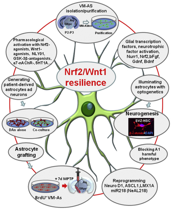
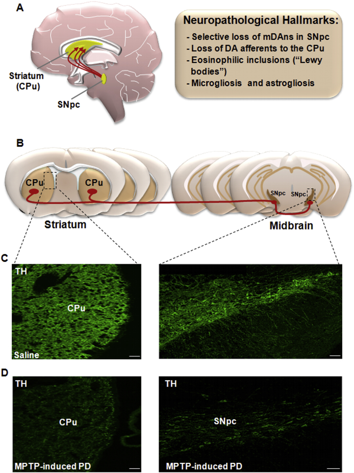
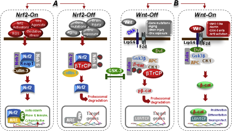
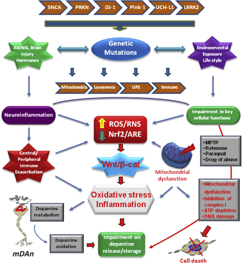
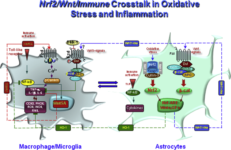
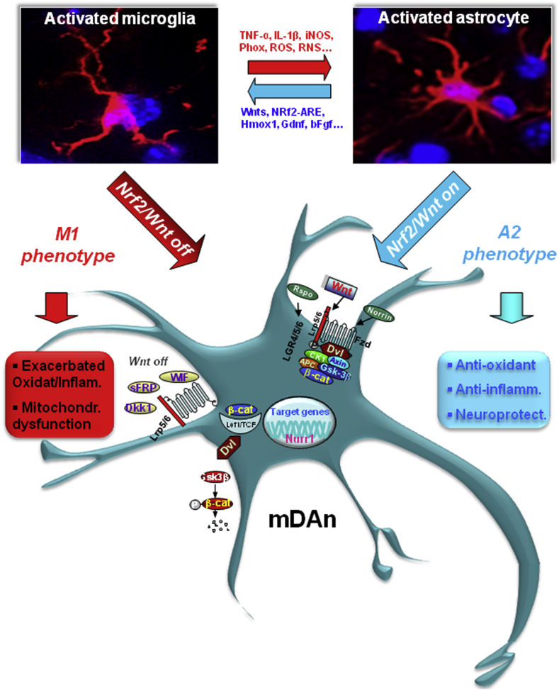
Oxidative stress and inflammation have long been recognized to contribute to Parkinson's disease (PD), a common movement disorder characterized by the selective loss of midbrain dopaminergic neurons (mDAn) of the substantia nigra pars compacta (SNpc). The causes and mechanisms still remain elusive, but a complex interplay between several genes and a number of interconnected environmental factors, are chiefly involved in mDAn demise, as they intersect the key cellular functions affected in PD, such as the inflammatory response, mitochondrial, lysosomal, proteosomal and autophagic functions. Nuclear factor erythroid 2 -like 2 (NFE2L2/Nrf2), the master regulator of cellular defense against oxidative stress and inflammation, and Wingless (Wnt)/β-catenin signaling cascade, a vital pathway for mDAn neurogenesis and neuroprotection, emerge as critical intertwinned actors in mDAn physiopathology, as a decline of an Nrf2/Wnt/β-catenin prosurvival axis with age underlying PD mutations and a variety of noxious environmental exposures drive PD neurodegeneration. Unexpectedly, astrocytes, the so-called "star-shaped" cells, harbouring an arsenal of "beneficial" and "harmful" molecules represent the turning point in the physiopathological and therapeutical scenario of PD. Fascinatingly, "astrocyte's fil rouge" brings back to Nrf2/Wnt resilience, as boosting the Nrf2/Wnt resilience program rejuvenates astrocytes, in turn (i) mitigating nigrostriatal degeneration of aged mice, (ii) reactivating neural stem progenitor cell proliferation and neuron differentiation in the brain and (iii) promoting a beneficial immunomodulation via bidirectional communication with mDAns. Then, through resilience of Nrf2/Wnt/β-catenin anti-ageing, prosurvival and proregenerative molecular programs, it seems possible to boost the inherent endogenous self-repair mechanisms. Here, the cellular and molecular aspects as well as the therapeutical options for rejuvenating glia-neuron dialogue will be discussed together with major glial-derived mechanisms and therapies that will be fundamental to the identification of novel diagnostic tools and treatments for neurodegenerative diseases (NDs), to fight ageing and nigrostriatal DAergic degeneration and promote functional recovery.

Keywords: Ageing; Astrocyte therapies; Gene-environment interactions; Glia-neuron crosstalk; Nrf2 signaling; Oxidative/inflammatory stress; Parkinson's disease; Wnt signaling.


Figures

Image 1
Graphical abstract
Fig. 1
Fig. 1
Nigrostriatal dopaminergic pathway and neuropathological hallmarks of Parkinson's disease (PD). A. In the left hand-side, a sagittal schematic view of nigrostriatal dopaminergic (DAergic) neurons originating (in red) in the subtantia nigra pars compacta (SNpc) of the mesesencephalon, and projecting to the corpus striatum (CPu), which includes the caudate and putamen nuclei. The major neuropathological hallmarks of PD are boxed on the right-hand side. B. Schematic drawing of coronal brain sections at the level of the striatum and SNpc showing the trajectory (in red) of the nigrostriatal DAergic pathway. C-D: Confocal laser scanning microcoscopic images of Cpu and SNpc in coronal brain sections stained with the dopamine marker tyrosine hydroxylase (TH, in green) in intact, saline-treated mice (C) and after exposure to the PD neurotoxin, MPTP (D). Note the severe loss of TH+ fibers in Cpu and of TH+ cell bodies in SNpc, occurring in MPTP-induced PD (D). Scale bars, panel C (Striatum: 25 μm; SNpc: 100 μm), panel D (Striatum: 25 μm; SNpc: 100 μm).
Fig. 2
Fig. 2
The Nrf2-ARE and Wnt/β-catenin/GSK-3β intertwined signaling cascades. A. In normal conditions, Nrf2 is inactive (Nrf2-Off”) and resides in the cytoplasm bound to Keap1. In response to oxidative stress and inflammation, the modification of Keap1 cysteine residues leads to inhibition of Nrf2 ubiquitylation and stabilization of Nrf2, allowing Nrf2 to accumulate in the cytosol and then to translocate into the nucleus where it binds to a small Maf protein and activates transcription of genes containing antioxidant response elements (AREs) in their regulatory regions (Nrf2-On”) [[76], [77], [78]]. In addition to its interaction with Nrf2, Keap1 also binds Cullin 3 (Cul3), which forms a core E3 ubiquitin ligase complex through an association with Ring-box1 protein (Rbx1, also called Roc1) [[76], [77], [78]]. Besides Keap1-mediated regulation, two other E3 ubiquitin ligases have been found to regulate the protein level of Nrf2. Nrf2 is controlled by two distinct β-TrCP recognition motifs in its Neh6 domain, one of which can be modulated by glycogen synthase kinase 3β (GSK-3β) activity phosphorylating a group of Ser residues in the Neh6 domain of Nrf2 [81, 82, see text]. B. In Wnt/β-catenin pathway, Wnt signal activation is tightly controlled by a dynamic signaling complex, constituted by class Frizzled (Fzd) of the G-protein-coupled receptor (GPCRs) superfamily, the LDL receptor-related protein (LRP) 5/6 coreceptors and Dishevelled (Dvl) and Axin adapters [75]. In the absence of a Wnt ligand, (Wnt-off) the signaling cascade is inhibited. Cytoplasmic β-catenin is phosphorylated and degraded via proteasome mediated destruction, which is controlled by the “destruction complex”, consisting of GSK3β, casein kinase 1α (CK1α), the scaffold protein AXIN, and the tumor suppressor adenomatous polyposis coli (APC) [75]. As a result, the translocation into nucleus is inhibited. Interruption of Wnt/β-catenin signaling also occurs in the presence of the Dkk’ and secreted FZD-related proteins (sFRPs) families of Wnt-antagonists, or Wnt inhibitory protein, WIF. Conversely, Wnt ligand binding to Fzd receptors at the surface of target cells (Wnt-on) triggers a chain of events aimed at disrupting the degradation complex via Dvl phosphorylation [75]. Then β-catenin is separated from the destruction complex, resulting in its accumulation and stabilization in the cytoplasm. Subsequently, β-catenin is imported into the nucleus where it can interact with the TCF/LEF family of transcription factors and recruit transcriptional co-activators, p300 and/or CBP (CREB-binding protein), as well as other components to transcribe a panel of downstream target genes. Conditions that can direct to Nrf2/Wnt-On (Nrf2-activators, GSK-3-antagonists, Wnt1-agonists.) or to Nrf2/Wnt-/Off (PD gene mutations, ageing, inflammation, environmental toxins.) are indicated. Because GSK-3β crosstalk with both Nrf-ARE and canonical Wnt-signaling, inhibition of GSK-3β activity by molecular compounds and various enzymes represents a potential means to activate the anti-oxidant, anti-inflammatory, prosurvial, neuroprotective and neurogenic downstream Nrf2/Wnt gene cascades (for details, see the text).
Fig. 3
Fig. 3
NRf2/Wnt/β-catenin interconnected pathways and gene-environment interactions in Parkinson's disease (PD). Scheme of the reciprocal gene-environment interactions impacting on nuclear factor erythroid 2 -like 2 (NFE2L2/Nrf2) and Wnt/β-catenin signaling cascades in PD. The expression of SNCA, PRKN, PINK1, DJ-1, and LRRK2 in astrocytes and microglial cells affect the inflammatory response, endoplassmic reticulum (ER) stress, mitochondrial, lysosomal, ubiquitin-proteasome system (UPS), autophagic and Wnt signaling functions. Genetic mutations powerfully interact with a panel of environmental factors, including ageing, neurotoxic exposures (i.e., rotenone, paraquat, MPTP, drugs of abuse), the hormonal background (the stress and reproductive hormones) and life style. Central to the dopaminotoxic cascades, is the dysfunction of Nf2/Wnt signaling axis, critically involved in providing anti-oxidant and anti-inflammatory self-defenses, and promoting the survival and protection of the vulnerable midbrain dopamine neurons (mDAns) via bidirectional astrocyte-neuron crosstalk. In light of the intrinsic vulnerability of mDAns as a result of DA oxidative metabolism associated to the specific microglial environment within the SNpc, a combination of genetic and environmental factors, leading to astrocyte and microglia overactivation, and consequent generation of a panel of cytotoxic mediators, further exacerbates inflammation and oxidative stress. PD mutations via their impact in astrocyte and microglia cells biology, their inter-relations with mitochondrial Nrf2 and Wnt/β-catenin/GSK-β signaling predispose the brain to reach a critical threshold of inflammation and mitochondrial dysfunction, in turn acting as a driving force to exacerbate the progression of inflammation-mediated neurodegeneration of PD.
Fig. 4
Fig. 4
Wnt1 is a novel actor in astrocyte-neuron crosstalk. A: scheme of ventral midbrain astrocytes (VM-AA) isolation, purification and direct (co-culture) or indirect (AS-conditioned medium, ACM) culture paradigms with purified primary mesencephalic dopaminergic neurons (mDAn), in the absence or the presence of the PD neurotoxin, MPP+. Survival, dopamine (DA) uptake and caspase 3 assays are used to monitor mDAn death and functionality. B:In situ hybridization histochemistry coupled to confocal laser scanning microscopy and dual immunofluorescent staining with the astrocyte cytoskeleton marker, glial fibrillary acidic protein (GFAP, in red) and Wnt1 mRNA (in green) showing the expression of Wnt1 in primary GFAP+ cells (orange-yellow). C: Astrocyte-neuron crosstalk with primary mDAns. Confocal image of TH+ neurons (in green) in co-culture with VM-AS (in red), showing TH+ neurons with long and branched TH+ neuronal processes, interacting with GFAP+ star-shaped astrocytes. Fig. 4. Scale bars, panel B: 25 μm, Box: 10 μm, panel C: 50 μm.
Fig. 5
Fig. 5
Dopaminergic neurorepair upon MPTP injury is directed by glial fibrillary acid protein (GFAP+) astrocytes. A reconstruction of representative confocal images of midbrain coronal sections at the SNpc level, stained with tyrosine hydroxylase positive neurons (TH+, in green) and GFAP (in red) from MPTP-treated mice, 65 days post injury is shown. Note the robust TH neurorepair, as revealed by fluorescence immunohistochemistry. Within the rescued SNpc, bright TH+ neurons extending long processes can be observed running together with bright reactive GFAP+ astrocytes, coursing intermingled with TH+ neurons (boxed magnification) and seemingly guiding the dopaminergic neurorestorative process. VTA: ventral tegmental area. Scale bar:100 μm.
Fig. 6
Fig. 6
Nrf2/Wnt/immune crosstalk in oxidative stress and inflammation in PD. Schematic illustration of astrocyte-microglia crosstalk. Upon activation by neurotoxins, endotoxins, brain injury and ageing, macrophage/microglia produce a panel of pro-inflammatory cytokines (TNF-α and IL-1β) and chemokines (CCL3, CXCl10 and CXCL11). Up-regulation of microglial PHOX-derived ROS, iNOS-derived NO, and GSK-3β, a known regulator of NF-kB-dependent gene transcription, further exacerbates microglia reaction. Wnt5a constitutes one part of a self-perpetrating cycle, via autocrine Wnt5A/CamKII activation and paracrine stimulation of Th-1- cytokines, iNOS and COX2 [280–2282]. To restrain microglia exacerbation, up-regulation of astrocyte- Nrf2/HO-1 and Wnt1/β-catenin, mitigate the inflammatory milieu and favor a down-regulation of cytokines expression. NF-κB and the Wnt/β-catenin pathway also interact to differentially regulate inflammation, with GSK-3β playing a central role in between. While GSK-3β is a negative regulator of β-catenin, it positively regulates NF-κB by targeting IkB, the major inhibitor of NF-κB, to proteasomal degradation. On the other hand, β-catenin itself can form a complex with the p50 subunit of NF-κB, thereby preventing NF-κB transcriptional activity. Besides, HO-1 indirect modulation, Nrf2-NF-κB interplay contributes to the regulation of immune response under oxidative stress and inflammation aimed at counterbalancing the exacerbated inflammation. Then, astrocyte upregulation of Nrf2/HO-1 and Wnt1/β-catenin during oxidative stress and inflammation represent a critical regulatory level, whereby astrocytes can mitigate M1 exacerbated phenotype and the heighthened levels of proinflammatory cytokines.
Fig. 7
Fig. 7
Astrocyte-microglia interactions and Nf2/Wnt1/β-catenin resilience in mDAn neuroprotection. Major environmental factors including ageing, inflammation, neurotoxin exposure (MPTP/MPP+, 6-OHDA, pesticides), in synergy with genetic mutations results in dysfunctional astrocyte-microglial crosstalk associated to the exacerbated production of proinflammatory mediators. The glial switch to the A1/M1 harmful astrocyte and microglial phenotype is the result of the inhibition of Nrf2/Wnt/β-catenin signaling (“Nrf2/Wnt off”). In these conditions, reactive astrocytes no longer mount an efficient resilience program for the vulnerable mDAns. Hence, the crucial anti-oxidant and anti-inflammatory Nrf2/HO-1 and dopaminotrophic factors, namely Wnt1, are sharply inhibited. As a result, active GSK-3β is up-regulated in mDAns, leading to β-catenin degradation. Then, in the absence of an efficient Nrf2-ARE axis at play, the “frailty” of mDAns increases in turn leading to mDAn degeneration. By contrast, astrocyte upregulation of Nrf2/HO-1 and Wnt1/β-catenin during oxidative stress and inflammation represent a critical resilience program for DAns. Then, increased astrocyte-derived Wnt1 (and Wnt1-like agonists, such as Wnt1, Rspo or Norrin) activates Fzd-1 receptors (“Wnt on”), leading to the blockade of GSK-3β-induced phosphorylation (P) and proteosomal degradation of the neuronal pool of β-catenin. Stabilized β-catenin can translocate into the nucleus and associate with a family of transcription factors and regulate the expression of Wnt target genes involved in DA neuron survival/plasticity, neuroprotection and repair. β-catenin may also function as a pivotal defense molecule against oxidative stress, and can act as a coactivator for several nuclear receptors involved in the maintenance/protection of DA neurons. The hypothetical contribution of various endogenous Wnt agonists (Responding, Rspo, Norrin) or antagonists (Dkkopf, Dkk1, Wif, frizzled-related proteins, SFRp) are also indicated. Resilience of Nrf2/Wnt/β-catenin program can be activated by several treatments, including GSK-3β antagonists, Wnt1-like agonists, nitric-oxide–(NO)–anti-inflammatory non-steroidal drugs (NSAID). Different conditions/treatments can inhibit Nrf2/Wnt beneficial signaling cascades, including gene mutations, ageing, inflammation, endogenous Wnt-antagonist expression, leading mDAn degeneration (see the text for details).
Fig. 8
Fig. 8
Astrocyte's “fil rouge” targeting Nrf2/Wnt resilience cascades. Manipulating astrocytes provides a new approach for drug development in neurological diseases. Schematic representation of a panel of manipulations of astrocytes for DAn neurorepair and regeneration. The potential exists to revert some A1 age-dependent changes, including pharmacological correction of glial dysfunction harnessing astrocyte-derived Nrf2/Wnts and neurotrophic factors, or blocking A1 harmful phenotype with glucagon-like peptide-1 receptor agonist, NLY01; activating glial Nurr1, or activating astrocyte neurotransmitter receptors; antagonizing GSK-3β in either neurons and glial cells by GSK-3β-antagonists, as well as physical activity and exercising. Novel frontiers regard the use optogenetics to illuminate astrocytes, promoting their neuroprotective and proneurogenic functions. Additionally, genetic manipulation of astrocytes and co-grafting techniques to improve the injured microenvironment, activate dopaminergic neurogenesis and incite neurorepair are being studied. Derivation of astrocyte differentiated from NSCs or hiPSC sources; astrocyte reprogramming into neurons, represent some of these very challenging new research areas. Additionally, generating patient-specific astrocytes capable of recapitulate a patient's genetic background and disease phenotype and using co-culture techniques with PD-specific neurons, may help screening new molecules for drug discovery and therapeutical applications to treat neurological diseases.

Similar articles

Cited by

References

    1. McGeer P.L., Itagaki S., Boyes B.E. Reactive microglia are positive for HLA-DR in the substantia nigra of Parkinson's and Alzheimer's disease brains. Neurology. 1988;38:1285–1291. doi: 10.1212/wnl.38.8.1285. - DOI - PubMed
    1. Schapira A.H., Cooper J.M., Dexter D. Mitochondrial complex I deficiency in Parkinson's disease. Lancet. 1989;1:1269. doi: 10.1016/s0140-6736(89)92366-0. - DOI - PubMed
    1. Glinka Y., Tipton K.F., Youdim M.B. Mechanism of inhibition of mitochondrial respiratory complex I by 6-hydroxydopamine and its prevention by desferrioxamine. Eur. J. Pharmacol. 1998;351:121–129. doi: 10.1016/s0014-2999(98)00279-9. - DOI - PubMed
    1. Langston J.W., Forno L.S., Tetrud J. Evidence of active nerve cell degeneration in the substantia nigra of humans years after 1-methyl-4-phenyl-1,2,3,6-tetrahydropyridine exposure. Ann. Neurol. 1999;46:598–605. doi: 10.1002/1531-8249(199910)46:4<598::aid-ana7>3.0.co;2-f. - DOI - PubMed
    1. Nguyen M., Wong Y.C., Ysselstein D. Synaptic, mitochondrial, and lysosomal dysfunction in Parkinson's disease. Trends Neurosci. 2019;42:140–149. doi: 10.1016/j.tins.2018.11.001. - DOI - PMC - PubMed

Publication types

Substances