. 2020 Sep:36:101616.
doi: 10.1016/j.redox.2020.101616. Epub 2020 Jun 24.

Asbestos conceives Fe(II)-dependent mutagenic stromal milieu through ceaseless macrophage ferroptosis and β-catenin induction in mesothelium

Affiliations

Asbestos conceives Fe(II)-dependent mutagenic stromal milieu through ceaseless macrophage ferroptosis and β-catenin induction in mesothelium

Fumiya Ito et al. Redox Biol. 2020 Sep.

Abstract

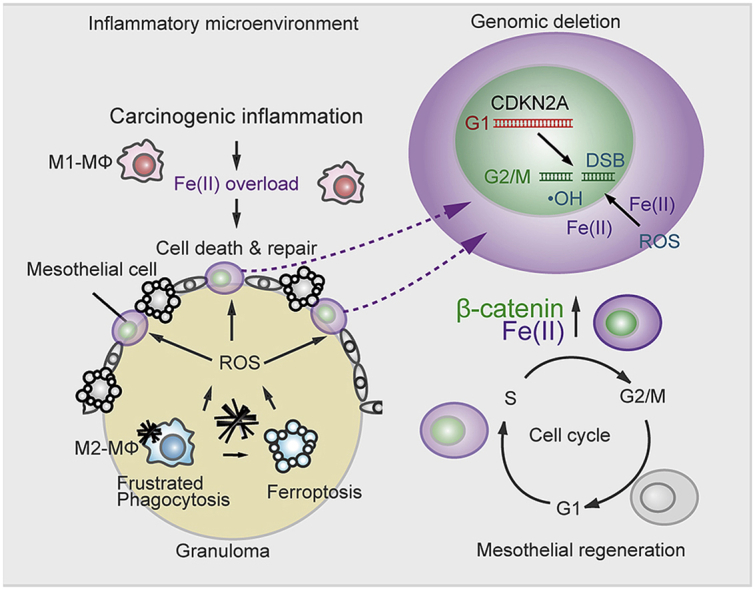
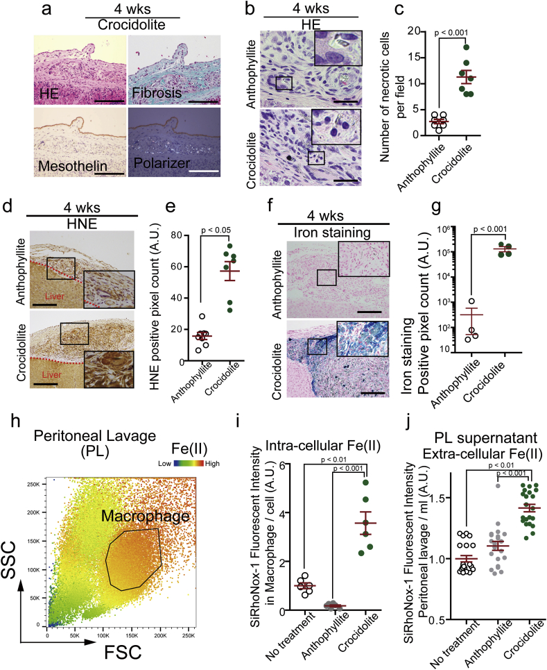
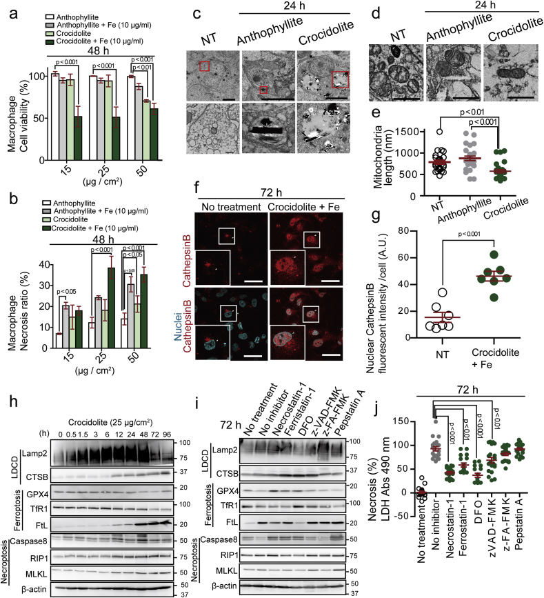
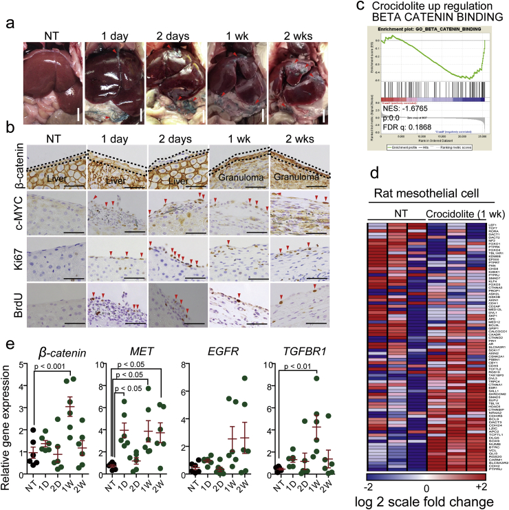
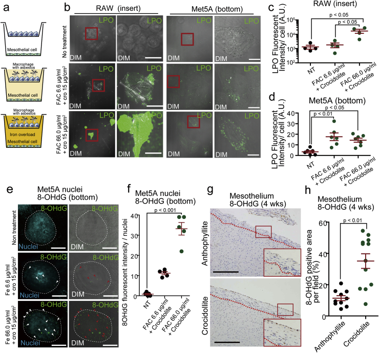
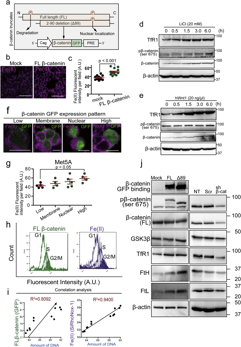
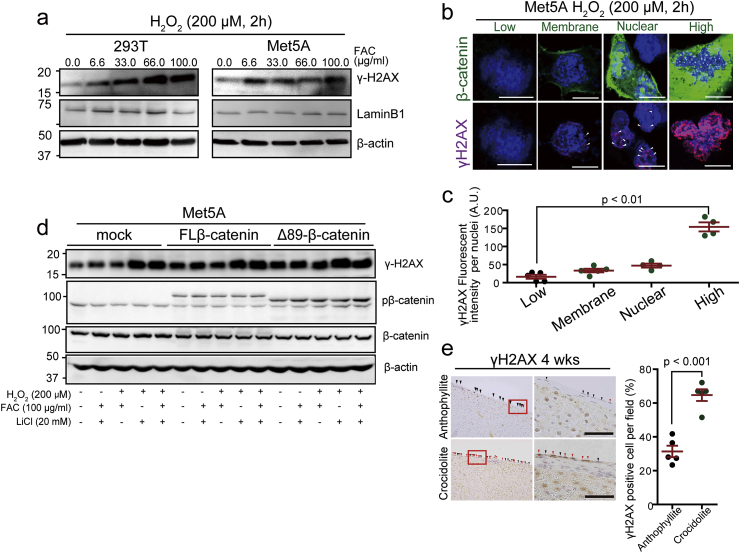
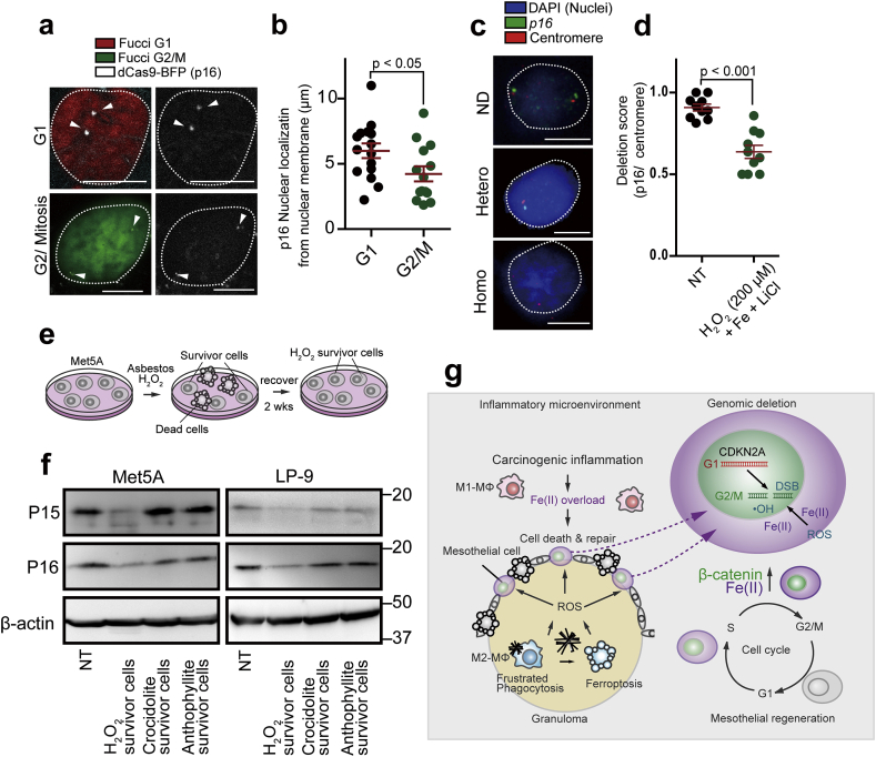
Asbestos is still a social burden worldwide as a carcinogen causing malignant mesothelioma. Whereas recent studies suggest that local iron reduction is a preventive strategy against carcinogenesis, little is known regarding the cellular and molecular mechanisms surrounding excess iron. Here by differentially using high-risk and low-risk asbestos fibers (crocidolite and anthophyllite, respectively), we identified asbestos-induced mutagenic milieu for mesothelial cells. Rat and cell experiments revealed that phagocytosis of asbestos by macrophages results in their distinctive necrotic death; initially lysosome-depenent cell death and later ferroptosis, which increase intra- and extra-cellular catalytic Fe(II). DNA damage in mesothelial cells, as assessed by 8-hydroxy-2'-deoxyguanosine and γ-H2AX, increased after crocidolite exposure during regeneration accompanied by β-catenin activation. Conversely, β-catenin overexpression in mesothelial cells induced higher intracellular catalytic Fe(II) with increased G2/M cell-cycle fraction, when p16INK4A genomic loci localized more peripherally in the nucleus. Mesothelial cells after challenge of H2O2 under β-catenin overexpression presented low p16INK4A expression with a high incidence of deletion in p16INK4A locus. Thus, crocidolite generated catalytic Fe(II)-rich mutagenic environment for mesothelial cells by necrotizing macrophages with lysosomal cell death and ferroptosis. These results suggest novel molecular strategies to prevent mesothelial carcinogenesis after asbestos exposure.

Keywords: Asbestos; Ferroptosis; Iron; Lysosomal cell death; p16(CDKN2A).


Conflict of interest statement

The authors declare no conflict of interest to present.

Figures

Image 1
Graphical abstract
Fig. 1
Fig. 1
Only high-risk asbestos generates iron-rich stromal milieu via macrophage necrosis. (a) Peritoneal histology of rat model 4 wks after intraperitoneal injection of crocidolite, a high-risk asbestos; HE, hematoxylin & eosin staining; Masson trichrome staining; polarizer detects asbestos fibers (bar = 50 μm). (b, c) Macropahge necrosis is observed only in granuloma by crocidolite but not by anthophyllite, a low-risk asbestos (bar = 20 μm). (d, e) Deposition of 4-hydroxy-2-nonenal (HNE)-modified proteins is significantly higher in the peritoneal stroma after crocidolite exposure than anthophyllite exposure. (f, g) Iron deposition by Berlin blue staining is significantly higher after crocidolite exposure than anthophyllite exposure. (h, i, j) Macrophage fraction in peritoneal lavage as well as lavage fluid shows significantly higher catalytic Fe(II) 4 wks after crocidolite exposure than anthophyllite exposure. SSC, side scatter; FSC, forward scatter; area by black line, macrophage fraction; PL, peritoneal lavage. Catalytic Fe(II) was detected by SiRhoNox-1 (means ± SEM; N ≧ 4). Refer to text for details. (For interpretation of the references to color in this figure legend, the reader is referred to the Web version of this article.)
Fig. 2
Fig. 2
Crocidolite induces macrophage necrosis via lysosome-dependent cell death at an early phase and later via ferroptosis. (a, b) Cell viability (WST) and necrosis ratio (LDH) assays reveal higher toxicity of crocidolite than anthophyllite for THP1 macropahge in the presence of excess iron. Crocidolite or anthophyllite asbestos was exposed to THP1 macrophage cells in the presence of 10 μg/ml ferric ammonium citrate (FAC) for 48 h. (c) Transmission electron microscopy of M0-state THP1 macrophage exposed to asbestos for 24 h. Crocidolite causes lysosomal rupture whereas anthopyllite fibers are confined inside lysosome (bar = 5 μm). (d, e) Transmission electron microscopy of mitochondrial damage in THP1 after crocidolite exposure. Significantly shorter mitochondria are observed (bar = 200 nm). NT, no-treatment. (f, g) Nuclear localization of lysosomal cathepsin B (CTSB) in IL-4 stimulated THP1 cells after exposure to crocidolite (25 μg/cm2) and 100 μg/ml FAC. White arrowhead, nucleus; cathepsin B, red; nucleus, cyan (bar = 50 μm). (h) Time-course immunoblot analysis reveals the involvement of ferroptosis. IL-4 stimulated THP1 was exposed to crocidolite (25 μg/cm2). (i) Immunoblot analysis of IL-4 stimulated THP1 72 h after crocidolite exposure (25 μg/cm2) in the presence of various inhibitors. Necrostatin-1; (necroptosis inhibitor, 40 μM), Ferrostain-1; DFO, deferoxamine (ferroptosis inhibitors; 500 nM and 400 μM, respectively), zVAD-FMK (apoptosis inhibitor, 184 μM), z-FA-FMK (lysosome-dependent cell death inhibitor, 20 μM), Pepstatin-A (autophagic cell death inhibitor, 20 μg/ml). (J) LDH assay in IL-4 stimulated THP1 exposed to crocidolite in the same condition as in (I) (means ± SEM; N ≧ 6). Refer to text for details. . (For interpretation of the references to color in this figure legend, the reader is referred to the Web version of this article.)
Fig. 3
Fig. 3
Mesothelial regeneration after crocidolite-induced damage accompanies activation of Wnt/β-catanin signaling pathway. (a) Intraperitoneal administration of crocidolite induces progressive peritonitis in rats. Arrowhead, crocidolite fibers. (b) Corresponding histology of peritoneum to (a) with immunohistochemistry (β-catenin, c-Myc, Ki-67 and BrdU). (c, d) Collected rat mesothelial cells were used for microarray analysis. Gene-set enrichment analysis (GSEA) revealed significant upregulation of β-catenin signaling pathway. (e) qRT-PCR analysis of selected regeneration-associated genes (β-catenin, MET, EGFR and TGFβR1) in rat mesothelial cells collected from mesentery (means ± SEM; N ≧ 4). Refer to text for details. (For interpretation of the references to color in this figure legend, the reader is referred to the Web version of this article.)
Fig. 4
Fig. 4
Macrophage ferroptosis via crocidolite causes oxidative damage in distant mesothelial cells. (a) Scheme of coculture system for Met5A mesothelial (bottom) and RAW264 macropahge (top) cells. (b–d) Macrophage ferroptosis via crocidolite in the presence of iron (FAC) induces lipid peroxidation (LPO; green color by Click-iT Lipid peroxidation kit) iron dose-dependently, which further causes lipid peroxidation in distant mesothelial cells. (e, f) In the same co-culture system, nuclear 8-hydroxy-2′-deoxyguanosine (8-OHdG) increased in an iron dose-dependent manner (8-OHdG, green; Hoechst33342, cyan; DIM, differential interference contrast microscope). (g, h) Significantly increased nuclear 8-OHdG signals in mesothelial cells in rats 4 wks after crocidolite i.p. injection in comparison to anthophyllite (means ± SEM; N ≧ 4). Refer to text for details. . (For interpretation of the references to color in this figure legend, the reader is referred to the Web version of this article.)
Fig. 5
Fig. 5
β-Catenin overexpression increases catalytic Fe(II) in mesothelial cell by modifying iron metabolism and cell cycle. (a) Scheme of β-catenin overexpression system as a fusion protein with GFP. FL, full-length; Δ89-β-catenin, N-terminal 89-amino acids were deleted for stabilization of β-catenin. PRE, post-transcriptional regulatory element. (b, c) Catalytic Fe(II) (SiRhoNox-1 fluorescent probe, magenta) was significantly increased in β-catenin overexpressed Met5A mesothelial cell. (d, e) Immunoblot analysis of iron metabolism and β-catenin; LiCl (20 mM) and hWnt1 (20 ng/ml) for β-catenin activation. (f, g) Imaging for catalytic Fe(II) and β-catenin in FL-β-catenin (GFP) overexpressed Met5A cells. Expression pattern of β-catenin was classified into 4 types as follows from low to high; low expression, (plasma) membrane localization, nuclear localization and high expression. Catalytic Fe(II) levels were β-catenin dose-dependent in (g). (h, i) FACS analysis of β-catenin (GFP; green) and catalytic Fe(II) (SiRhoNox-1, magenta). Cell cycle was separated by DNA amount (Hoechst33342, blue) into G1, S and G2/M phases in FACS analysis. G2/M phase reveals high β-catenin expression and high catalytic Fe(II). (j) Immunoblotting analysis for the association of β-catenin levels with iron metabolism in Met5A cells. β-Catenin overexpression on the left increases intracellular iron and its knockdown on the right decreases it (NT, no treatment; Scr, scrambled; sh β-cat: β-catenin knock down; means ± SEM; N ≧ 4). Refer to text for details. (For interpretation of the references to color in this figure legend, the reader is referred to the Web version of this article.)
Fig. 6
Fig. 6
β-Catenin overexpression promotes induction of DNA double-strand breaks in oxidatively stressed mesothelial cells. (a) DNA double-strand breaks evaluated by γ-H2AX in Met5A and 293T cells exposed to oxidative stress for 2 h by Fenton reaction (0–100 μg/ml FAC and 200 μM H2O2). (b, c) Association of FL-β-catenin expression with γ-H2AX in Met5A cells under exposure to H2O2 (200 μM, 2 h; green, β-catenin-GFP; magenta, γH2AX; bar = 5 μm). (d) Immunoblot analysis of γH2AX in various conditions to modify β-catenin expression and oxidative stress. Transfected Met5A cells are exposed to iron (FAC, 100 μg/ml, 2 h), LiCl (20 mM, 6 h) and/or H2O2 (200 μM, 2 h). (e) Immunohistochemistry of γH2AX in rat mesothelium on the granuloma after exposure either to anthophyllite or crocidolite (4 wks; red arrow heads, γH2AX-positive; black arrow head: γH2AX-negative; bar = 50 μm). Quantification of γH2AX positive cells on the right side (means ± SEM; N ≧ 4). . (For interpretation of the references to color in this figure legend, the reader is referred to the Web version of this article.)
Fig. 7
Fig. 7
p16INK4A loci are more nuclear peripheral at G2/M phase and its allele is lost accompanied by low p16INK4A expression after oxidative stress under high β-catenin expression. (a, b) dCas FISH analysis for p16INK4A loci in Met5A cells shows their more peripheral presence at G2/M phase in comparison to G1 phase; red, G1 phase; green, G2/M phase; white, p16INK4A locus (bar = 5 μm). (c, d) FISH analysis in rat peritoneal mesothelial cell reveals hemizygous deletion 24 h after incubation with FAC (100 μg/ml, 2 h), LiCl (20 mM, 6 h) and H2O2 (200 μM, 2 h). p16INK4A, green (FITC); chromosome No. 5 centromere, red (Dyelight 594) are detected in the nuclei (blue, DAPI). Each FISH signals were counted for deletion score; no deletion, p16INK4A/centromere ratio = ~1.0, heterozygous deletion, signals of p16INK4A/centromere ratio = ~0.5. (bar = 5 μm) (e, f) Scheme of culture system for mesothelial survivor analysis using Met5A or LP-9 human mesothelial cells. Immunoblot analysis in Met5A and LP9 cells for p15INK4B and p16INK4A after exposure of H2O2 (25 μM), asbestos (25 μg/cm2) for 3 days. After 2 wks of recovery, survived cells were used for protein detection analysis. (g) Graphical abstract illustrating the key concepts on the iron-rich stromal mutagenic milieu after exposure to crocidolite (left). Mesothelial regeneration after exposure to crocidolite accompanies β-catenin overexpression, which increases the risk of p16INK4A deletion via multiple mechanisms, such as increased G2/M phase fraction, higher intracellular catalytic Fe(II) and peripheral transfer of p16INK4A genomic loci in the nucleus (means ± SEM; N ≧ 4). (For interpretation of the references to color in this figure legend, the reader is referred to the Web version of this article.)

Similar articles

Cited by

References

    1. Gualtieri A.F., Andreozzi G.B., Tomatis M., Turci F. Iron from a geochemical viewpoint. Understanding toxicity/pathogenicity mechanisms in iron-bearing minerals with a special attention to mineral fibers. Free Radic. Biol. Med. 2019;133:21–37. - PubMed
    1. WHO, IARC . IARC Monographs on the Evaluation of Carcinogenic Risks to Humans. A Review of Human Carcinogens; Part C: Arsenic, Metals, Fibres, and Dusts, Lyon, France. 2012. Asbestos (chrysotile, amosite, crocidolite, tremolite, actinolite, and anthophyllite) pp. 219–309. - PMC - PubMed
    1. Allen L.P., Baez J., Stern M.E.C., Takahashi K., George F. Trends and the economic effect of asbestos bans and decline in asbestos consumption and production worldwide. Int. J. Environ. Res. Publ. Health. 2018;15(3):531. - PMC - PubMed
    1. Toyokuni S. Iron addiction with ferroptosis-resistance in asbestos-induced mesothelial carcinogenesis: toward the era of mesothelioma prevention. Free Radic. Biol. Med. 2019;133:206–215. - PubMed
    1. Toyokuni S. Mechanisms of asbestos-induced carcinogenesis. Nagoya J. Med. Sci. 2009;71(1-2):1–10. - PMC - PubMed

Publication types