. 2020 Sep:36:101643.
doi: 10.1016/j.redox.2020.101643. Epub 2020 Jul 13.

Metabolic reprogramming sustains cancer cell survival following extracellular matrix detachment

Affiliations

Metabolic reprogramming sustains cancer cell survival following extracellular matrix detachment

Hitoshi Endo et al. Redox Biol. 2020 Sep.

Abstract

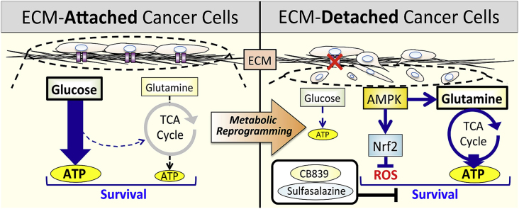
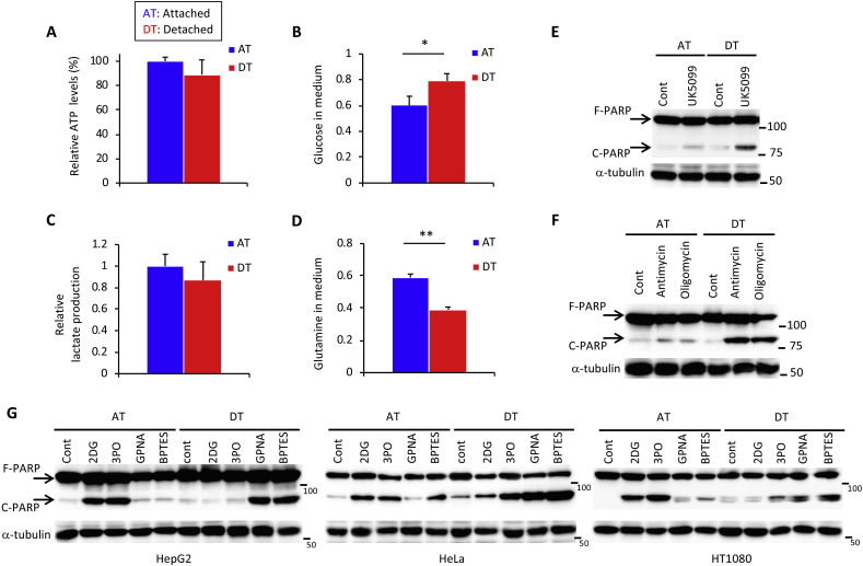
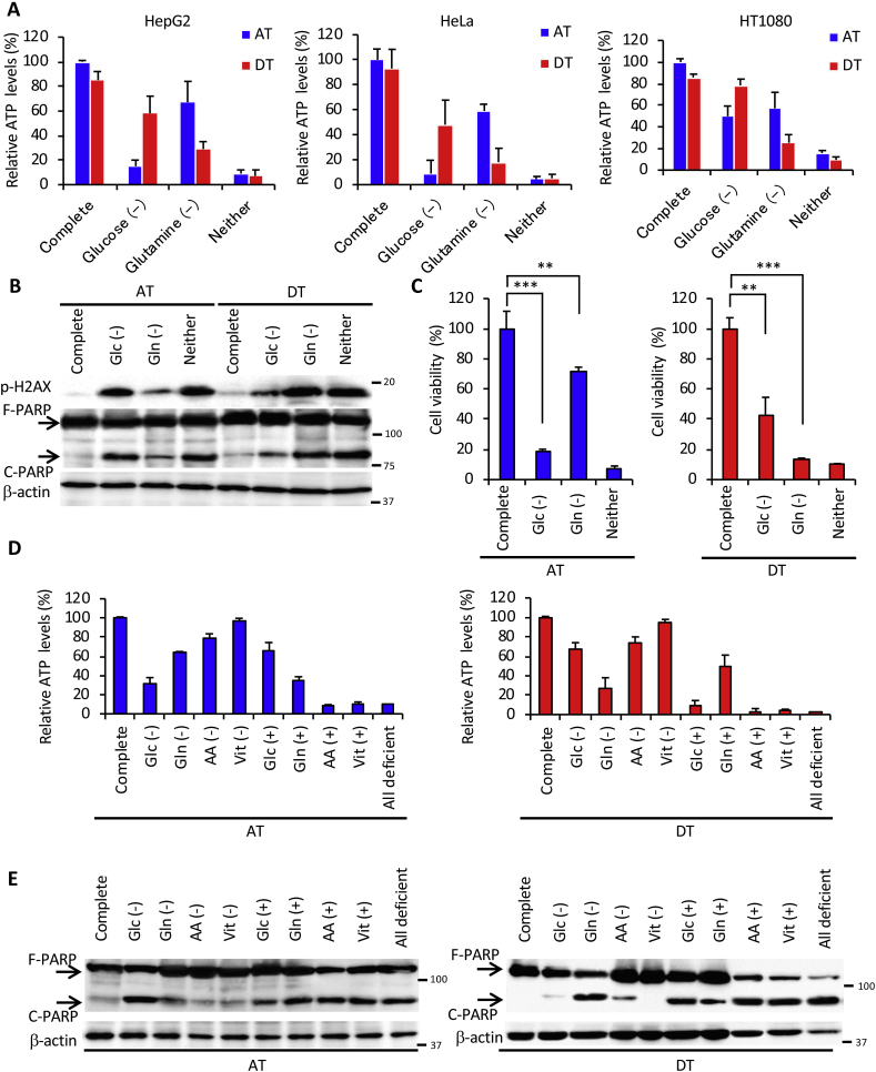
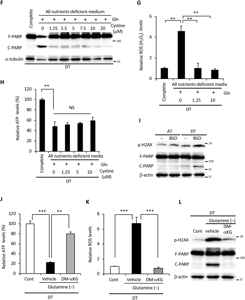
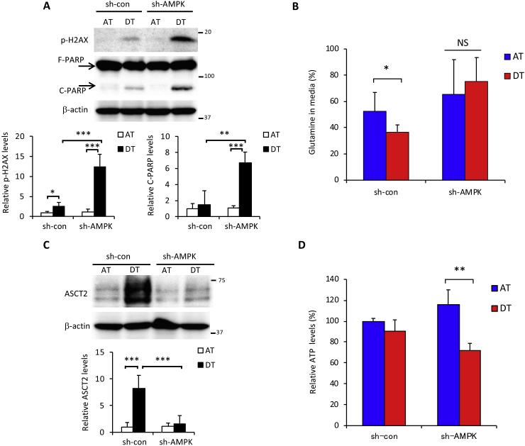
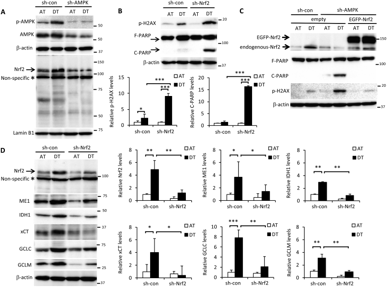
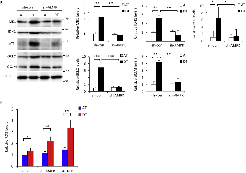
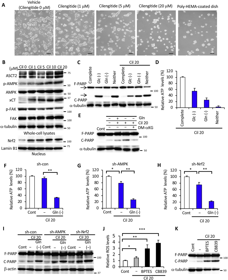
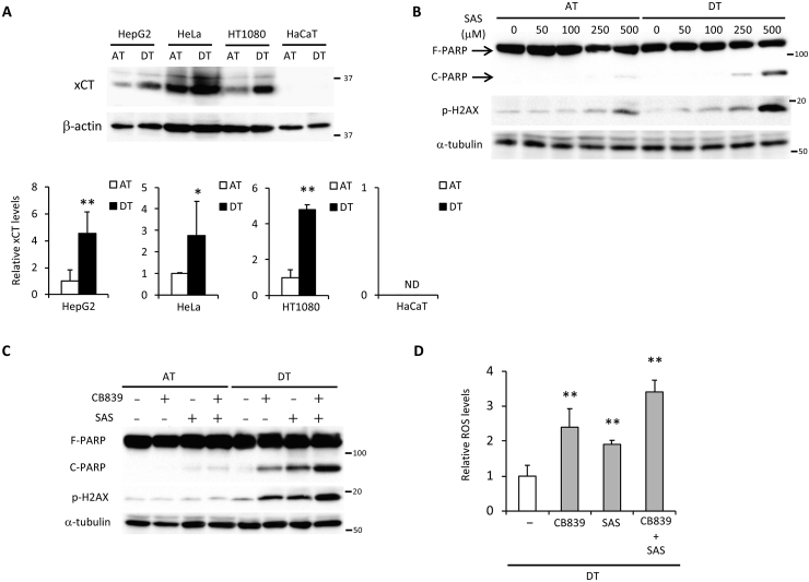
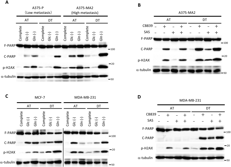
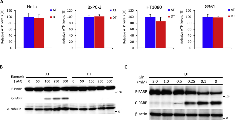
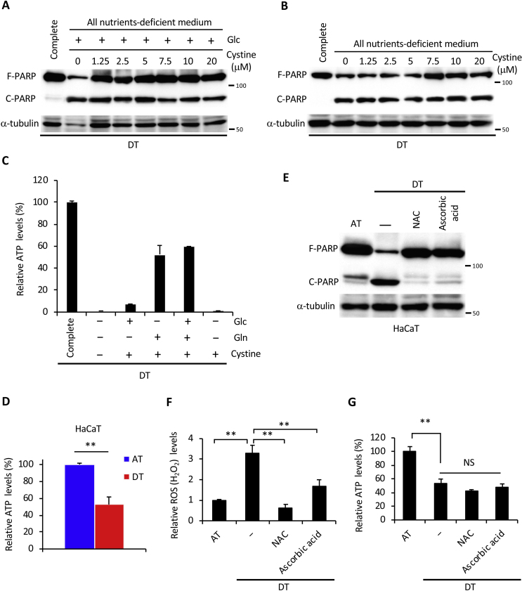
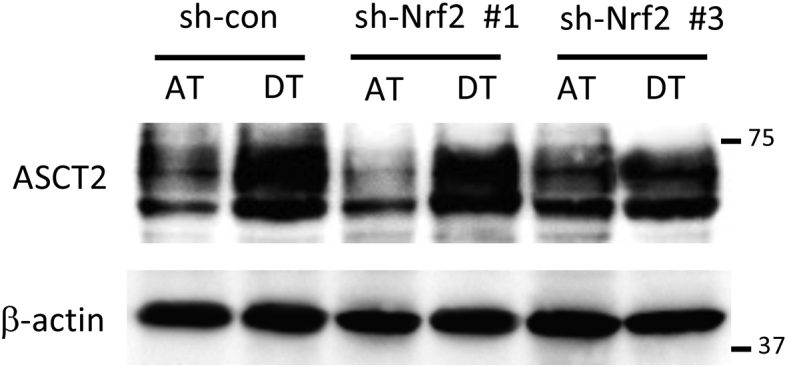
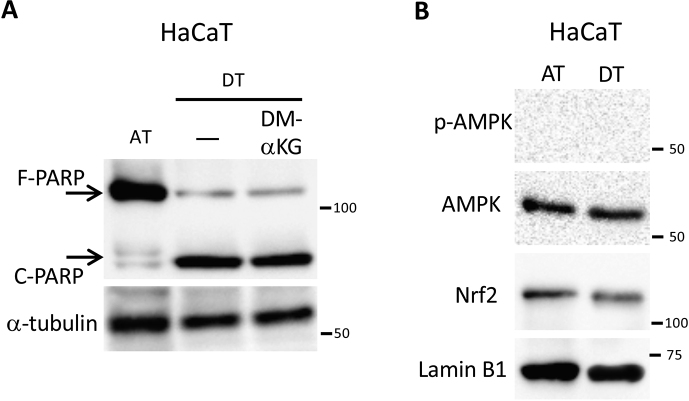
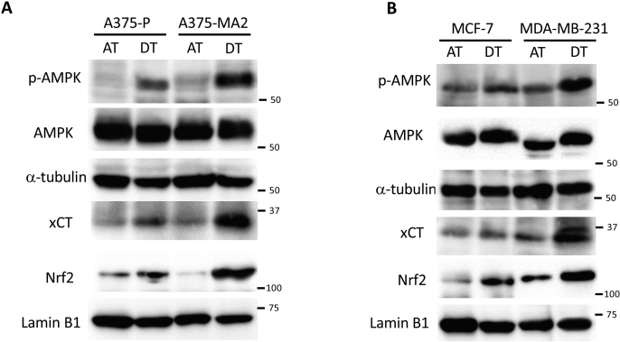
Epithelial cells require attachment to a support, such as the extracellular matrix, for survival. During cancer progression and metastasis, cancerous epithelial cells must overcome their dependence on adhesion signals. Dependence on glucose metabolism is a hallmark of cancer cells, but the nutrient requirements of cancer cells under anchorage-deficient conditions remain uncharacterized. Here, we report that cancer cells prioritize glutamine-derived tricarboxylic acid cycle energy metabolism over glycolysis to sustain anchorage-independent survival. Moreover, glutamine-dependent metabolic reprogramming is required not only to maintain ATP levels but also to suppress excessive oxidative stress through interaction with cystine. Mechanistically, AMPK, a central regulator of cellular responses to metabolic stress, participates in the induction of the expression of ASCT2, a glutamine transporter, and enhances glutamine consumption. Most interestingly, AMPK activation induces Nrf2 and its target proteins, allowing cancer cells to maintain energy homeostasis and redox status through glutaminolysis. Treatment with an integrin inhibitor was used to mimic the alterations in cell morphology and metabolic reprogramming caused by detachment. Under these conditions, cells were vulnerable to glutamine starvation or glutamine metabolism inhibitors. The observed preference for glutamine over glucose was more pronounced in aggressive cancer cell lines, and treatment with the glutaminase inhibitor, CB839, and cystine transporter inhibitor, sulfasalazine, caused strong cytotoxicity. Our data clearly show that anchorage-independent survival of cancer cells is supported mainly by glutaminolysis via the AMPK-Nrf2 signal axis. The discovery of new vulnerabilities along this route could help slow or prevent cancer progression.

Keywords: Anoikis; Extracellular matrix detachment; Glutaminolysis; Metabolic reprogramming; Metastasis.


Conflict of interest statement

The authors declare no conflict of interest.

Figures

Image 1
Graphical abstract
Fig. 1
Fig. 1
ECM detachment causes a switch from glucose-to glutamine-dependent survival mechanisms. (A) Intracellular ATP levels in HepG2 cells 24 h after plating in adherent (AT) or non-adherent (poly-HEMA-coated) (DT) plates. (B–D) Changes in glucose, lactate, and glutamine concentration in the medium of HepG2 cells after 24 h under attached or detached conditions. Data are presented as mean ± SD of three independent experiments. Full-length PARP (F-PARP) and cleaved PARP (C-PARP) expression in protein extracts obtained from HepG2 cells incubated in the presence of (E) 50 μM UK5099 and (F) 1 μM antimycin A1 or oligomycin, or in their absence (Cont) for 24 h under attached or detached conditions. (G) F-PARP and C-PARP expression in protein extracts obtained from three different cancer cell lines (HepG2, HeLa, and HT1080) incubated with 25 mM 2DG, 20 μM 3PO, 1 mM GPNA, or 20 μM BPTES, or without these chemicals (Cont) for 24 h under attached or detached conditions. α-tubulin served as a loading control. Representative western blots are shown. *P < 0.05; **P < 0.01.
Fig. 2
Fig. 2
Glutamine is indispensable for cancer cell survival under ECM detachment. (A) ATP levels in HepG2, HeLa, and HT1080 cells cultured in complete medium, glucose-deficient (Glc (-)), glutamine-deficient (Gln (-)), or glucose and glutamine-deficient (Neither) medium for 24 h under attached (AT) or detached (DT) conditions. (B) Full-length PARP (F-PARP) and cleaved PARP (C-PARP) expression in HepG2 cells cultured as indicated in panel A for 24 h. β-actin served as a loading control. (C) Viability of HepG2 cells cultured for 24 h in the indicated medium on adherent or poly-HEMA-coated plates. (D) ATP levels in HepG2 cells cultured in the indicated nutrient-deficient or supplemented medium on attachment or detachment plates for 24 h. Complete: DMEM-based complete nutrient medium; AA: DMEM-amino acid; Vit: DMEM-vitamin; All deficient: deficient for all nutrients. (E) F-PARP and C-PARP expression in HepG2 cells cultured in the indicated nutrient-deficient or supplemented medium on attachment or detachment plates for 24 h. (F) F-PARP or C-PARP expression in HepG2 cells cultured in complete medium or in all nutrients-free medium supplemented with 2 mM glutamine and the indicated doses of cystine for 24 h under detached conditions. α-tubulin served as a loading control. (G) ROS (H2O2) and (H) ATP levels in HepG2 cells cultured in complete medium or all nutrients-free medium supplemented with 2 mM glutamine and the indicated dose of cystine for 24 h in detached conditions. (I) H2AX phosphorylation (p-H2AX) and PARP expression in whole-cell extracts of HepG2 cells incubated with or without 1 mM BSO for 24 h under attached or detached conditions. β-actin served as a loading control. (J) ATP levels, (K) ROS levels, (L) H2AX phosphorylation and PARP expression in HepG2 cells cultured in glutamine-free medium in the presence (Cont) or absence (-) of glutamine, with or without 4 mM DM-αKG under detached conditions. β-actin served as a loading control. Representative western blots are shown. Data are presented as mean ± SD of three independent sets of experiments. NS, not significant; **P < 0.01; ***P < 0.001.
Fig. 2
Fig. 2
Glutamine is indispensable for cancer cell survival under ECM detachment. (A) ATP levels in HepG2, HeLa, and HT1080 cells cultured in complete medium, glucose-deficient (Glc (-)), glutamine-deficient (Gln (-)), or glucose and glutamine-deficient (Neither) medium for 24 h under attached (AT) or detached (DT) conditions. (B) Full-length PARP (F-PARP) and cleaved PARP (C-PARP) expression in HepG2 cells cultured as indicated in panel A for 24 h. β-actin served as a loading control. (C) Viability of HepG2 cells cultured for 24 h in the indicated medium on adherent or poly-HEMA-coated plates. (D) ATP levels in HepG2 cells cultured in the indicated nutrient-deficient or supplemented medium on attachment or detachment plates for 24 h. Complete: DMEM-based complete nutrient medium; AA: DMEM-amino acid; Vit: DMEM-vitamin; All deficient: deficient for all nutrients. (E) F-PARP and C-PARP expression in HepG2 cells cultured in the indicated nutrient-deficient or supplemented medium on attachment or detachment plates for 24 h. (F) F-PARP or C-PARP expression in HepG2 cells cultured in complete medium or in all nutrients-free medium supplemented with 2 mM glutamine and the indicated doses of cystine for 24 h under detached conditions. α-tubulin served as a loading control. (G) ROS (H2O2) and (H) ATP levels in HepG2 cells cultured in complete medium or all nutrients-free medium supplemented with 2 mM glutamine and the indicated dose of cystine for 24 h in detached conditions. (I) H2AX phosphorylation (p-H2AX) and PARP expression in whole-cell extracts of HepG2 cells incubated with or without 1 mM BSO for 24 h under attached or detached conditions. β-actin served as a loading control. (J) ATP levels, (K) ROS levels, (L) H2AX phosphorylation and PARP expression in HepG2 cells cultured in glutamine-free medium in the presence (Cont) or absence (-) of glutamine, with or without 4 mM DM-αKG under detached conditions. β-actin served as a loading control. Representative western blots are shown. Data are presented as mean ± SD of three independent sets of experiments. NS, not significant; **P < 0.01; ***P < 0.001.
Fig. 3
Fig. 3
AMPK is involved in glutamine metabolism upon ECM detachment. HepG2 cells stably expressing control-shRNA (sh-con) or AMPKα1-shRNA (sh-AMPK) were cultured in attachment (AT) or detachment (DT) dishes for 24 h. (A) Full-length PARP (F-PARP) and cleaved PARP (C-PARP), phosphorylated H2AX (p-H2AX) expression, and (C) ASCT2 expression in protein extracts of the above cells. β-actin served as a loading control. (B) Glutamine in culture medium and (D) intracellular ATP levels in the above cells. Representative western blots are shown. The expression levels of p-H2AX, C-PARP, and ASCT2 were measured by densitometric analysis. Data are presented as mean ± SD of three independent sets of experiments. NS, not significant; *P < 0.05; **P < 0.01; ***P < 0.001.
Fig. 4
Fig. 4
Glutamine metabolism-mediated cancer cell survival is regulated by AMPK-Nrf2 under ECM detachment. (A, B, D, E) HepG2 cells expressing sh-con or sh-AMPK were cultured in attachment (AT) or poly-HEMA-coated (DT) dishes for 24 h. Western blot analysis was performed on protein extracts of these cells with antibodies against the indicated proteins, with either β-actin or lamin B1 as loading controls. (A, D) The asterisk indicates a nonspecific band. The expression levels of protein were measured by densitometric analysis. (C) Cells stably expressing sh-con were transfected with an empty vector. Cells stably expressing sh-AMPK were transfected with either an empty vector or the EGFP-Nrf2 expression vector. Western blot analysis was performed on protein extracts of these cell lines with antibodies against the indicated proteins after culturing in adherent or poly-HEMA-coated dishes for 24 h. Representative image of western blots are shown. (F) ROS levels in HepG2 cells stably expressing sh-con, sh-AMPK, or sh-Nrf2 for 24 h after plating in adherent or poly-HEMA-coated pates for 24 h. Data are presented as mean ± SD of three independent experiments. *P < 0.05; **P < 0.01; ***P < 0.001.
Fig. 4
Fig. 4
Glutamine metabolism-mediated cancer cell survival is regulated by AMPK-Nrf2 under ECM detachment. (A, B, D, E) HepG2 cells expressing sh-con or sh-AMPK were cultured in attachment (AT) or poly-HEMA-coated (DT) dishes for 24 h. Western blot analysis was performed on protein extracts of these cells with antibodies against the indicated proteins, with either β-actin or lamin B1 as loading controls. (A, D) The asterisk indicates a nonspecific band. The expression levels of protein were measured by densitometric analysis. (C) Cells stably expressing sh-con were transfected with an empty vector. Cells stably expressing sh-AMPK were transfected with either an empty vector or the EGFP-Nrf2 expression vector. Western blot analysis was performed on protein extracts of these cell lines with antibodies against the indicated proteins after culturing in adherent or poly-HEMA-coated dishes for 24 h. Representative image of western blots are shown. (F) ROS levels in HepG2 cells stably expressing sh-con, sh-AMPK, or sh-Nrf2 for 24 h after plating in adherent or poly-HEMA-coated pates for 24 h. Data are presented as mean ± SD of three independent experiments. *P < 0.05; **P < 0.01; ***P < 0.001.
Fig. 5
Fig. 5
Loss of integrin regulates glutaminolysis in cancer cells. (A) Images obtained by phase-contrast microscopy of HepG2 cells cultured in the presence of different cilengitide (Cil) doses for 24 h in adherent culture dishes. Control cells were cultured using a poly-HEMA-coated dish without cilengitide treatment. Scale bar = 100 μm. (B) Western blot analysis of HepG2 cells treated with various doses (0–20 μM) of cilengitide for 24 h under attached conditions (unless otherwise indicated) and probed with antibodies against the indicated proteins, with either α-tubulin or lamin B1 as loading controls. (C) Full-length PARP (F-PARP) and cleaved PARP (C-PARP) expression in whole-cell lysates of HepG2 cells cultured in complete medium, glucose-free medium (Glc (-)), glutamine-free medium (Gln (-)), glucose- and glutamine-free medium (Neither), and the latter supplemented with either glucose (Glc (+)) or glutamine (Gln (+)) in the absence or presence of 20 μM cilengitide for 24 h. α-tubulin served as loading control. (D) ATP levels in HepG2 cells cultured in complete medium, glucose-deficient (Glc (-)), glutamine-deficient (Gln (-)), or glucose- and glutamine-deficient (Neither) medium in the presence of 20 μM cilengitide for 24 h. (E) F-PARP and C-PARP expression in whole-cell lysates of HepG2 cells cultured in glutamine-free medium (-) or control medium (+) for 24 h in the presence or absence of 20 μM cilengitide and 4 mM DM-αKG. α-tubulin served as loading control. (F–H) ATP levels and (I) F-PARP and C-PARP expression in HepG2 cells stably expressing sh-con, sh-AMPK, or sh-Nrf2 incubated in vehicle control medium (Cont) or glutamine-free medium (-) supplemented with 20 μM cilengitide for 24 h. β-actin served as a loading control. (J) ROS levels and (K) F-PARP and C-PARP expression in HepG2 cells treated or not with 20 μM BPTES or 10 μM CB839 in the presence of 20 μM cilengitide or in vehicle control medium (Cont) for 24 h. α-tubulin served as a loading control. Representative western blots are shown. Data are presented as mean ± SD of three independent experiments. *P < 0.05; **P < 0.01.
Fig. 6
Fig. 6
A combination of sulfasalazine and CB839 causes cell damage by increasing oxidative stress in cancer cells following ECM detachment. (A) xCT expression in HepG2, HeLa, HT1080, and HaCaT cells cultured for 24 h under attached (AT) or detached (DT) conditions. β-actin served as loading control. The expression levels of xCT were measured by densitometric analysis. (B) Full-length PARP (F-PARP), cleaved PARP (C-PARP), and phosphorylated H2AX (p-H2AX) expression in HepG2 cells treated with various doses (0–500 μM) of sulfasalazine (SAS) for 24 h under attached or detached conditions. α-tubulin served as a loading control. (C) F-PARP, C-PARP, and p-H2AX expression and (D) ROS levels in HepG2 cells treated with either 10 μM CB839 or 250 μM sulfasalazine, or both. α-tubulin served as a loading control. Representative western blots are shown. Data are presented as mean ± SD of three independent experiments. *P < 0.05; **P < 0.01.
Fig. 7
Fig. 7
Cancer cells with high metastatic potential have an increased dependence on glutamine metabolism under detached conditions. (A, C) Full-length PARP (F-PARP), cleaved PARP (C-PARP), and phosphorylated H2AX (p-H2AX) expression in whole-cell lysates of A375P, A375-MA2, MCF-7, and MDA-MB-231 cells cultured in complete medium, glucose-deficient (Glc (-)), glutamine-deficient (Gln (-)), or glucose and glutamine-deficient (Neither) medium for 24 h under attached (AT) or detached (DT) conditions. (B, D) F-PARP, C-PARP, and p-H2AX expression in whole-cell lysates of HepG2 cells treated with either 10 μM CB839 or 250 μM sulfasalazine, or both for 24 h. α-tubulin served as loading control. Data are representative of three independent sets of experiments.
figs1
figs1
figs2
figs2
figs3
figs3
figs4
figs4
figs5
figs5

References

    1. Steeg P.S. Tumor metastasis: mechanistic insights and clinical challenges. Nat. Med. 2006;12:895–904. doi: 10.1038/nm1469. - DOI - PubMed
    1. Hanahan D., Weinberg R.A. Hallmarks of cancer: the next generation. Cell. 2011;144:646–674. doi: 10.1016/j.cell.2011.02.013. - DOI - PubMed
    1. Sethi N., Kang Y. Unravelling the complexity of metastasis - molecular understanding and targeted therapies. Nat. Rev. Canc. 2011;11:735–748. doi: 10.1038/nrc3125. - DOI - PMC - PubMed
    1. Wirtz D., Konstantopoulos K., Searson P.C. The physics of cancer: the role of physical interactions and mechanical forces in metastasis. Nat. Rev. Canc. 2011;11:512–522. doi: 10.1038/nrc3080. - DOI - PMC - PubMed
    1. Guadamillas M.C., Cerezo A., Del Pozo M.A. Overcoming anoikis--pathways to anchorage-independent growth in cancer. J. Cell Sci. 2011;124:3189–3197. doi: 10.1242/jcs.072165. - DOI - PubMed

Publication types