. 2020 Sep:36:101642.
doi: 10.1016/j.redox.2020.101642. Epub 2020 Jul 9.

Angiogenic and Antiangiogenic mechanisms of high density lipoprotein from healthy subjects and coronary artery diseases patients

Affiliations

Angiogenic and Antiangiogenic mechanisms of high density lipoprotein from healthy subjects and coronary artery diseases patients

Hua-Ming Li et al. Redox Biol. 2020 Sep.

Abstract

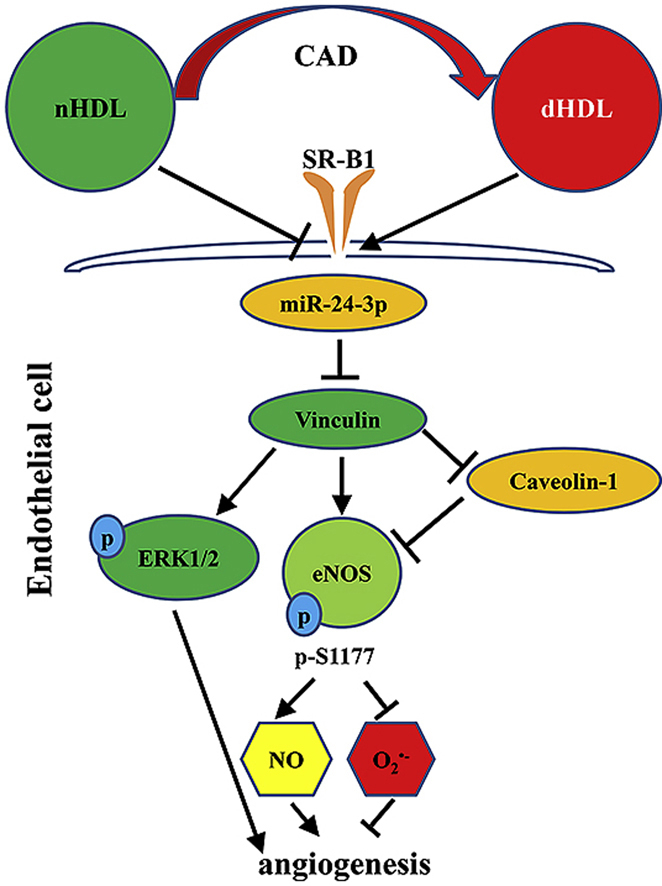
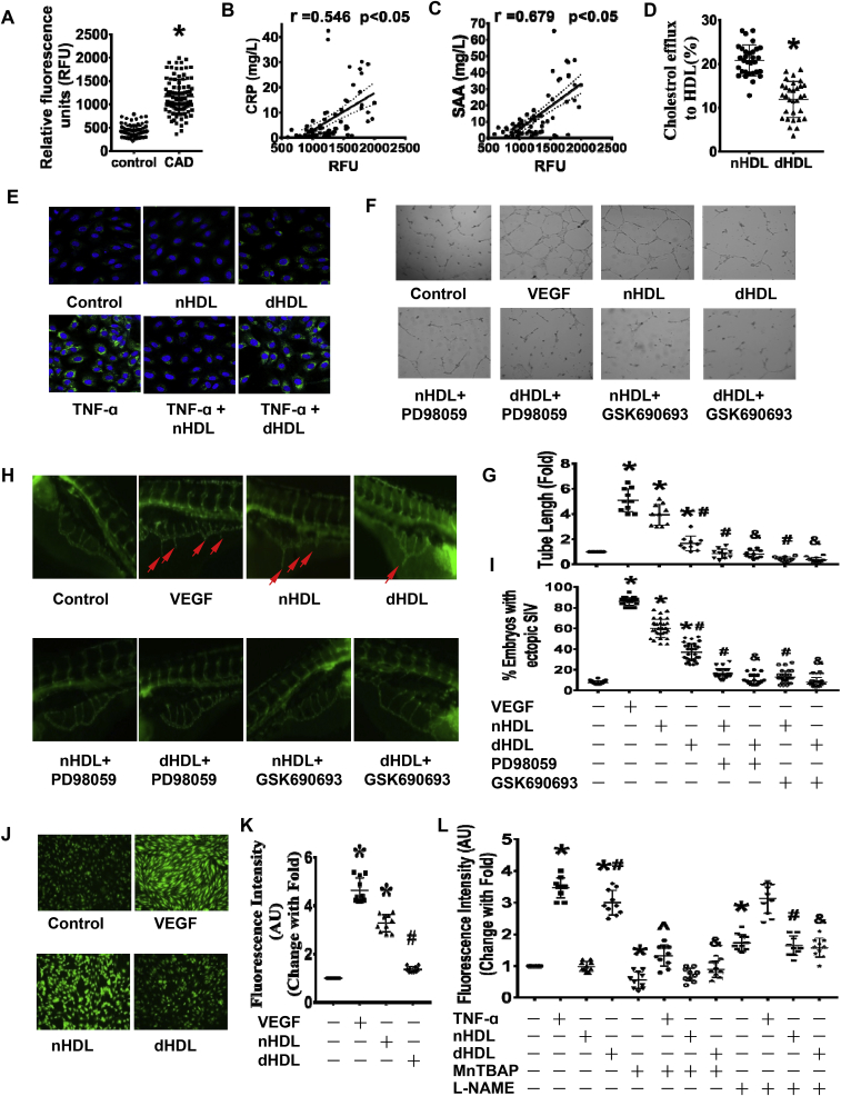
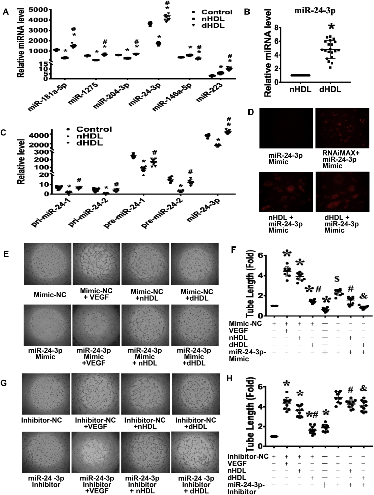
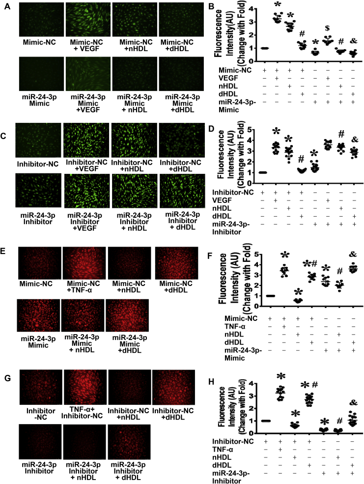
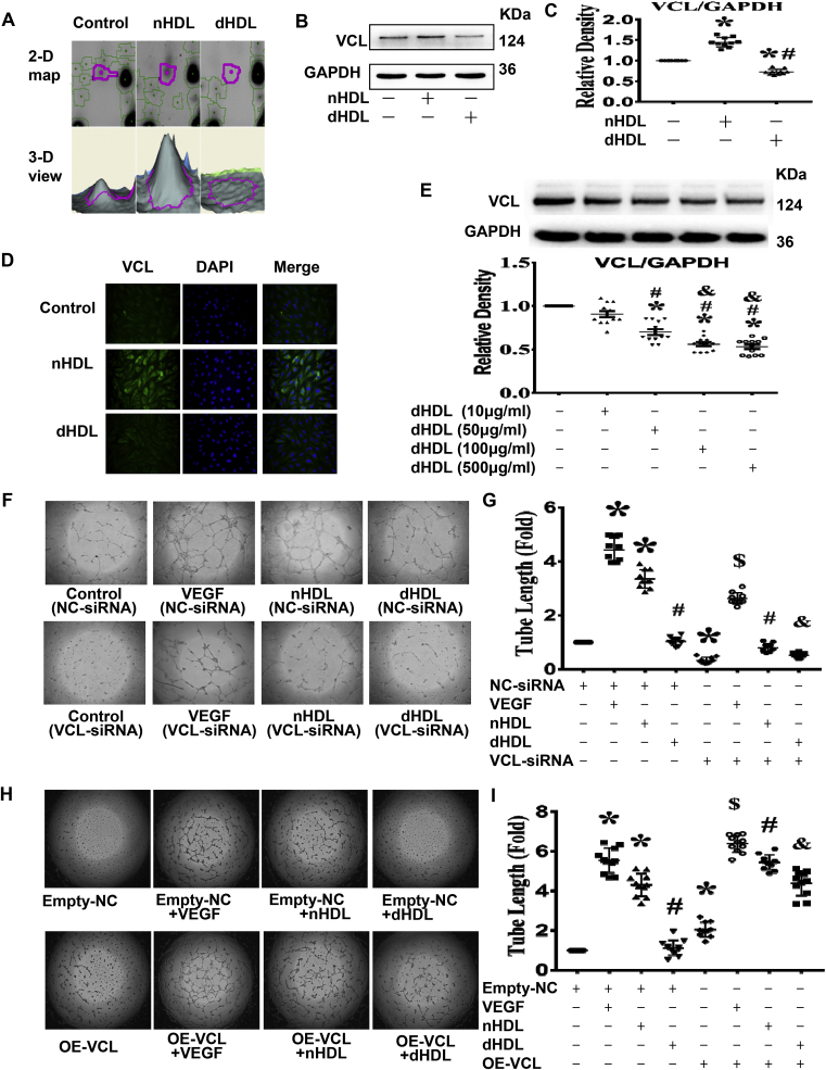
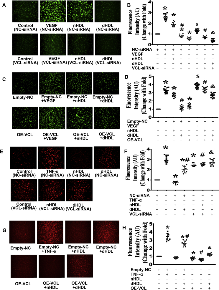
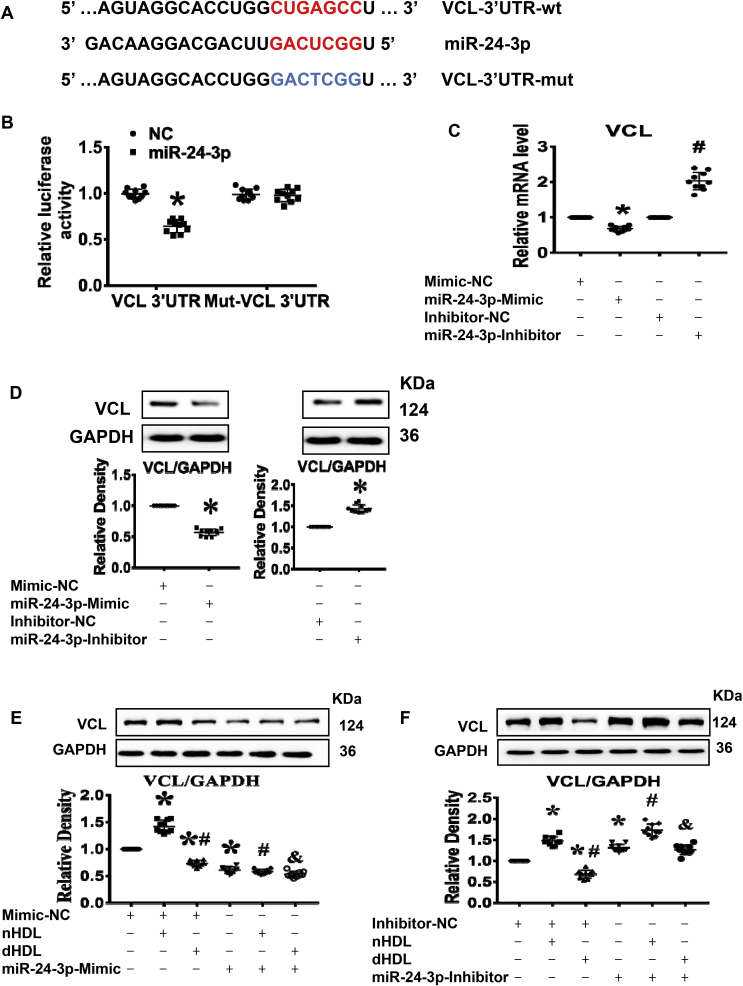
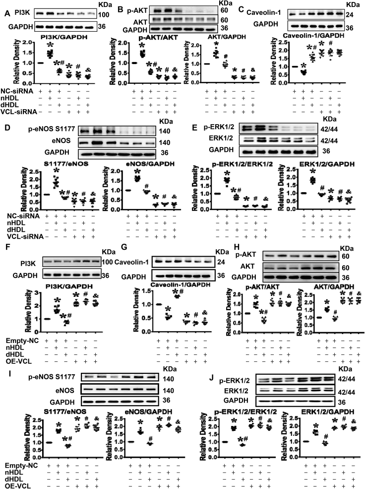
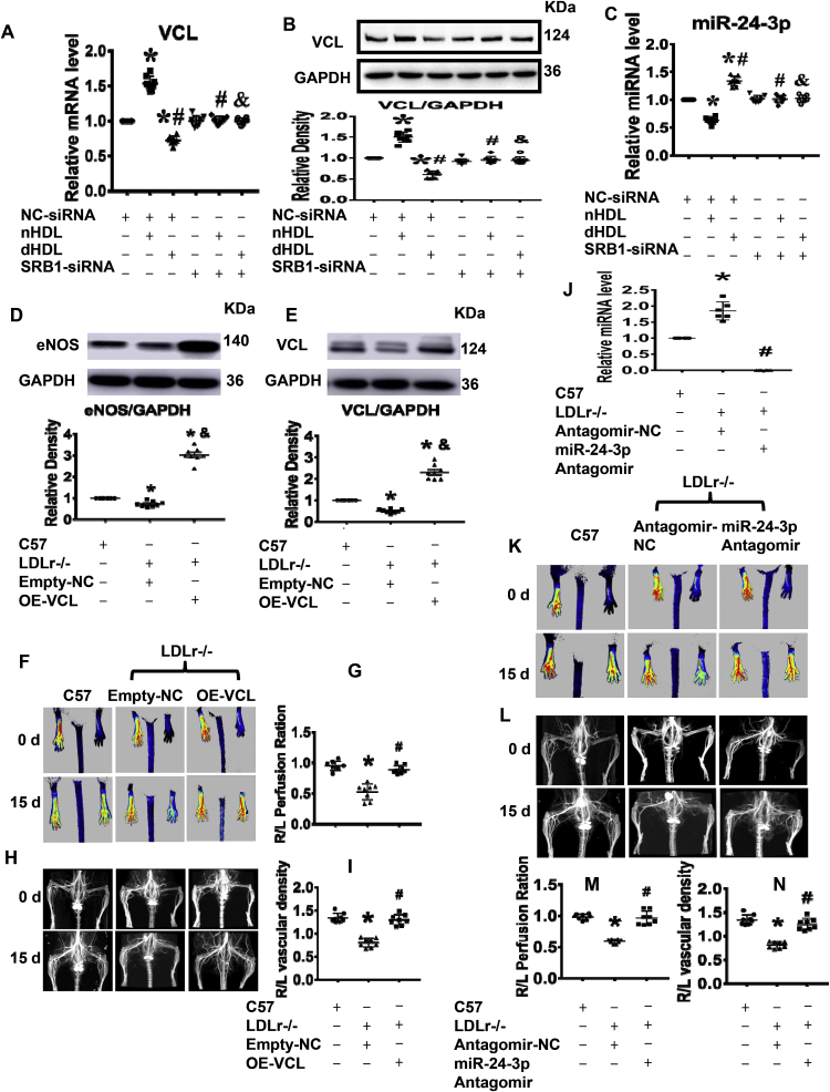
Normal high-density lipoprotein (nHDL) in normal, healthy subjects is able to promote angiogenesis, but the mechanism remains incompletely understood. HDL from patients with coronary artery disease may undergo a variety of oxidative modifications, rendering it dysfunctional; whether the angiogenic effect is mitigated by such dysfunctional HDL (dHDL) is unknown. We hypothesized that dHDL compromises angiogenesis. The angiogenic effects of nHDL and dHDL were assessed using endothelial cell culture, endothelial sprouts from cardiac tissue from C57BL/6 mice, zebrafish model for vascular growth and a model of impaired vascular growth in hypercholesterolemic low-density lipoprotein receptor null(LDLr-/-)mice. MiRNA microarray and proteomic analyses were used to determine the mechanisms. Lipid hydroperoxides were greater in dHDL than in nHDL. While nHDL stimulated angiogenesis, dHDL attenuated these responses. Protein and miRNA profiles in endothelial cells differed between nHDL and dHDL treatments. Moreover, nHDL suppressed miR-24-3p expression to increase vinculin expression resulting in nitric oxide (NO) production, whereas dHDL delivered miR-24-3p to inhibit vinculin expression leading to superoxide anion (O2•-) generation via scavenger receptor class B type 1. Vinculin was required for endothelial nitric oxide synthase (eNOS) expression and activation and modulated the PI3K/AKT/eNOS and ERK1/2 signaling pathways to regulate nHDL- and VEGF-induced angiogenesis. Vinculin overexpression or miR-24-3p inhibition reversed dHDL-impaired angiogenesis. The expressions of vinculin and eNOS and angiogenesis were decreased, but the expression of miR-24-3p and lipid hydroperoxides in HDL were increased in the ischemic lower limbs of hypercholesterolemic LDLr-/- mice. Overexpression of vinculin or miR-24-3p antagomir restored the impaired-angiogenesis in ischemic hypercholesterolemic LDLr-/- mice. Collectively, nHDL stimulated vinculin and eNOS expression to increase NO production by suppressing miR-24-3p to induce angiogenesis, whereas dHDL inhibited vinculin and eNOS expression to enhance O2•- generation by delivering miR-24-3p to impair angiogenesis, and that vinculin and miR-24-3p may be therapeutic targets for dHDL-impaired angiogenesis.

Keywords: Angiogenesis; Coronary artery disease; Endothelial nitric oxide synthase; High-density lipoprotein; Vinculin; miRNA.


Conflict of interest statement

The authors have declared that no conflict of interest exists.

Figures

Image 1
Graphical abstract
Fig. 1
Fig. 1
The characteristics and functions of HDL from healthy subjects and patients with coronary artery disease (nHDL and dHDL, respectively) and their effects on angiogenesis.(A) The levels of lipid hydroperoxide in nHDL and dHDL. *p < 0.05 (n = 95). (B and C) The correlation between levels of lipid hydroperoxide in dHDL and C reactive protein (CRP) and serum amyloid A (SAA). *p < 0.05 (n = 78). (D) The cholesterol efflux capacities of nHDL and dHDL(100 μg/ml). *p < 0.05 (n = 31). (E) Immunofluorescence assays showing the effects of nHDL and dHDL with or without pretreatment with TNF-α on VCAM-1 expression (n = 6). (FG) Images and dot plot show that nHDL stimulated endothelial cell (ECs) tube formation. The dHDL had less effect on EC tube formation. Both the ERK1/2 and AKT inhibitors impaired nHDL and dHDL-induced EC tube formation. * vs. control; # vs. nHDL; & vs. dHDL, p < 0.05 (n = 10). (H–I) The image and dot plot show that nHDL promoted angiogenesis in zebrafish. The dHDL had a lower effect on angiogenesis. Both ERK1/2 and AKT inhibitors decreased nHDL and dHDL-induced angiogenesis. * vs. control; # vs. nHDL; & vs. dHDL, p < 0.05 (n = 30). (JK) Image and dot plot show that nHDL increased EC nitric oxide (NO) production. The dHDL had a lower effect on EC NO production. AU, arbitrary units. * vs. control; # vs. nHDL, p < 0.05 (n = 10). (L) The dot plot shows that dHDL, but not nHDL, was enhanced endothelial O2 generation. l-NAME inhibited dHDL-induced O2 generation. AU, arbitrary units. * vs. control; # vs. nHDL; & vs. dHDL; vs. TNF-α, p < 0.05 (n = 10).
Fig. 2
Fig. 2
MiRNA microarray revealed that nHDL suppressed, but dHDL enhanced expression of miR-24-3p in endothelial cells (ECs), which affected tube formation.(A) The qRT-PCR confirmed that four miRNAs were downregulated, whereas one miRNA was upregulated by nHDL. In contrast, these four miRNAs were upregulated, whereas miR-146a-5p was downregulated by dHDL. dHDL increase miR-223 more than nHDL, but the levels of miR-223 in ECs is hundred folds less than other miRNAs * vs. control; # vs. nHDL, p < 0.05 (n = 8–10). (B) The level of miR-24-3p was higher in dHDL than in nHDL. *p < 0.05 (n = 20). (C) The nHDL inhibited, but dHDL slightly decreased, the expressions of pri-miR-24-1, pri-miR-24-2, pre-miR-24-1 and pre-miR-24-2 in cultured ECs. * vs. control; # vs. nHDL, p < 0.05 (n = 6–10). (D) The image showed that miR-24-3p mimic alone can not enter into ECs, but Lipofectamine® RNAiMAX, nHDL and dHDL can deliver Cy3-labeled miR-24-3p mimic to ECs. (EH) The miR-24-3p mimic inhibited EC tube formation, whereas the miR-24-3p inhibitor enhanced EC tube formation. The miR-24-3p mimic decreased VEGF and nHDL-induced EC tube formation, whereas the miR-24-3p inhibitor increased dHDL-induced EC tube formation. The miR-24-3p inhibitor further enhanced nHDL-induced EC tube formation. NC, negative control, * vs. Mimic-NC or Inhibitor-NC; # vs. Mimic-NC + nHDL or Inhibitor-NC + nHDL; & vs. Mimic-NC + dHDL or inhibitor-NC + dHDL, $ vs. Mimic-NC + VEGF, p < 0.05 (n = 10–14).
Fig. 3
Fig. 3
MiR 24-3p affected endothelial cell (EC) NO and O2•-generation induced by nHDL and dHDL. (AD) The miR-24-3p mimic inhibited EC NO production whereas the miR-24-3p inhibitor enhanced EC NO production in all groups. (EH) The miR-24-3p mimic enhanced EC O2•- generation, whereas the miR-24-3p inhibitor inhibited EC O2•- generation in all groups. TNF-α was the positive control. NC, negative control. AU, arbitrary Units. * vs. Mimic-NC or Inhibitor-NC; # vs. Mimic-NC or Inhibitor-NC + nHDL; $ vs. Mimic-NC or inhibitor-NC + VEGF; & vs. Mimic-NC or inhibitor-NC + dHDL, p < 0.05 (n = 10–14).
Fig. 4
Fig. 4
Proteomics study revealed that nHDL were increased, but dHDL decreased the expressions of vinculin (VCL) in endothelial cells (ECs), which affected tube formation.(A) Enlarged images of a 2D protein profile and 3D view in the proteomics study revealed that nHDL increased the expression of vinculin, but dHDL decreased the expression of vinculin in cultured ECs (n = 3). (B and C) Western blotting show that nHDL elevated the expression of vinculin, but dHDL decreased the expression of vinculin in cultured ECs. * vs. control; # vs nHDL, p < 0.05 (n = 10). (D) Immunofluorescence assays show that nHDL increased the expression of vinculin, but dHDL reduced the expression of vinculin in cultured ECs. The cells were positively stained with vinculin. DAPI was used to show the position of nuclei. (n = 10). (E) dHDL decreased the expression of vinculin (VCL) in a dose-dependent manner in ECs. (F–I) Images and dot plots show that the knockdown of vinculin by siRNA inhibited nHDL-induced EC tube formation. Knockdown of vinculin also inhibited EC tube formation in control- and dHDL-treated groups. Overexpression of vinculin increased EC tube formation in all group. NC, negative control; OE, overexpression. * vs. control (NC-siRNA) or Empty-NC; # vs. nHDL (NC-siRNA) or Empty-NC + nHDL; & vs. dHDL (NC-siRNA) or Empty-NC + dHDL, p < 0.05 (n = 10–14).
Fig. 5
Fig. 5
Vinculin (VCL) affected NO and O2•-generation, which were induced by nHDL and dHDL.(AD) Images and dot plots show that knockdown of vinculin inhibited NO production induced by nHDL in endothelial cells (ECs). Knockdown of vinculin impaired EC NO production in control and dHDL-treated groups. Overexpression of vinculin increased EC NO production in VEGF-, nHDL-, and dHDL-treated groups. AU, arbitrary units. * vs. control (NC-siRNA) or Empty-NC; # vs. nHDL (NC-siRNA) or Empty-NC + nHDL; & vs. dHDL (NC-siRNA) or Empty-NC + dHDL, $ vs. VEGF (NC-siRNA) or Empty-NC + VEGF, p < 0.05 (n = 10). (EH) Image and dot plots show that knockdown of vinculin enhanced EC O2•- generation in control-, nHDL- and dHDL-treated groups. Overexpression of vinculin inhibited EC O2•- generation in nHDL- and dHDL-treated groups. AU, arbitrary units; NC, negative control; OE, overexpression. * vs. control (NC-siRNA) or Empty-NC; # vs. nHDL (NC-siRNA) or Empty-NC + nHDL; & vs. dHDL (NC-siRNA) or Empty-NC + dHDL, p < 0.05 (n = 8–14).
Fig. 6
Fig. 6
Vinculin (VCL) was the direct target of miR-24-3p, which affected the expression of VCL induced by nHDL and dHDL. (A) A potential human miR-24-3p binding site in the vinculin. (B) Luciferase activity confirmed that vinculin was the direct target of miR-24-3p. NC, control 3’UTR. *vs. NC, p < 0.05 (n = 10). (C and D) The expression of vinculin was decreased by the miR-24-3p mimic but was increased by the miR-24-3p inhibitor. * vs. Mimic-NC or Inhibitor-NC, p < 0.05 (n = 10). (E and F) The miR-24-3p mimic inhibited nHDL-induced the expression of vinculin, whereas the miR-24-3p inhibitor enhanced dHDL-inhibited the expression of vinculin in cultured ECs. The miR-24-3p mimic decreased the expression of vinculin in dHDL-treated EC, whereas the miR-24-3p inhibitor further increased the expression of vinculin in nHDL-treated EC. NC, negative control; * vs. Mimic-NC or Inhibitor-NC; # vs. Mimic-NC or inhibitor-NC + nHDL; & vs. Mimic-NC or inhibitor-NC + dHDL, p < 0.05 (n = 10).
Fig. 7
Fig. 7
Vinculin (VCL) affected PI3K/AKT/eNOS and ERK1/2 signaling pathways.(AE) Immunoblots and dot plots show that knockdown of vinculin inhibited the expressions of PI3K, AKT, eNOS and ERK1/2 as well as the phosphorylations of AKT, eNOS, and ERK1/2 in cultured endothelial cells (ECs) in all groups, but increased the expression of caveolin-1 in cultured ECs in control- and nHDL-treated groups. NC, negative control, * vs. control (NC-siRNA); # vs. nHDL (NC-siRNA); & vs. dHDL (NC-siRNA), p < 0.05 (n = 10). (FJ) Immunoblots and dot plots show that overexpression of vinculin increased the expressions of PI3K, AKT, eNOS and ERK1/2 as well as the phosphorylations of AKT, eNOS and ERK1/2, but decreased the expression of caveolin-1 in cultured ECs in all groups. NC, negative control; OE, overexpression. * vs. Empty-NC; # vs. Empty-NC + nHDL; & vs. Empty-NC + dHDL, p < 0.05 (n = 6–8).
Fig. 8
Fig. 8
Vinculin and miR-24-3p affectedin vivovascular growth, and SRB1 was important for HDL-mediated expression of vinculin (VCL) and miR-24-3p.(AC) Silencing SRB1 blocked the effects of nHDL and dHDL on the expressions of vinculin and miR-24-3p on cultured human umbilical vein endothelial cells (HUVECs). *vs. control (NC-siRNA); # vs. nHDL (NC-siRNA); & vs. dHDL (NC-siRNA), p < 0.05 (n = 8). (DE) The expressions of vinculin and eNOS were decreased in gastrocnemius muscle of ischemic lower limbs of hypercholesterolemic LDLr-/- mice, but increased after overexpression of vinculin on day 15 after ischemia surgery. OE, overexpression. *vs. C57, # vs. LDLr-/-+ Empty-NC, p < 0.05 (n = 8). (F–I) Overexpression of vinculin increased blood flow and collateral in the ischemic lower limbs of hypercholesterolemic LDLr-/- mice. *vs. C57, # vs. LDLr−/− Empty-NC, p < 0.05 (n = 8). (J) The expression of miR-24-3p was increased in gastrocnemius muscle of ischemic lower limbs of hypercholesterolemic LDLr-/- mice, but decreased after injection with the miR-24-3p antagomir on day 15 after surgery for ischemia. *vs. C57, # vs. LDLr−/−, p < 0.05 (n = 8). (K–N) The miR-24-3p antagomir increased blood flow and collateral in the ischemic lower limbs of hypercholesterolemic LDLr-/- mice. *vs. C57, # vs. LDLr−/− + antagomir-NC, p < 0.05 (n = 8).

References

    1. Ansell B.J., Navab M., Hama S. Inflammatory/antiinflammatory properties of high-density lipoprotein distinguish patients from control subjects better than high-density lipoprotein cholesterol levels and are favorably affected by simvastatin treatment. Circulation. 2003;108:2751–2756. - PubMed
    1. Honda H., Hirano T., Ueda M. Associations among apolipoproteins, oxidized high-density lipoprotein and cardiovascular events in patients on hemodialysis. PLoS One. 2017;12 - PMC - PubMed
    1. Miura S., Fujino M., Matsuo Y., Kawamura A., Tanigawa H., Nishikawa H., Saku K. High density lipoprotein-induced angiogenesis requires the activation of ras/map kinase in human coronary artery endothelial cells. Arterioscler. Thromb. Vasc. Biol. 2003;23:802–808. - PubMed
    1. Seetharam D., Mineo C., Gormley A.K. High-density lipoprotein promotes endothelial cell migration and reendothelialization via scavenger receptor-b type i. Circ. Res. 2006;98:63–72. - PubMed
    1. Jin F., Hagemann N., Sun L., Wu J., Doeppner T.R., Dai Y., Hermann D.M. High-density lipoprotein (hdl) promotes angiogenesis via s1p3-dependent vegfr2 activation. Angiogenesis. 2018;21:381–394. - PubMed

Publication types