. 2020 Aug 26:20:407.
doi: 10.1186/s12935-020-01501-7. eCollection 2020.

Circ_0000140 restrains the proliferation, metastasis and glycolysis metabolism of oral squamous cell carcinoma through upregulating CDC73 via sponging miR-182-5p

Affiliations

Circ_0000140 restrains the proliferation, metastasis and glycolysis metabolism of oral squamous cell carcinoma through upregulating CDC73 via sponging miR-182-5p

Jia Guo et al. Cancer Cell Int. .

Abstract

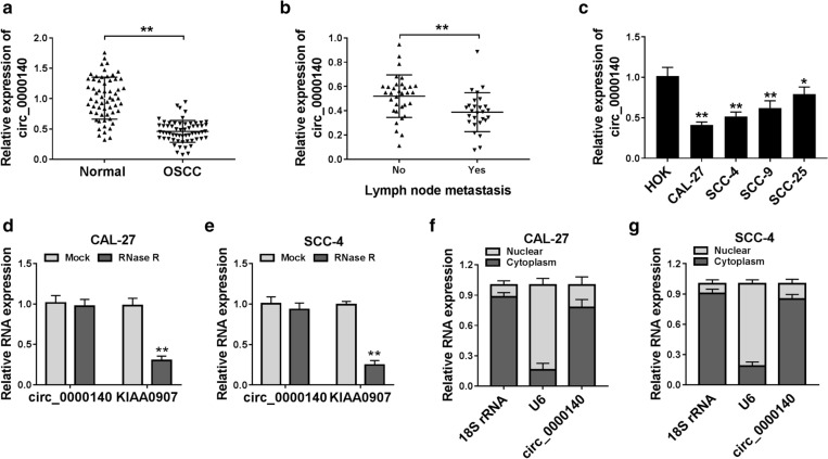
Background: Oral squamous cell carcinoma (OSCC) is a more common cancer in the world. Emerging evidence suggests that circular RNAs (circRNAs) participate in the progression of OSCC. However, the role of circ_0000140 in OSCC is still unknown.

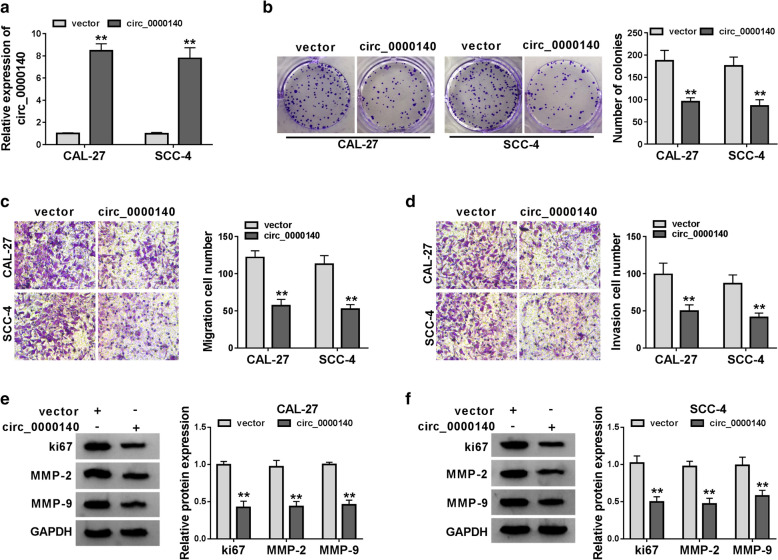
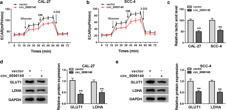
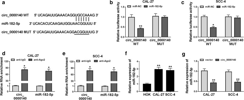
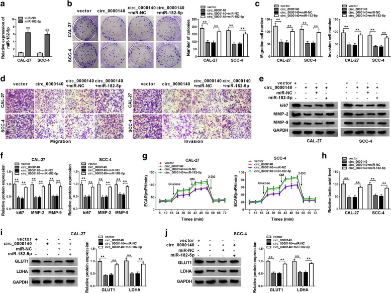
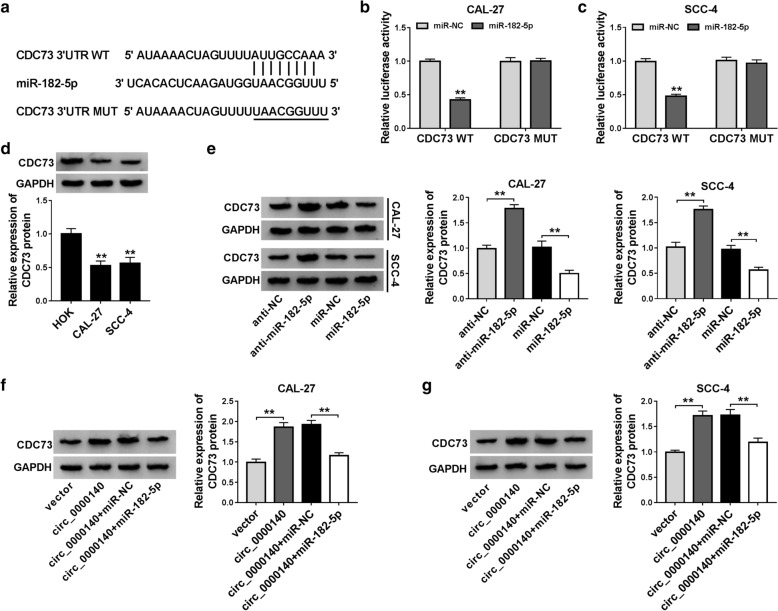
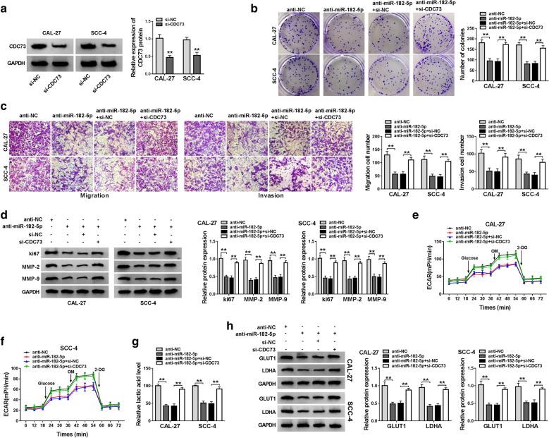
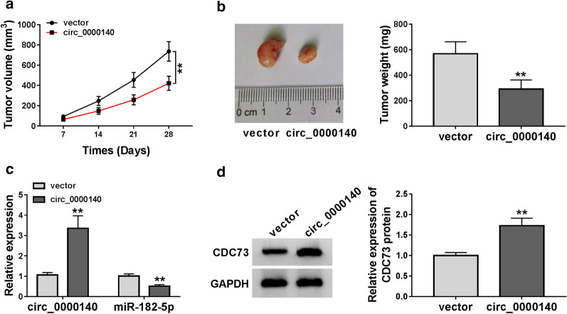
Methods: The expression of circ_0000140 and microRNA-182-5p (miR-182-5p) were assessed by quantitative real-time polymerase chain reaction (qRT-PCR). Also, cell proliferation, migration and invasion were measured by colony formation and transwell assays, respectively. Western blot (WB) analysis was used to test the levels of proliferation, metastasis and glycolysis metabolism-related proteins as well as cell division cycle 73 (CDC73) protein. Further, the extracellular acidification rate (ECAR) of cells was detected by the Seahorse XF Extracellular Flux Analyzer. The lactate acid level of cells was tested by Lactate Assay Kit. Moreover, dual-luciferase reporter was used to verify the interaction between miR-182-3p and circ_0000140 or CDC73, and RNA immunoprecipitation (RIP) assay was employed to further confirm the relationship between miR-182-3p and circ_0000140. In addition, mice xenograft models were built to measure the effect of circ_0000140 on OSCC tumor growth in vivo.

Results: Circ_0000140 was lowly expressed in OSCC, and its overexpression hindered proliferation, migration, invasion and glycolysis metabolism in OSCC cells. MiR-182-5p could be sponged by circ_0000140, and its mimic could invert the suppression of circ_0000140 overexpression on OSCC progression. CDC73 could be targeted by miR-182-3p, and its silencing could reverse the inhibition of miR-182-3p inhibitor on OSCC progression. Further, overexpressed circ_0000140 reduced the OSCC tumor growth in vivo.

Conclusions: Circ_0000140 might play an anti-cancer role in OSCC, which provided a novel target for clinical therapy of OSCC.

Keywords: CDC73; OSCC; circ_0000140; miR-182-5p.


Conflict of interest statement

Competing interestsThe authors declare that they have no competing interests.

Figures

Fig. 1
Fig. 1
The expression of circ_0000140 in OSCC tissues and cells. a The expression of circ_0000140 in OSCC tissues (OSCC) and adjacent normal mucosal tissues (Normal) was detected by qRT-PCR. b The circ_0000140 expression in OSCC tissues with or without lymph node metastasis (Yes or No) was measured by qRT-PCR. c qRT-PCR was used to test the circ_0000140 expression in OSCC cell lines (CAL-27, SCC-4, SCC-9 and SCC-25) and HOK cells. de The relative expression levels of circ_0000140 and KIAA0907 in CAL-27 and SCC-4 cells were assessed by qRT-PCR after treatment with RNase R. fg The relative expression levels of circ_0000140, U6 and 18 s rRNA in the nuclear and cytoplasmic of CAL-27 and SCC-4 cells were detected by qRT-PCR. * P < 0.05, ** P < 0.01
Fig. 2
Fig. 2
Effects of circ_0000140 overexpression on the proliferation, migration and invasion of OSCC cells. CAL-27 and SCC-4 cells were transfected with circ_0000140 overexpression vector or vector. a The expression of circ_0000140 in CAL-27 and SCC-4 cells was detected by qRT-PCR to evaluate transfection efficiency. b Colony formation assay was performed to measure the number of colonies in CAL-27 and SCC-4 cells. c, d The number of migrated and invaded CAL-27 and SCC-4 cells was determined by transwell assay. e, f WB analysis was performed to detect the protein levels of ki67, MMP-2 and MMP-9 in CAL-27 and SCC-4 cells. ** P < 0.01
Fig. 3
Fig. 3
Effects of circ_0000140 overexpression on the glycolysis metabolism of OSCC cells. CAL-27 and SCC-4 cells were transfected with circ_0000140 overexpression vector or vector. a, b The ECAR of CAL-27 and SCC-4 cells was measured by Seahorse XF Extracellular Flux Analyzer. c The lactate acid level of CAL-27 and SCC-4 cells was tested by Lactate Assay Kit. de The protein levels of GLUT1 and LDHA in CAL-27 and SCC-4 cells were detected by WB analysis. ** P < 0.01
Fig. 4
Fig. 4
Circ_0000140 could absorb miR-182-5p. a The sequences of circ_0000140 containing the miR-182-5p binding sites or mutant binding sites were presented. b, c Dual-luciferase reporter assay was used to detect the interaction between circ_0000140 and miR-182-5p in CAL-27 and SCC-4 cell. d, e The enrichment of circ_0000140 and miR-182-5p in anti-Ago2 or anti-IgG was measured by the RIP assay. f qRT-PCR was performed to measure the expression of miR-182-5p in OSCC cells (CAL-27 and SCC-4) and NOK cells. g The expression of miR-182-5p in CAL-27 and SCC-43 cells was assessed by qRT-PCR to assess the effect of circ_0000140 overexpression on miR-182-5p expression. * P < 0.05, ** P < 0.01
Fig. 5
Fig. 5
Effects of miR-182-5p mimic on the progression of OSCC cells. a The expression of miR-182-5p in CAL-27 and SCC-4 cells was measured by qRT-PCR to evaluate the transfection efficiency of miR-182-5p mimic. CAL-27 and SCC-4 cells were co-transfected with circ_0000140 overexpression vector and miR-182-5p mimic. b The number of colonies in CAL-27 and SCC-4 cells was detected by colony formation assay. c, d The number of migrated and invaded CAL-27 and SCC-4 cells was tested by transwell assay. e, f The protein levels of ki67, MMP-2 and MMP-9 in CAL-27 and SCC-4 cells were assessed by WB analysis. g Seahorse XF Extracellular Flux Analyzer was used to evaluate the ECAR of CAL-27 and SCC-4 cells. h The lactate acid level of CAL-27 and SCC-4 cells was determined by Lactate Assay Kit. i, j WB analysis was performed to measure the protein levels of GLUT1 and LDHA in CAL-27 and SCC-4 cells. ** P < 0.01
Fig. 6
Fig. 6
MiR-182-5p could target CDC73. a The sequences of CDC73 3′UTR containing the miR-182-5p binding sites or mutant binding sites were shown. b, c The interaction between miR-182-5p and CDC73 in CAL-27 and SCC-4 cells was assessed by dual-luciferase reporter assay. d The protein level of CDC73 in OSCC cells (CAL-27 and SCC-4) and HOK cells was determined by WB analysis. e WB analysis was used to measure the CDC73 protein level in CAL-27 and SCC-4 cells to evaluate the miR-182-5p expression on CRC73 expression. f, g The CDC73 protein level in CAL-27 and SCC-4 cells was detected by WB analysis to evaluate the circ_0000140 and miR-182-5p expression on CDC73 expression. ** P < 0.01
Fig. 7
Fig. 7
Effects of CDC73 silencing on the progression of OSCC cells. a The protein level of CDC73 in CAL-27 and SCC-4 cells was measured by WB analysis to evaluate the transfection efficiency of si-CDC73. CAL-27 and SCC-4 cells were co-transfected with anti-miR-182-5p and si-CDC73. b Colony formation assay was performed to assess the number of colonies in CAL-27 and SCC-4 cells. c The number of migrated and invaded CAL-27 and SCC-4 cells was detected by transwell assay. d WB analysis was used to measure the protein levels of ki67, MMP-2 and MMP-9 in CAL-27 and SCC-4 cells. e, f Seahorse XF Extracellular Flux Analyzer was employed to assess the ECAR of CAL-27 and SCC-4 cells. g The lactate acid level of CAL-27 and SCC-4 cells was detected by Lactate Assay Kit. h The protein levels of GLUT1 and LDHA in CAL-27 and SCC-4 cells were tested by WB analysis. ** P < 0.01
Fig. 8
Fig. 8
Effects of circ_0000140 overexpression on the tumor growth of OSCC in vivo. a Tumor volume was calculated at the indicated time points (7 d, 14 d, 21 d, and 28 d). b Tumor weight was measured after removed the tumors from mice. c The expression levels of circ_0000140 and miR-182-5p were tested by qRT-PCR. d WB analysis was used to assess the protein level of CDC73 in the tumors. ** P < 0.01

Similar articles

Cited by

References

    1. Sankaranarayanan R, Swaminathan R, Brenner H, Chen K, Chia KS, Chen JG, Law SC, Ahn YO, Xiang YB, Yeole BB, et al. Cancer survival in Africa, Asia, and Central America: a population-based study. Lancet Oncol. 2010;11(2):165–73. doi: 10.1016/S1470-2045(09)70335-3. - DOI - PubMed
    1. Lala M, Chirovsky D, Cheng JD, Mayawala K. Clinical outcomes with therapies for previously treated recurrent/metastatic head-and-neck squamous cell carcinoma (R/M HNSCC): a systematic literature review. Oral Oncol. 2018;84:108–20. doi: 10.1016/j.oraloncology.2018.07.005. - DOI - PubMed
    1. Brocklehurst PR, Baker SR, Speight PM. Oral cancer screening: what have we learnt and what is there still to achieve? Fut Oncol. 2010;6(2):299–304. doi: 10.2217/fon.09.163. - DOI - PubMed
    1. Sasahira T, Kirita T, Kuniyasu H. Update of molecular pathobiology in oral cancer: a review. Int J Clin Oncol. 2014;19(3):431–6. doi: 10.1007/s10147-014-0684-4. - DOI - PubMed
    1. Dos Reis PP, Bharadwaj RR, Machado J, Macmillan C, Pintilie M, Sukhai MA, Perez-Ordonez B, Gullane P, Irish J, Kamel-Reid S. Claudin 1 overexpression increases invasion and is associated with aggressive histological features in oral squamous cell carcinoma. Cancer. 2008;113(11):3169–80. doi: 10.1002/cncr.23934. - DOI - PubMed

LinkOut - more resources