. 2020 Oct;20(4):116.
doi: 10.3892/ol.2020.11977. Epub 2020 Aug 12.

Pectolinarigenin inhibits cell viability, migration and invasion and induces apoptosis via a ROS-mitochondrial apoptotic pathway in melanoma cells

Affiliations

Pectolinarigenin inhibits cell viability, migration and invasion and induces apoptosis via a ROS-mitochondrial apoptotic pathway in melanoma cells

Yuanle Deng et al. Oncol Lett. 2020 Oct.

Abstract

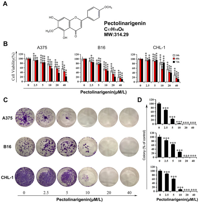
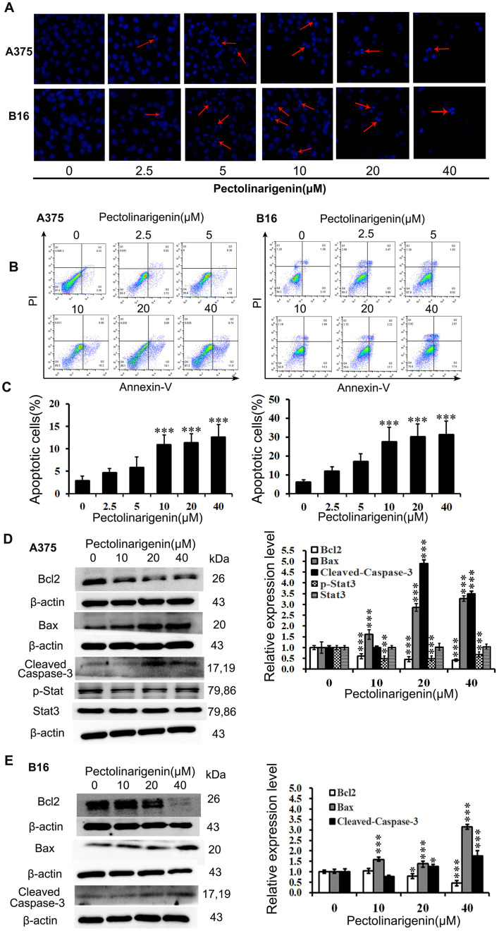
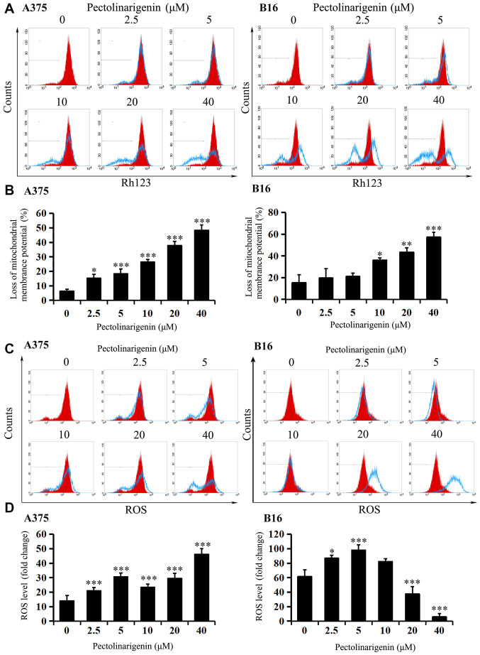
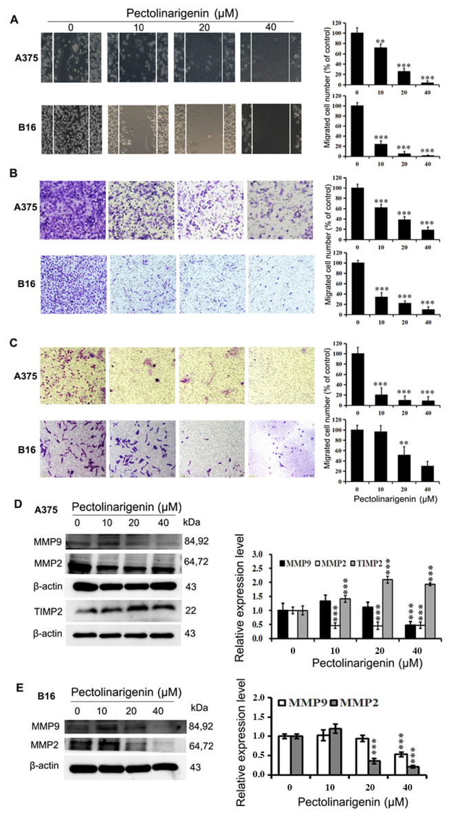
Pectolinarigenin a plant secondary metabolite that has various biological effects, including the inhibition of melanogenesis and tumor growth. Melanoma has a high degree of malignancy, with rapid metastasis and severe drug resistance, explaining the need for new candidate drugs that inhibit tumor growth and metastasis. However, the pharmacological action and mechanism of pectolinarigenin on the growth and metastasis of melanoma remain elusive. Thus, the present study aimed to investigate the role of pectolinarigenin in melanoma cell proliferation, apoptosis, migration and invasion. Apoptotic and metastasis-associated proteins were analyzed using western blotting. The results demonstrated that pectolinarigenin treatment resulted in growth inhibition and apoptosis induction in melanoma cells, arising from the loss of mitochondrial transmembrane potential, reactive oxygen species and the altered expression of apoptosis-associated proteins. In addition, wound-healing and Transwell assays demonstrated the potential of pectolinarigenin to impair the migration and invasion of melanoma cells in accordance with the changes in the expression of the associated proteins. Therefore, the results of the present study suggested that pectolinarigenin may serve a pivotal role in promoting melanoma cell apoptosis and reducing metastasis, and may thus be a promising potential candidate for an anti-melanoma treatment strategy.

Keywords: apoptosis; invasion; melanoma cell; migration; pectolinarigenin.


Figures

Figure 1.
Figure 1.
Inhibitory effects of pectolinarigenin on melanoma cell viability and colony formation. (A) Chemical structure of Pectolinarigenin. (B) Melanoma cells (A375, B16 and CHL-1) were treated with pectolinarigenin for 24, 48 and 72 h, and cell viability was tested by an MTT assay. (C) Effects of pectolinarigenin on colony formation in melanoma cell lines A375, B16 and CHL-1. (D) Statistical analyses of the inhibition in the colony formation assays are presented as the percentage of surviving colonies relative to the untreated control group. Data are presented as the mean ± SD of at least three independent experiments. *P<0.05, **P<0.01 and ***P<0.001 vs. 0 µM pectolinarigenin.
Figure 2.
Figure 2.
Pectolinarigenin induces apoptosis in A375 and B16 cells. (A) A375 and B16 cells stained with Hoechst 33258 were observed under fluorescence microscopy (×20 magnification). Red arrows indicate bright blue regions, which represent the apoptotic bodies. (B and C) The cells were exposed to the indicated concentrations of pectolinarigenin for 48 h. Apoptosis was assessed by a flow cytometric Annexin V/PI labeling assay. (D and E) Effects of pectolinarigenin on the expression levels of apoptosis-associated proteins in A375 and B16 cells were determined by western blotting. The protein band and the loading control that follows were from the different parts of the same gel, and the loading controls and protein bands were grouped together in the figure from different gels. Data are presented as the mean ± SD of at least three independent experiments. *P<0.05 and ***P<0.001 vs. 0 µM pectolinarigenin. PI, propidium iodide.
Figure 3.
Figure 3.
Changes in mitochondrial membrane potential and ROS induced by pectolinarigenin treatment in A375 and B16 cells. (A and B) A375 and B16 cells were treated with pectolinarigenin for 48 h. The changes in mitochondrial membrane potential were determined by staining with Rh123, which was detected by flow cytometry. Red represents the control group. (C and D) A375 and B16 cells treated with various concentrations of pectolinarigenin for 48 h were incubated with DCFH-DA, and the ROS levels were analyzed by flow cytometry. Red represents the control group. The results are presented as the mean ± SD of at least three independent experiments. *P<0.05, **P<0.01 and ***P<0.001 vs. 0 µM pectolinarigenin. ROS, reactive oxygen species; Rh123, rhodamine 123.
Figure 4.
Figure 4.
Pectolinarigenin decreases A375 and B16 cell migration and invasion. (A) Representative images from a light microscope (×10 magnification) of the wound healing assay using A375 and B16 cells treated with pectolinarigenin for 48 h; The lines represent the area occupied by the initial scraping, and migrated cells were counted. (B) Transwell migration assay of A375 and B16 cells upon treatment with the indicated concentration of pectolinarigenin, after which the migrated cells were stained, photographed and quantified (×20 magnification). (C) Transwell invasion assay of A375 and B16 cells upon treatment with the indicated concentration of pectolinarigenin, after which the invading cells were stained, photographed and quantified (×20 magnification). (D and E) Expression of MMP2, MMP9, and TIMP2 were determined by western blotting with β-actin as the internal control. The protein band and the loading control that follows were from the different parts of the same gel, and the loading controls and protein bands were grouped together in the figure from different gels. Data are presented as the means ± SD of at least three independent experiments. **P<0.01 and ***P<0.001 vs. 0 µM pectolinarigenin. MMP, matrix metalloproteinase; TIMP2, tissue inhibitor or metalloproteinases 2; p, phosphorylated.

References

    1. Bray F, Ferlay J, Soerjomataram I, Siegel RL, Torre LA, Jemal A. Global cancer statistics 2018: GLOBOCAN estimates of incidence and mortality worldwide for 36 cancers in 185 countries. CA Cancer J Clin. 2018;68:394–424. doi: 10.3322/caac.21492. - DOI - PubMed
    1. Siegel RL, Miller KD, Jemal A. Cancer statistics, 2018. CA Cancer J Clin. 2018;68:7–30. doi: 10.3322/caac.21442. - DOI - PubMed
    1. Zheng Y, Wang K, Wu Y, Chen Y, Chen X, Hu CW, Hu F. Pinocembrin induces ER stress mediated apoptosis and suppresses autophagy in melanoma cells. Cancer Lett. 2018;431:31–42. doi: 10.1016/j.canlet.2018.05.026. - DOI - PubMed
    1. Xiong J, Wang Y, Gu Y, Xue Y, Dang L, Li Y. CDK5RAP1 targeting NF-κB signaling pathway in human malignant melanoma A375 cell apoptosis. Oncol Lett. 2018;15:4767–4774. - PMC - PubMed
    1. Slominski A, Zmijewski MA, Pawelek J. L-tyrosine and L-dihydroxyphenylalanine as hormone-like regulators of melanocyte functions. Pigment Cell Melanoma Res. 2012;25:14–27. doi: 10.1111/j.1755-148X.2011.00898.x. - DOI - PMC - PubMed

LinkOut - more resources