. 2019;10(3):177-204.
doi: 10.1080/19475411.2019.1631899. Epub 2019 Jul 3.

Advanced 4D Bioprinting Technologies for Brain Tissue Modeling and Study

Affiliations

Advanced 4D Bioprinting Technologies for Brain Tissue Modeling and Study

Timothy J Esworthy et al. Int J Smart Nano Mater. 2019.

Abstract

Although the process by which the cortical tissues of the brain fold has been the subject of considerable study and debate over the past few decades, a single mechanistic description of the phenomenon has yet to be fully accepted. Rather, two competing explanations of cortical folding have arisen in recent years; known as the axonal tension and the differential tangential expansion models. In the present review, these two models are introduced by analyzing the computational, theoretical, materials-based, and cell studies which have yielded them. Then Four-dimensional bioprinting is presented as a powerful technology which can not only be used to test both models of cortical folding de novo, but can also be used to explore the reciprocal effects that folding associated mechanical stresses may have on neural development. Therein, the fabrication of "smart" tissue models which can accurately simulate the in vivo folding process and recapitulate physiologically relevant stresses are introduced. We also provide a general description of both cortical neurobiology as well as the cellular basis of cortical folding. Our discussion also entails an overview of both 3D and 4D bioprinting technologies, as well as a brief commentary on recent advancements in printed central nervous system tissue engineering.

Keywords: 4D Bioprinting; Brain; Cortical folding; Foliation; Gyrification; Organoids; Smart materials.


Conflict of interest statement

Disclosure Statement The authors declare no conflicts of interest.

Figures

Figure 1:
Figure 1:
Anisotropic growth mechanisms of tissue expansion and tension model of cortical folding. (a) Diagram of a neuroepithelial sheet which depicts preferential tangential expansion over radial expansion. This hypothetical anisotropic model supposes that radial rigidity, which arises from cellular processes being under tension, constrains vertical growth. (b) A proposed swelling balloon-like model neuroepithelium which sees tangential expansion further arising from outward pressure exerted from ventricular fluid and compressive surface tensions about the radial axis. (c) Neural cells (small black circles) migrate along radial glia (red circles with line-like processes) and begin to extend axonal processes. The hypothetical model proposes that as these processes reach localized targets, adjacent regions become more connected, while more remote regions drift further apart (less connected). Local tensions are hypothesized to become stronger than tensions at length between distant regions, and thus would theoretically promote tangential expansion mediated folding. Adapted with permission from [7].
Figure 2:
Figure 2:
Surface swellability leads to different cortical folding patterns. (a) Sinusoidal folding occurs when the upper layer (gray matter) is stiffer than the lower layer (white matter) of a growing bi-layer cortical model. (b) Cupsed folding occurs when the upper layer is softer than the lower layer. (c) Distinctive gyri and sulci arise when both the upper and lower layers have similar stiffnesses. (d-f) show the folding patterns of bi-layer gels which arise from differential swelling. The resultant patterns demonstrate the sinusoidal, cupsed, and gyri/sulci folding predicted by (a)-(c) respectively. (g) Elastomer model of the brain folding constructed by making a core hemisphere of radius (r) which has a shear modulus of (μ0c). The hemispheric core is coated in a thin layer of polymer with a thickness (T0) which exhibits a shear modulus of (μ0t). The top and core (or upper and lower) layers have a combined radius (R). The completed model is then submeregd in solvent and allowed to swell for time (t). When the moduli of both the top and cor layers are similar (moduli ratio μt/μc 1) the distinct gyri/sulci folding pattern from (c) arises. Adapted with permission from [17].
Figure 3:
Figure 3:
Cortical expansion simulated by swellable brain model and comparison to computer simulation of folding pattern of in vivo brain. (a) 3D printed model of the smooth human brain at gestational week 22. The 3D printed smooth brain was used to make a silicone mold, which served as a cast for the gel-brain model. The gel model was then coated with a thin, swellable layer. (b) Folding patterns arising from differential swelling of the outer layer of the gel-brain model at times t0=0 mins, t1=4 mins, t2=9 mins, t3=16 mins post-submersion in a hexane solution. (c) Computer simulation of cortical folding which arises from tangential expansion at gestational weeks 22, 26, 29, 34, and 40, and yields the stereotyped wrinkled patterns observed by adulthood. The model folding patterns at times t0-t3 in (b) highly resemble the simulated cortical wrinkling at gestational weeks 22, 26, 29, and 34 respectively in (c). Adapted with permission from [18].
Figure 4:
Figure 4:
Brain organoid-on-a-chip model of cortical folding. (a) Schematic of neural organoid-on-a-chip system where the organoid is compressed between a coverslip and a media permeable membrane. (t) represents the organoid’s thickness while (h) represents the distance between the coverslip and the membrane, which is equal to 150μm. (b) Side-view (Z-stack) of organoid with Lifeact-GFP stained actin (green) and H2B-mCherry stained nuclei (red). (c) Development of organoid wrinkling from days 3–11 (arrows denote initial surface instability which yielded further formation of wrinkles). (d) Top-down view of neural organoid showing bi-polar morphology and nuclear distribution. Organoid has an inner surface (r=0) and an outer surface (r=t). (e) Outer layer wrinkling arises as the nuclear density (ρ) exceeds a critical density threshold (ρc), ρ>ρc. (f) Linear relationship between thickness (t) and wrinkle wavelength (λ). (g) Relationship between nuclear density ρ<a> and Wrinkling index. A critical nuclear density ρc=0.85±0.1<a> shows a notable increase in wrinkle formation. Adapted with permission from [22].
Figure 5:
Figure 5:
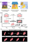
A 4D printed thermally sensitive natural soybean oil epoxidized acrylate (SOEA) constructs developed in our lab. (a) A tandem photolithography-stereolithography process to fabricate heart-shaped constructs from novel soybean oil epoxidized acrylate. (b) Schematic illustration of the 4D shape memory process triggered by temperature. (c) Rolled heart-shaped SOEA constructs can be affixed into flat temporary shapes at −18°C and can fully recover their original shape at 37 °C. Scale bar, 2 mm. Adapted with permission from [80].
Figure 6:
Figure 6:
Extrusion-based bioprinted 6-layered model of the Cerebral cortex in RGD-peptide modified Gellan-gum (GG). (a) Solidworks design of 6-layered Cerebral cortex model. (b) Layer-by-layer extrusion printing of cortical model using RGD-peptide modified Gellan Gum as a bioink. (c) Distribution of printed cortical neurons throughout the layers of the construct. Construct layers alternate having incorporated cells or containing no cells. (d) Blow-out image of square area highlighted in (c) which shows an axonal projection penetrating into a cell-devoid layer. For (c) & (d), the scale bar is 100μm. Adapted with permission from [59].
Figure 7:
Figure 7:
4D bioprinting novel SOEA constructs. (a) Bird-like architectures fabricated from SOEA modified with graphene can achieve a “flying” shape change by varying the graphene concentration (ranges tested 0–0.8%). (b) 4D bioprinted nerve guidance conduits with and without the addition of graphene. (c) Schematic of the self-entubulating nerve conduit being grafted onto the terminals of a damaged nerve (I-IV). The nerve conduit is placed over the damaged nerve stumps in its flattened temporary shape, but will fully cover the nerve in a self-entubulation/ wrapping process at 37°C. Nerve damage model ensheathed by the 4D printed nanohybrid conduit. (d) Immunofluorescence staining of hMSCs cultured on both the nanohybrid and uv cured nerve conduits. The printed conduit demonstrated significantly greater cell alignment than the non-printed UV cured conduit. (e) Photo images of a reversible shape change process with 4D printed flower structure which can open in ethanol and close in water. (f) Beyond 4D printing – shape memory effect with the 4D printed flower structure. Scale bar, 2 mm. Adapted with permission from [78]

Similar articles

Cited by

References

    1. Armstrong E, et al. (1995). “The Ontogeny of Human Gyrification.” Cerebral Cortex 5(1): 56–63. - PubMed
    1. Sekiguchi M, et al. (1991). “Abnormalities of foliation and neuronal position in the cerebellum of NZB/BINJ mouse.” Developmental Brain Research 64(1): 189–195. - PubMed
    1. Wallace Gregory L.; Robustelli Briana; Dankner Nathan; Kenworthy Lauren; Giedd Jay N.; Martin Alex (2013-June-01). “Increased gyrification, but comparable surface area in adolescents with autism spectrum disorders”. Brain. 136 (6): 1956–1967. doi:10.1093/brain/awt106. ISSN 0006–8950. PMC 3673467 Freely accessible. PMID 23715094. - DOI - PMC - PubMed
    1. Das T, Borgwardt S, Hauke DJ, et al. Disorganized Gyrification Network Properties During the Transition to Psychosis. JAMA Psychiatry. 2018;75(6):613–622. doi:10.1001/jamapsychiatry.2018.0391 - DOI - PMC - PubMed
    1. Harris JM, et al. (2007). “Increased Prefrontal Gyrification in a Large High-Risk Cohort Characterizes Those Who Develop Schizophrenia and Reflects Abnormal Prefrontal Development.” Biological Psychiatry 62(7): 722–729. - PubMed

LinkOut - more resources