. 2020 Aug 17:9:140.
doi: 10.1038/s41377-020-00375-8. eCollection 2020.

Dynamic full-field optical coherence tomography: 3D live-imaging of retinal organoids

Affiliations

Dynamic full-field optical coherence tomography: 3D live-imaging of retinal organoids

Jules Scholler et al. Light Sci Appl. .

Abstract

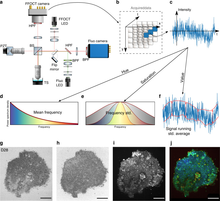
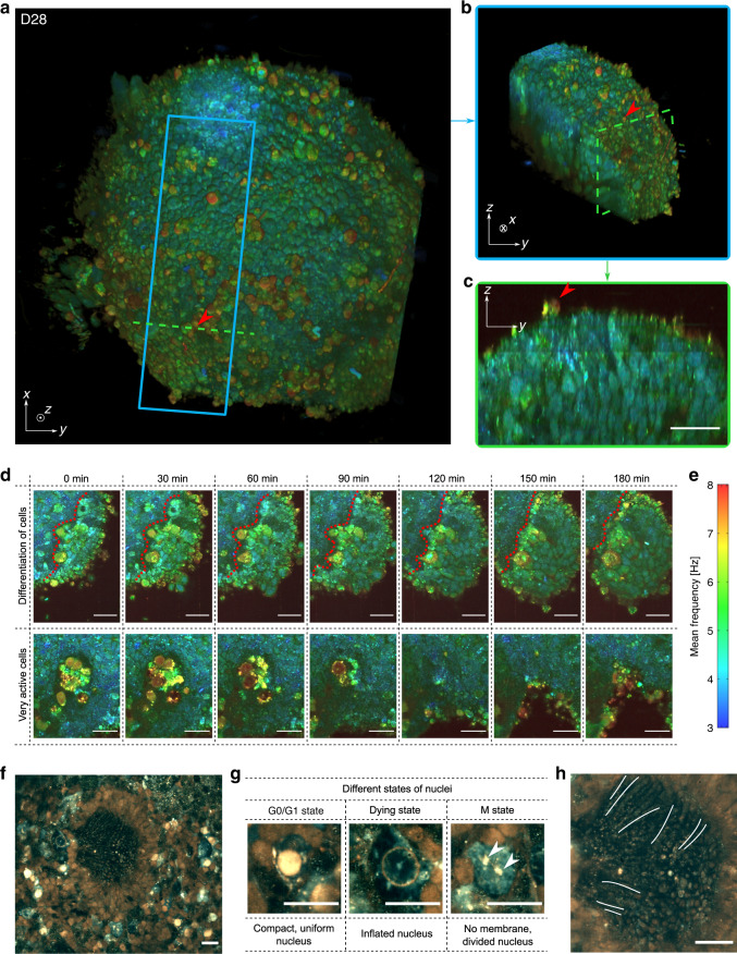
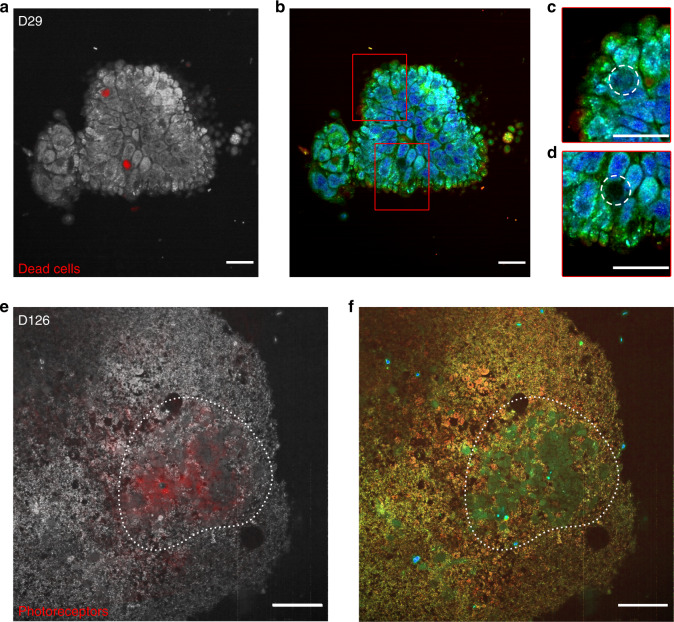
Optical coherence tomography offers astounding opportunities to image the complex structure of living tissue but lacks functional information. We present dynamic full-field optical coherence tomography as a technique to noninvasively image living human induced pluripotent stem cell-derived retinal organoids. Coloured images with an endogenous contrast linked to organelle motility are generated, with submicrometre spatial resolution and millisecond temporal resolution, creating a way to identify specific cell types in living tissue via their function.

Keywords: Imaging and sensing; Interference microscopy; Wide-field fluorescence microscopy.


Conflict of interest statement

Conflict of interestThe authors declare that they have no conflict of interest.

Figures

Fig. 1
Fig. 1. Experimental setup and post-processing schematic.
Acquisition of images a, Full-field OCT setup combined with wide-field fluorescence microscopy (side view). PZT piezoelectric translation, TS translation stage, BPF bandpass filter, HPF high pass filter. As fluorescence is recorded in the same spectral range as that used for D-FFOCT, D-FFOCT and fluorescence images could not be recorded at the same time; for this purpose, a flip mirror was added to switch from one modality to another. b 3D cube of data (x, y, t) acquired before processing. Each time evolution of a pixel is processed independently. c An intensity trace is plotted for a pixel inside a living retinal organoid. Post-processing steps df. Dynamic images are computed in the HSV colour space. d Hue is computed with the mean frequency, from blue (low temporal frequencies) to red (high temporal frequencies). e Saturation is computed as the inverse of the frequency bandwidth; as a consequence, a signal with a broader bandwidth (e.g., white noise) appears dull, whereas a signal with narrow bandwidth appears vivid. f The value is computed as the running standard deviation. Bottom row is a D29 retinal organoid. g Computation of the mean frequency (hue); h, frequency bandwidth (saturation); and i, dynamic (value) before j, reconstruction. Scale bar: 50 µm
Fig. 2
Fig. 2. Imaging hiPSC-derived retinal organoids with D-FFOCT.
a 3D reconstruction of the spherical D28 retinal organoid, composed of cells ~5 µm in diameter. Red arrows highlight surface cells exhibiting fast dynamics. b Image represents a sub-volume of a (blue square). c Image represents a cross-section in (a) (green dashed line) in which one can see the organization of the layers inside the retinal organoid. d High-magnification images of two different areas of the organoid during a 3h time-lapse acquisition: magnified images in the top row show the change in dynamic profile that could reflect a differentiation process (the boundary between the two types of cells is represented by a red dotted line); images in the bottom row show a very active zone composed of cells exhibiting fast and high dynamics, possibly undergoing apoptosis, in the centre of the organoid. e Colour bar of the D-FFOCT images for the 3D and time-lapse acquisitions with a consistent colormap for (ad). High-temporal-resolution imaging performed on a D147 retinal organoid. f Part of the retinal organoid revealing fusiform structures corresponding to emerging photoreceptor outer segments in the centre of the rosette. g Magnified view of nuclei in three different states around the rosette: (i) a nucleus in a normal state with a compact, uniform shape and is very bright (i.e., exhibiting a high activity); (ii) an seemingly dying, inflated nucleus, exhibiting almost no activity; and (iii) a nucleus undergoing division with no defined nuclear membrane in the cytoplasm, and two distinct parts (white arrows) of the content of a nucleus (suggesting mitosis of the nucleus with chromosomes already divided, with the same subcellular activity level as the “normal” nucleus). h Magnified image of the photoreceptor outer segment-like structures imaged side-on; three of them are marked with a white line. Scale bar: 20 µm
Fig. 3
Fig. 3. Fluorescence validation of D-FFOCT imaging.
D-FFOCT images are depicted in colour (bd, f), D-FFOCT images overlaid with wide-field fluorescence images are depicted in greyscale (D-FFOCT) and red (fluorescence) (a, e). ad D29 retinal organoid labelled with dye targeting the nuclei of dead cells. a One can see two dead cells marked by the red spots, corresponding to the two dark zones on (b), the D-FFOCT image, in which there is no dynamic signal in these zones. c, d Magnified images of the two dark zones (highlighted by a white dashed line, corresponding to the two red spots of fluorescence). e, f D126 retinal organoid derived from a fluorescent cone rod homeobox (CRX) reporter iPSC line exclusively labelling photoreceptors in red (mCherry). e Overlaid image on which the photoreceptor fluorescence matches the blue-green cells of (f), the D-FFOCT image. These areas are highlighted by a white dotted line. These precursors of photoreceptors have their own particular dynamic signature, which allows them to be distinguished from the surrounding cells by D-FFOCT alone. Scale bar: (ad) 10 µm, (e, f) 50 µm

References

    1. Hajdu SI. The first use of the microscope in medicine. Ann. Clin. Lab. Sci. 2002;32:309–310. - PubMed
    1. Mazzarello P. A unifying concept: the history of cell theory. Nat. Cell Biol. 1999;1:E13–E15. doi: 10.1038/8964. - DOI - PubMed
    1. Jedrzejczak-Silicka, M. History of cell culture in New Insights into Cell Culture Technology. ed. Sivakumar Joghi Thatha Gowder, IntechOpen, (2017).
    1. Hynds RE, Giangreco A. Concise review: the relevance of human stem cell-derived organoid models for epithelial translational medicine. Stem Cells. 2013;31:417–422. doi: 10.1002/stem.1290. - DOI - PMC - PubMed
    1. Lancaster MA, Knoblich JA. Organogenesis in a dish: modeling development and disease using organoid technologies. Science. 2014;345:1247125. doi: 10.1126/science.1247125. - DOI - PubMed