. 2019 Mar;3(1):167-185.
doi: 10.1146/annurev-cancerbio-030617-050407. Epub 2018 Nov 28.

Aberrant RNA Splicing in Cancer

Affiliations

Aberrant RNA Splicing in Cancer

Luisa Escobar-Hoyos et al. Annu Rev Cancer Biol. 2019 Mar.

Abstract

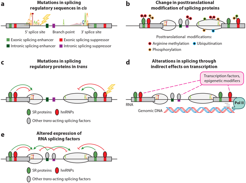
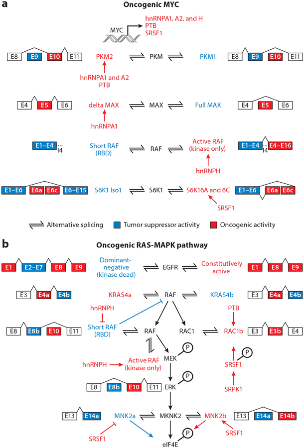
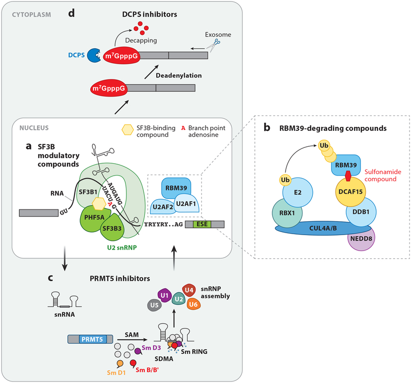
RNA splicing, the enzymatic process of removing segments of premature RNA to produce mature RNA, is a key mediator of proteome diversity and regulator of gene expression. Increased systematic sequencing of the genome and transcriptome of cancers has identified a variety of means by which RNA splicing is altered in cancer relative to normal cells. These findings, in combination with the discovery of recurrent change-of-function mutations in splicing factors in a variety of cancers, suggest that alterations in splicing are drivers of tumorigenesis. Greater characterization of altered splicing in cancer parallels increasing efforts to pharmacologically perturb splicing and early-phase clinical development of small molecules that disrupt splicing in patients with cancer. Here we review recent studies of global changes in splicing in cancer, splicing regulation of mitogenic pathways critical in cancer transformation, and efforts to therapeutically target splicing in cancer.

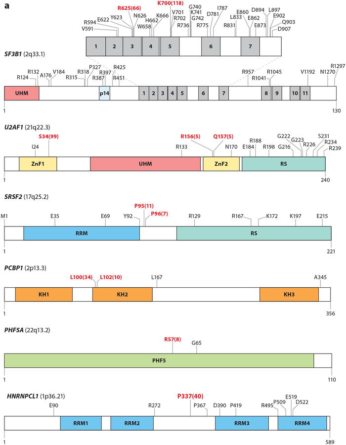
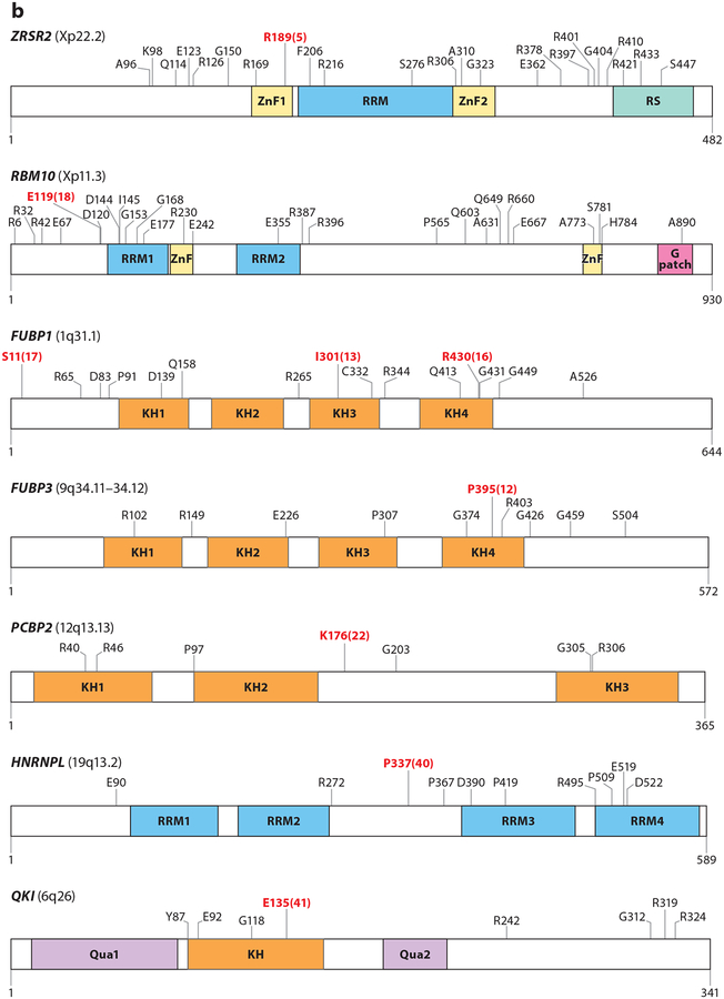
Keywords: RNA; SF3B1; SRSF2; U2AF1; ZRSR2; splicing.


Figures

Figure 1
Figure 1
Means by which RNA splicing is altered in cancer. As shown, genetic alterations affecting (a) critical sequences required for splicing of genes in cis have been described at splice sites, branch points, and splicing enhancers and silencers within exons and introns. In addition, altered (b) posttranslational modification of splicing proteins as well as (c) change-of-function and loss-of-function mutations in RNA splicing factors themselves occur in a variety of cancers. Given that splicing occurs cotranscriptionally, processes that (d) modify the transcription rate of RNA polymerase II (Pol II) may modify splicing. Finally, (e) altered expression of RNA splicing factors has been demonstrated to play a pathogenic role in a variety of cancers. Abbreviations: hnRNPs, heterogeneous nuclear ribonucleoproteins; SR proteins, serine- and arginine-rich proteins.
Figure 2
Figure 2
Mutations in RNA splicing factors in cancer. For each protein, mutations occurring in at least three samples are annotated, with the exceptions of FUBP1 and RBM10, where mutations occurring in at least four samples are annotated. Residues in red represent the most frequently reported hot spot mutations, and the total numbers of observed occurrences are displayed in parentheses. Residue and frequency data were mined from cBioPortal (Cerami et al. 2012) and the image was created with DOG 1.0 (Ren et al. 2009). Mutations in proteins in panel a are commonly thought to induce change-of-function mutations, whereas mutations in proteins in panel b typically result in loss-of-function mutations. Colored regions within each protein diagram represent known domains.
Figure 2
Figure 2
Mutations in RNA splicing factors in cancer. For each protein, mutations occurring in at least three samples are annotated, with the exceptions of FUBP1 and RBM10, where mutations occurring in at least four samples are annotated. Residues in red represent the most frequently reported hot spot mutations, and the total numbers of observed occurrences are displayed in parentheses. Residue and frequency data were mined from cBioPortal (Cerami et al. 2012) and the image was created with DOG 1.0 (Ren et al. 2009). Mutations in proteins in panel a are commonly thought to induce change-of-function mutations, whereas mutations in proteins in panel b typically result in loss-of-function mutations. Colored regions within each protein diagram represent known domains.
Figure 3
Figure 3
Alternative splicing regulation of the oncogenic MYC and RAS-MAPK pathways. (a) MYC increases the expression of splicing regulators PTB, SRSF1, and hnRNPA1, A2, and H, which in turn change the expression of isoforms of PKM, RAF, MAX, and S6K1. hnRNPA1, A2, and PTB promote the expression of PKM2, a variant of PKM that promotes aerobic glycolysis. hnRNPA1 has also been found to promote the expression of delta MAX, an isoform of MAX that further promotes MYC-dependent transformation and glycolytic gene expression. hnRNPH, under a MYC oncogenic background, promotes the expression of active oncogenic RAF while repressing the short RAF containing only the RBD, which inhibits RAS. The splicing factor SRSF1 can affect the splicing of S6K1, inducing oncogenic short isoforms of this kinase (h6A and h6C), which bind mTOR and enhance 4E-BP1 phosphorylation and cap-dependent translation.(b) Multiple splicing factor regulators change the expression of oncogenic isoforms of proteins involved in the RAS-MAPK pathway. Receptor tyrosine kinases, such as EGFR, are alternatively spliced to generate truncated isoforms, which act in a dominant-negative manner, or constitutively active isoforms (EGFRvIII), which are active regardless of ligand binding. RAS can be alternatively spliced to generate RAS4A, an isoform commonly found in cancers. RAS activates RAF, which can be alternatively spliced to generate a short isoform with RBDs that inhibit RAS or constitutively active isoforms containing only the kinase domain. hnRNPH inhibits the production of dominant-negative RAF isoforms. RAF phosphorylates MEK, which in turn phosphorylates ERK. ERK can phosphorylate MNK2, which is alternatively spliced and regulated by SRSF1. SRSF1 upregulates a pro-oncogenic MNK2B isoform and reduces the MNK2A isoform. Figure adapted from Siegfried et al. (2013). Abbreviations: E, exon; hnRNP, heterogeneous nuclear ribonucleoprotein; RBD, RAS-binding domain.
Figure 4
Figure 4
Means to pharmacologically target splicing in cancer. (a) SF3B modulatory compounds bind to the SF3B complex and block its interaction with the branch point adenosine, a process essential for RNA splicing. (b) Recently, a series of molecules known as anticancer sulfonamides were described (Han et al. 2017, Uehara et al. 2017) that bridge the cellular CUL4-DDB1-DDA1 E3 ubiquitin ligase complex to RBM39 via the adaptor protein DCAF15, resulting in polyubiquitination and subsequent proteosomal degradation of RBM39. (c) Arginine methylation of a variety of splicing proteins by protein arginine methyltransferases (PRMTs) is required for normal spliceosome assembly and function. Symmetric dimethylation of arginines (SDMA) on Sm proteins is required for assembly of the small nuclear ribonucleoprotein (snRNP) complexes. (d) It has recently been shown that small-molecule inhibitors of the scavenger messenger (mRNA)-decapping enzyme DCPS [required for removal of the N7-methylated guanine cap (m7GpppG) on mRNAs] result in a change in dose-dependent effects on splicing (Yamauchi et al. 2018).

References

    1. Agrawal AA, Yu L, Smith PG, Buonamici S. 2018. Targeting splicing abnormalities in cancer. Curr. Opin. Genet. Dev 48:67–74 - PubMed
    1. Agrawal S, Eng C. 2006. Differential expression of novel naturally occurring splice variants of PTEN and their functional consequences in Cowden syndrome and sporadic breast cancer. um. Mol. Genet 15:777–87 - PubMed
    1. Alsafadi S, Houy A, Battistella A, Popova T, Wassef M, et al. 2016. Cancer-associated SF3B1 mutations affect alternative splicing by promoting alternative branchpoint usage. Nat. Commun 7:10615. - PMC - PubMed
    1. Anczuków O, Rosenberg AZ, Akerman M, Das S, Zhan L, et al. 2012. The splicing factor SRSF1 regulates apoptosis and proliferation to promote mammary epithelial cell transformation. Nat. Struct. Mol. Biol 19:220–28 - PMC - PubMed
    1. Arber DA, Orazi A, Hasserjian R, Thiele J, Borowitz MJ, et al. 2016. The 2016 revision to the World Health Organization classification of myeloid neoplasms and acute leukemia. Blood 127:2391–405 - PubMed

LinkOut - more resources