. 2020 Sep;9(18):e2000530.
doi: 10.1002/adhm.202000530. Epub 2020 Aug 31.

Self-Folding 3D Silk Biomaterial Rolls to Facilitate Axon and Bone Regeneration

Affiliations

Self-Folding 3D Silk Biomaterial Rolls to Facilitate Axon and Bone Regeneration

Yimin Huang et al. Adv Healthc Mater. 2020 Sep.

Abstract

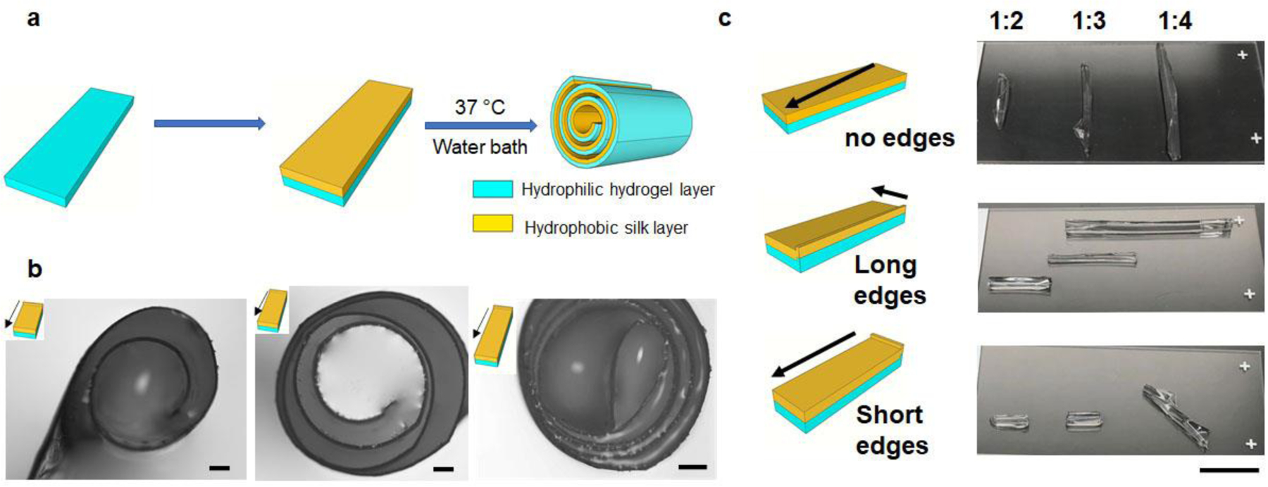
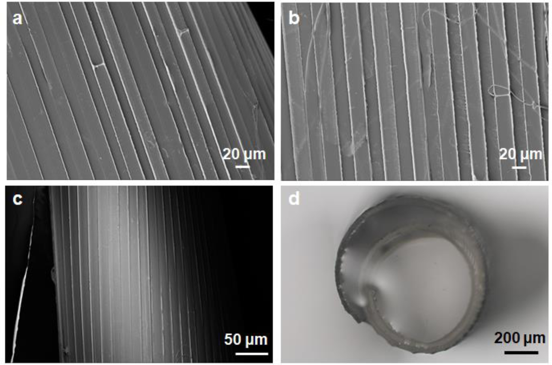
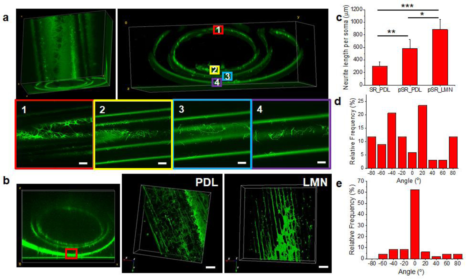
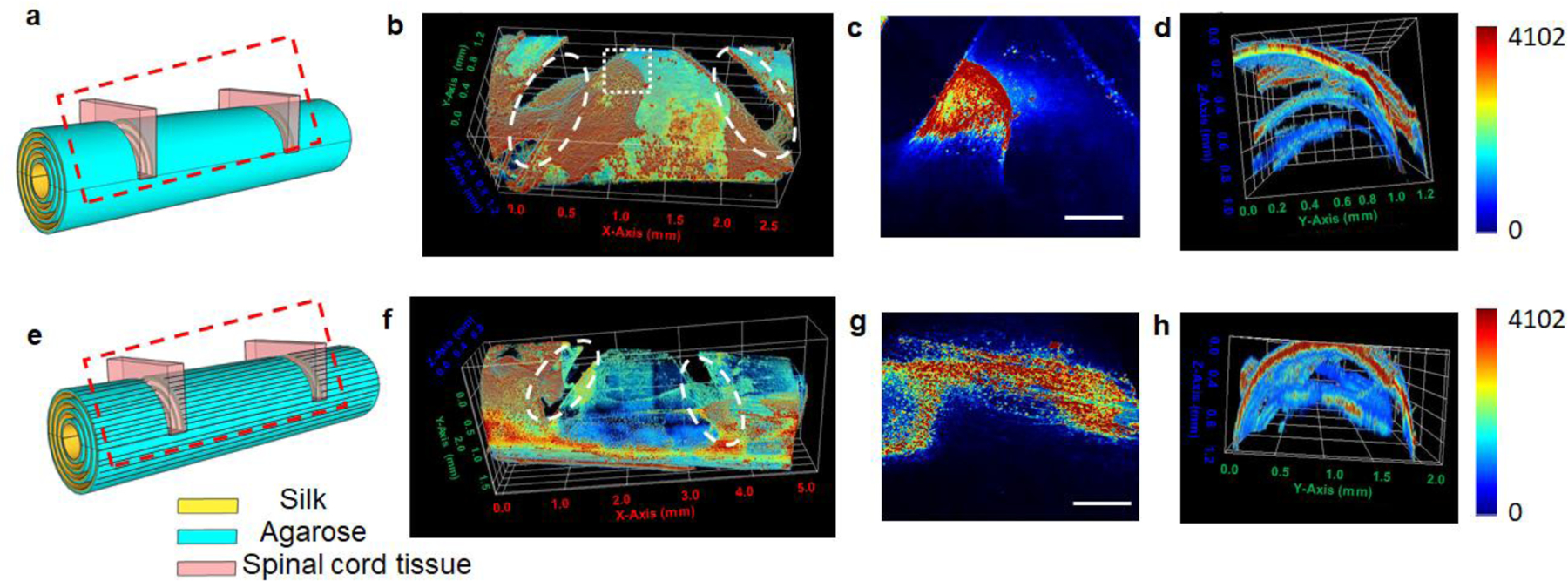
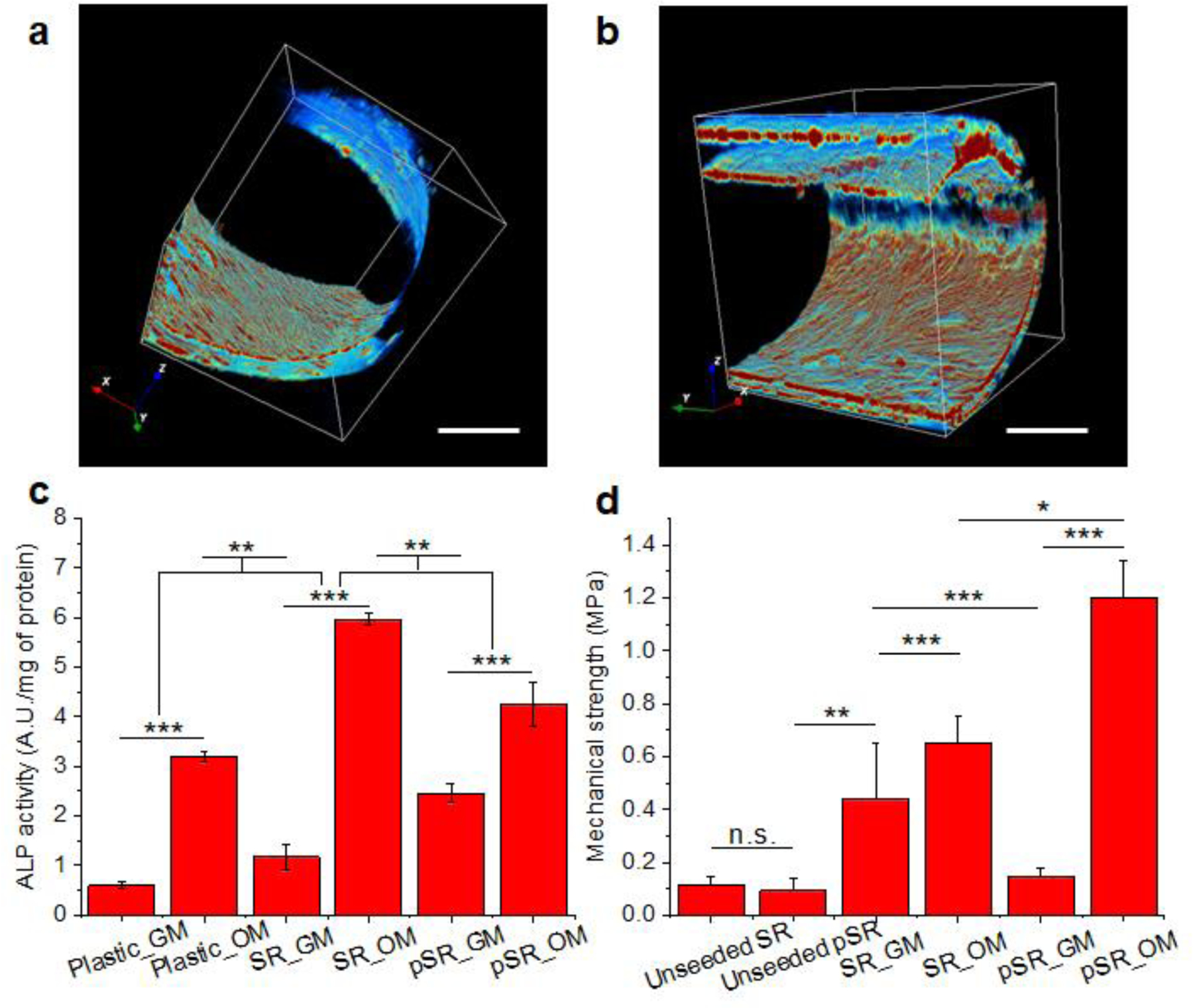
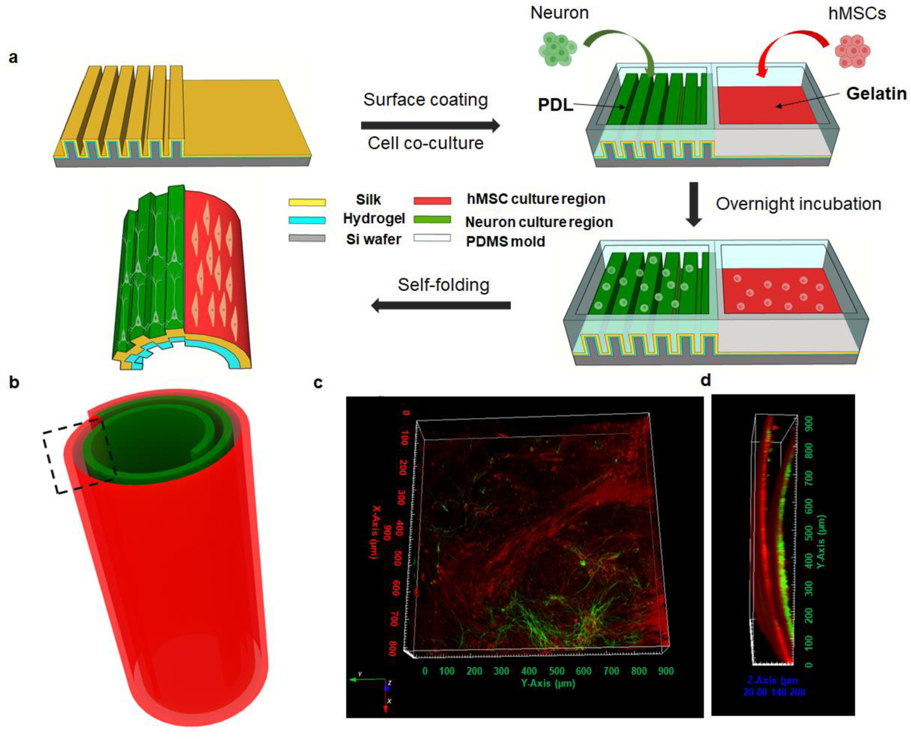
Biomaterial scaffold designs are needed for self-organizing features related to tissue formation while also simplifying the fabrication processes involved. Toward this goal, silk protein-based self-folding scaffolds to support 3D cell culture, while providing directional guidance and promotion of cell growth and differentiation, are reported. A simple and robust one-step self-folding approach is developed using bilayers consisting of a hydrogel and silk film in aqueous solution. The 3D silk rolls, with patterns transferred from the initially prepared 2D films, guide the directional outgrowth of neurites and also promote the osteogenic differentiation of human mesenchymal stem cells (hMSCs). The osteogenic outcomes are further supported by enhanced biomechanical performance. By utilizing this self-folding method, cocultures of neurons and hMSCs are achieved by patterning cells on silk films and then converting these materials into a 3D format with rolling, mimicking aspects of the structure of osteons and providing physiologically relevant structures to promote bone regeneration. These results demonstrate the utility of self-folded silk rolls as efficient scaffold systems for tissue regeneration, while exploiting relatively simple 2D designs programmed to form more complex 3D structures.

Keywords: 3D cell cultures; heterogeneous cell cultures; neuron regeneration; osteogenesis; self-folding biomaterials.


Figures

Figure 1.
Figure 1.. Self-folding strategy to control folding direction and number of layers.
(a) Schematic of the self-folding process. (b) Layer number control by tuning the aspect ratio of the bilayer film with 1:1, 1:2 and 1:3, respectively. Scale bar: 200 μm. (c) Folding direction control by edge effects. Scale bar: 1 cm.
Figure 2.
Figure 2.. Pattern design on self-folding silk rolls.
(a,b) SEM images of the patterned transferred onto agarose layer (a) and silk layer (b). (c) SEM images of the patterned silk rolls. (d) Optical images of patterned silk rolls.
Figure 3.
Figure 3.. Silk rolls for neuron directional growth.
(a) Reconstructed confocal images of neurons cultured on silk rolls within each layer. (b) Reconstructed confocal images (left) of neurons cultured on patterned silk rolls within PDL (middle) and LMN coating (right). Scale bars: 20 μm. (c) Neurite length measurement (N=36 for SR PDL coated group, N=55 for pSR PDL coated group and N=60 for pSR LMN coated group, respectively ). (PDL: poly-d-lysine, LMN: laminin; SR: silk roll; pSR: patterned silk roll. ) (d,e) Neurite angular analysis on silk roll (d) and patterned silk roll (e). (n.s.: non-significant, p > 0.5; *: p < 0.5; **: p < 0.01; ***: p < 0.001)
Figure 4.
Figure 4.. Spinal cord tissue slices cultured in the silk rolls.
(a,e) Schematic of culturing spinal cord tissue slices in SRs (a) and pSRs (e) (N=3). Red dash box: region imaged by confocal microscopy. (b-d) Reconstructed confocal images of spinal cord tissues cultured on SRs with a top view (b), zoomed-in view at the top layer (c, indicated by the white dash box) and cross-section view (d). White dash circles indicated the location of the spinal cord tissue slices placed in SRs and pSRs. (f-h) Reconstructed confocal images of spinal cord tissues cultured on pSRs with a top view (f), zoomed-in view (g, indicated by the white dash box) and cross-section view (h). Scale bars: 500 μm.
Figure 5.
Figure 5.. Silk rolls as osteoconductive substrates for hMSCs.
(a,b) 3D reconstructed confocal images of hMSCs cultured with SRs (a) and pSRs (b). Scale bars: 500 μm. (c) ALP activity. (d) Biomechanical measurements of silk rolls. (SR: silk rolls, pSR: patterned silk rolls; GM: growth medium; OM: osteogenic medium. N=5 samples for each group. n.s.: non-significant, p > 0.5; *: p < 0.5; **: p < 0.01; ***: p < 0.001)
Figure 6.
Figure 6.. Silk rolls as scaffolds for functional bone segments with nerve and osteogenesis.
(a) Schematic of heterogeneous cell seeding and co-culture. (b) Schematic of heterogeneous cell culture in the self-folding silk roll. (c,d) Reconstructed confocal images of co-culture in the silk roll, indicated in the black dashed region. (c) top view and (d) cross-section view.

References

    1. Griffin DR, Weaver WM, Scumpia PO, Di Carlo D, Segura T, Nature Materials 2015, 14, 737–744; - PMC - PubMed
    2. Kato-Negishi M, Morimoto Y, Onoe H, Takeuchi S, Advanced Healthcare Materials 2013, 2, 1564–1570. - PubMed
    1. Inzana JA, Olvera D, Fuller SM, Kelly JP, Graeve OA, Schwarz EM, Kates SL, Awad HA, Biomaterials 2014, 35, 4026–4034; - PMC - PubMed
    2. Murphy SV, Atala A, Nature Biotechnology 2014, 32, 773–785; - PubMed
    3. Do AV, Khorsand B, Geary SM, Salem AK, Advanced Healthcare Materials 2015, 4, 1742–1762. - PMC - PubMed
    1. Kolesky DB, Truby RL, Gladman AS, Busbee TA, Homan KA, Lewis JA, Advanced Materials 2014, 26, 3124–3130; - PubMed
    2. Colosi C, Costantini M, Barbetta A, Dentini M, Methods in Molecular Biology 2017, 1612, 369–380. - PubMed
    1. Hanson Shepherd JN, Parker ST, Shepherd RF, Gillette MU, Lewis JA, Nuzzo RG, Advanced Functional Materials 2011, 21, 47–54. - PMC - PubMed
    1. Ionov L, Soft Matterials 2011, 7, 6786–6791.

Publication types

LinkOut - more resources