

Adverse Initial CT Findings Associated with Poor Prognosis of Coronavirus Disease
- PMID: 32864912
- PMCID: PMC7458853
- DOI: 10.3346/jkms.2020.35.e316
Adverse Initial CT Findings Associated with Poor Prognosis of Coronavirus Disease
Abstract
Background: The predictors of poor prognosis in patients with coronavirus disease 2019 (COVID-19) using computed tomography (CT) have not been investigated in a large cohort. Therefore, the purpose of this study was to investigate the adverse initial CT features to predict poor prognosis in COVID-19.
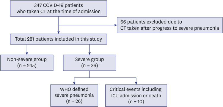
Methods: From February to April 2020, 281 COVID-19 patients who underwent CT at the time of admission were included. We divided the patients into the severe and non-severe disease groups. The severe group included patients with severe pneumonia or critical events. Intensive care unit admission or death were the critical events in this study. We compared the clinical and CT findings between the severe and non-severe groups and investigated the prognostic factors and critical events of the severe group using the regression analysis.
Results: Among the 281 patients, 36 (12.8%) patients were in the severe group and 245 (87.2%) patients were in the non-severe group. Critical events occurred in 10 patients (3.6%). In the severe group, patients showed significantly more pneumonia with consolidation, crazy-paving appearance, pleural effusion, and higher CT scores than those in the non-severe group (all, P < 0.05). In the multivariate regression, pleural effusion (odds ratio [OR], 8.96; 95% confidence interval [CI], 1.81-44.42; P = 0.007), CT score > 5 (OR, 3.70; 95% CI, 1.44-9.53; P = 0.007), old age (> 77 years, OR, 9.96; 95% CI, 3.78-26.28; P < 0.001), and elevated C-reactive protein (OR, 4.15; 95% CI, 1.62-10.6; P = 0.003) were significant prognostic factors of severe disease. CT score > 5 (OR, 7.29; 95% CI, 1.37-38.68; P = 0.020), pleural effusion (OR, 5.67; 95% CI, 1.04-30.8; P = 0.045) and old age (OR, 8.6; 95% CI, 1.80-41.0; P = 0.007) were also significant predictors of critical events.
Conclusion: Pleural effusion and the extent of pneumonia on initial CT scans are associated with poor prognosis in patients with COVID-19.
Keywords: COVID-19; Computed Tomography; Coronavirus; Pneumonia.
© 2020 The Korean Academy of Medical Sciences.
Conflict of interest statement
The authors have no potential conflicts of interest to disclose.
Figures


Similar articles
-
Prognostic Implication of Volumetric Quantitative CT Analysis in Patients with COVID-19: A Multicenter Study in Daegu, Korea.Korean J Radiol. 2020 Nov;21(11):1256-1264. doi: 10.3348/kjr.2020.0567. Epub 2020 Aug 4. Korean J Radiol. 2020. PMID: 32767868 Free PMC article.
-
CT lung lesions as predictors of early death or ICU admission in COVID-19 patients.Clin Microbiol Infect. 2020 Oct;26(10):1417.e5-1417.e8. doi: 10.1016/j.cmi.2020.07.030. Epub 2020 Jul 24. Clin Microbiol Infect. 2020. PMID: 32717417 Free PMC article.
-
Well-aerated Lung on Admitting Chest CT to Predict Adverse Outcome in COVID-19 Pneumonia.Radiology. 2020 Aug;296(2):E86-E96. doi: 10.1148/radiol.2020201433. Epub 2020 Apr 17. Radiology. 2020. PMID: 32301647 Free PMC article.
-
Clinical and radiological features of novel coronavirus pneumonia.J Xray Sci Technol. 2020;28(3):391-404. doi: 10.3233/XST-200687. J Xray Sci Technol. 2020. PMID: 32538893 Free PMC article. Review.
-
Similarities and Differences of Early Pulmonary CT Features of Pneumonia Caused by SARS-CoV-2, SARS-CoV and MERS-CoV: Comparison Based on a Systemic Review.Chin Med Sci J. 2020 Sep 30;35(3):254-261. doi: 10.24920/003727. Chin Med Sci J. 2020. PMID: 32972503 Free PMC article.
Cited by
-
Longitudinal investigation of severe acute respiratory syndrome coronavirus 2 (SARS-CoV-2) infection in older patients in the province of Palermo (Southern Italy) during the early wave of the pandemic.Arch Med Sci. 2021 Mar 18;18(6):1488-1497. doi: 10.5114/aoms/134024. eCollection 2022. Arch Med Sci. 2021. PMID: 36457987 Free PMC article.
-
Impact of Mediastinal Lymphadenopathy on the Severity of COVID-19 Pneumonia: A Nationwide Multicenter Cohort Study.J Korean Med Sci. 2022 Jun 6;37(22):e78. doi: 10.3346/jkms.2022.37.e78. J Korean Med Sci. 2022. PMID: 35668683 Free PMC article.
-
Influence of threshold selection strategy on the prognostic accuracy of chest CT severity score for mortality prediction of COVID-19 patients.Heart Lung. 2022 Nov-Dec;56:74-75. doi: 10.1016/j.hrtlng.2022.06.021. Epub 2022 Jun 28. Heart Lung. 2022. PMID: 35792344 Free PMC article. No abstract available.
-
Mortality and Survival Factors in Patients with Moderate and Severe Pneumonia Due to COVID-19.Healthcare (Basel). 2023 Mar 23;11(7):932. doi: 10.3390/healthcare11070932. Healthcare (Basel). 2023. PMID: 37046859 Free PMC article.
-
Artificial intelligence algorithms based approach in evaluating COVID-19 patients and management.J Crit Care Med (Targu Mures). 2025 Jul 31;11(3):247-256. doi: 10.2478/jccm-2025-0032. eCollection 2025 Jul. J Crit Care Med (Targu Mures). 2025. PMID: 40765546 Free PMC article.
References
-
- World Health Organization. WHO Director-general's opening remarks at the media briefing on COVID-19. [Updated 2020]. [Accessed April 13, 2020]. https://www.who.int/docs/default-source/coronaviruse/clinical-management....
-
- Worldometer. COVID-19 coronavirus pandemic. [Updated 2020]. [Accessed May 20, 2020]. https://www.worldometers.info/coronavirus/
MeSH terms
Substances
Grants and funding
LinkOut - more resources
Full Text Sources
Research Materials
Miscellaneous
