

Gamma-delta T cells stimulate IL-6 production by pancreatic stellate cells in pancreatic ductal adenocarcinoma
- PMID: 32865617
- PMCID: PMC7679341
- DOI: 10.1007/s00432-020-03367-8
Gamma-delta T cells stimulate IL-6 production by pancreatic stellate cells in pancreatic ductal adenocarcinoma
Abstract
Introduction: The immunosuppressive tumor microenvironment promotes progression of pancreatic ductal adenocarcinoma (PDAC). γδ T cells infiltrate the pancreatic tumor stroma and support tumorigenesis through αβ T cell inhibition. Pancreatic stellate cell (PSC) activation contributes to pancreatic fibrosis in PDAC, limiting the delivery and efficacy of therapeutic agents. Whether γδ T cells have direct effects on PSC activation is unknown.
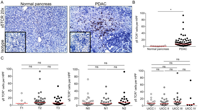
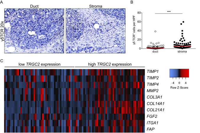
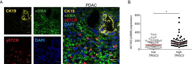
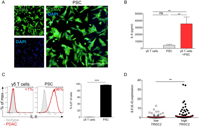
Methods: In this study, we analyzed tumor tissue from 68 patients with PDAC and determined the frequency and location of γδ T cells using immunohistochemistry and immunofluorescence. PDAC samples from the TCGA database with low and high TRGC2 expression were correlated with the expression of extracellular matrix genes. Further, PSCs were isolated from pancreatic tumor tissue and co-cultured with γδ T cells for 48 hours and cytokine production was measured using a cytometric bead array.
Results: γδ T cells infiltrated the pancreatic tumor stroma and were located in proximity to PSCs. A high infiltration of γδ T cells was associated with increased expression of several extracellular matrix genes in human PDAC. In vitro, γδ T cells stimulated IL-6 production by PDAC-derived PSCs.
Conclusion: γδ T cells activated PSCs and modulation of this interaction may enhance the efficacy of combinational therapies in human PDAC.
Keywords: Gamma-delta T cells; IL-6; Pancreatic cancer; Pancreatic stellate cells.
Conflict of interest statement
The authors have declared that no conflict of interest exists.
Figures
Similar articles
-
Galectin-3 Mediates Tumor Cell-Stroma Interactions by Activating Pancreatic Stellate Cells to Produce Cytokines via Integrin Signaling.Gastroenterology. 2018 Apr;154(5):1524-1537.e6. doi: 10.1053/j.gastro.2017.12.014. Epub 2017 Dec 21. Gastroenterology. 2018. PMID: 29274868
-
Global targetome analysis reveals critical role of miR-29a in pancreatic stellate cell mediated regulation of PDAC tumor microenvironment.BMC Cancer. 2020 Jul 13;20(1):651. doi: 10.1186/s12885-020-07135-2. BMC Cancer. 2020. PMID: 32660466 Free PMC article.
-
Pancreatic Stellate Cells Promote Tumor Progression by Promoting an Immunosuppressive Microenvironment in Murine Models of Pancreatic Cancer.Pancreas. 2020 Jan;49(1):120-127. doi: 10.1097/MPA.0000000000001464. Pancreas. 2020. PMID: 31856087
-
Pancreatic stellate cells and pancreas cancer: current perspectives and future strategies.Eur J Cancer. 2014 Oct;50(15):2570-82. doi: 10.1016/j.ejca.2014.06.021. Epub 2014 Aug 1. Eur J Cancer. 2014. PMID: 25091797 Review.
-
Pancreatic stellate cell: Pandora's box for pancreatic disease biology.World J Gastroenterol. 2017 Jan 21;23(3):382-405. doi: 10.3748/wjg.v23.i3.382. World J Gastroenterol. 2017. PMID: 28210075 Free PMC article. Review.
Cited by
-
The Cytotoxic Potential of Humanized γδ T Cells Against Human Cancer Cell Lines in In Vitro.Cells. 2025 Aug 4;14(15):1197. doi: 10.3390/cells14151197. Cells. 2025. PMID: 40801630 Free PMC article.
-
Research trends on immunotherapy for pancreatic cancer: A bibliometric analysis.Hum Vaccin Immunother. 2023 Dec 15;19(3):2269794. doi: 10.1080/21645515.2023.2269794. Epub 2023 Oct 26. Hum Vaccin Immunother. 2023. PMID: 37885280 Free PMC article.
-
The pancreatic tumor microenvironment of treatment-naïve patients causes a functional shift in γδ T cells, impairing their anti-tumoral defense.Oncoimmunology. 2025 Dec;14(1):2466301. doi: 10.1080/2162402X.2025.2466301. Epub 2025 Feb 13. Oncoimmunology. 2025. PMID: 39945298 Free PMC article.
-
LAG-3-Expressing Tumor-Infiltrating T Cells Are Associated with Reduced Disease-Free Survival in Pancreatic Cancer.Cancers (Basel). 2021 Mar 15;13(6):1297. doi: 10.3390/cancers13061297. Cancers (Basel). 2021. PMID: 33803936 Free PMC article.
-
Key players of immunosuppression in epithelial malignancies: Tumor-infiltrating myeloid cells and γδ T cells.Cancer Rep (Hoboken). 2024 May;7(5):e2066. doi: 10.1002/cnr2.2066. Cancer Rep (Hoboken). 2024. PMID: 38703051 Free PMC article. Review.
References
-
- Clark CE, Hingorani SR, Mick R, Combs C, Tuveson DA, Vonderheide RH (2007) Dynamics of the immune reaction to pancreatic cancer from inception to invasion. Cancer Res 67(19):9518–9527 - PubMed
MeSH terms
Substances
Grants and funding
LinkOut - more resources
Full Text Sources
