Review
. 2020 Dec;140(6):793-810.
doi: 10.1007/s00401-020-02215-w. Epub 2020 Aug 31.

Cerebral blood flow decrease as an early pathological mechanism in Alzheimer's disease

Affiliations
Review

Cerebral blood flow decrease as an early pathological mechanism in Alzheimer's disease

Nils Korte et al. Acta Neuropathol. 2020 Dec.

Abstract

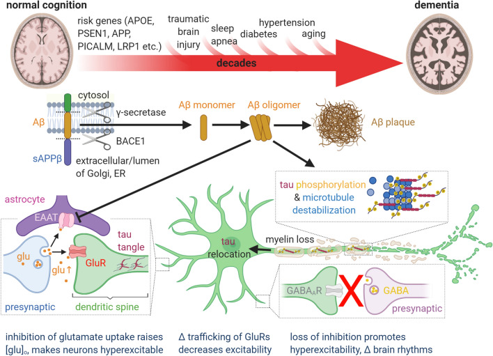
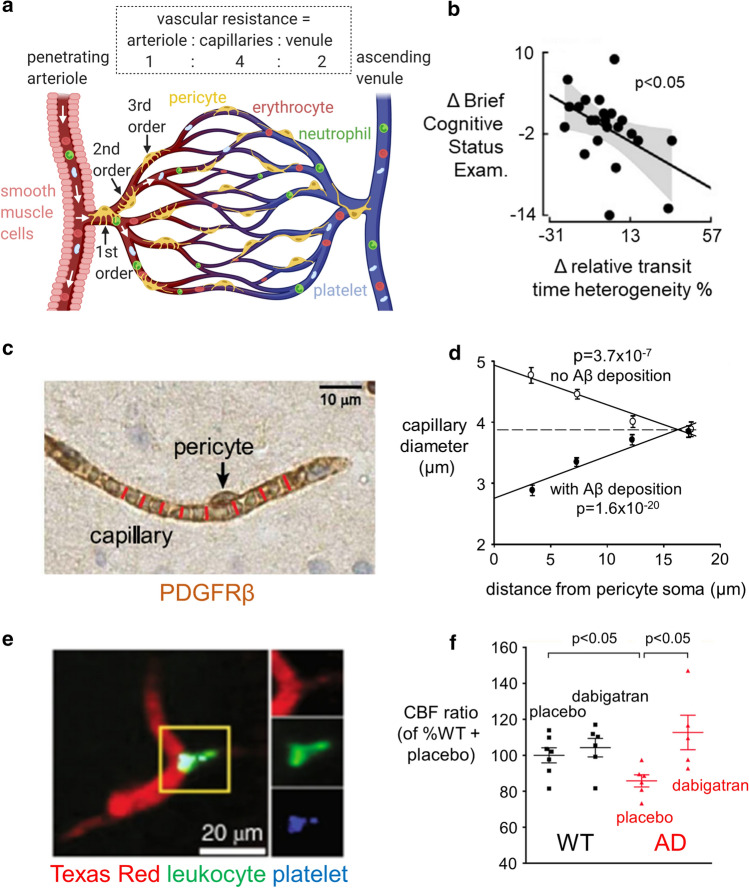
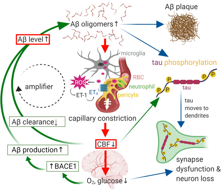
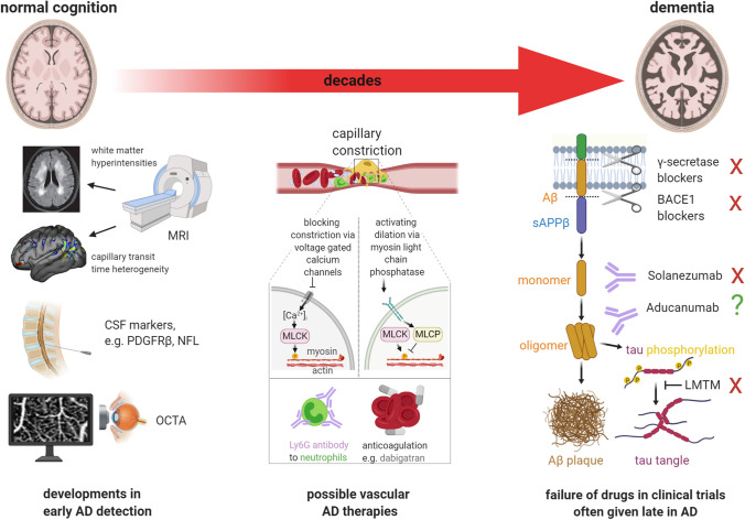
Therapies targeting late events in Alzheimer's disease (AD), including aggregation of amyloid beta (Aβ) and hyperphosphorylated tau, have largely failed, probably because they are given after significant neuronal damage has occurred. Biomarkers suggest that the earliest event in AD is a decrease of cerebral blood flow (CBF). This is caused by constriction of capillaries by contractile pericytes, probably evoked by oligomeric Aβ. CBF is also reduced by neutrophil trapping in capillaries and clot formation, perhaps secondary to the capillary constriction. The fall in CBF potentiates neurodegeneration by upregulating the BACE1 enzyme that makes Aβ and by promoting tau hyperphosphorylation. Surprisingly, therefore, CBF reduction may play a crucial role in driving cognitive decline by initiating the amyloid cascade itself, or being caused by and amplifying Aβ production. Here, we review developments in this area that are neglected in current approaches to AD, with the aim of promoting novel mechanism-based therapeutic approaches.

Keywords: Alzheimer’s; Amyloid β; Capillary; Cerebral blood flow; Neutrophil; Pericyte.


Figures

Fig. 1
Fig. 1
Current, generally held ideas about the pathology underlying Alzheimer’s disease (see main text for details). The transition from normal cognition to dementia, over decades, is promoted by the risk factors shown above the large red arrow. Aβ is produced from amyloid precursor protein (APP) by the action of the γ secretase and β secretase (BACE1) as monomers, but these can then form soluble oligomers, which ultimately form extracellular precipitates as amyloid plaques. Aβ oligomers inhibit astrocyte glutamate uptake (EAAT), thus potentiating the action of synaptically released glutamate (glu). This, together with a loss of GABAergic inhibition, leads to some neurons becoming hyperexcitable. Meanwhile, Aβ oligomers also induce hyperphosphorylation of axonal microtubule-associated tau, which leads to tau redistributing partly to dendrites where it disrupts trafficking of glutamate receptors and thus depresses excitation and neuronal firing. These synaptic effects, and Aβ- and/or tau-induced loss of axonal myelin, may induce cognitive dysfunction well before synapses are lost and neurons die. The levels of Aβ oligomers and hyperphosphorylated tau correlate better with cognitive decline than does the level of Aβ plaques
Fig. 2
Fig. 2
The role of pericytes in the physiology and Alzheimer’s-related pathology of the brain circulation. a Schematic diagram of the vascular bed (colour of blood represents oxygenation), indicating the relative resistance in the capillaries compared to penetrating arterioles and venules, for flow from the pial surface down an arteriole to layer 4, through the capillary bed, and returning to the pial surface through a venule [49]. Capillary diameter can be adjusted by a population of pericytes (yellow) that are contractile, which are located on at least the first four branch orders (see labels) of the capillary bed [56]. Blood flowing through capillaries with pericytes that are contracting to reduce the diameter will flow more slowly and so has a longer capillary transit time than blood flowing through capillaries with relaxed pericytes, thus generating capillary transit time heterogeneity (CTTH). b In patients with AD, CTTH (shown as a % change) increases as cognitive power (assessed with the Brief Cognitive Status Examination) declines (from Fig. 5A of [128], reproduced courtesy of John Wiley and Sons). c, d Capillary imaged in right frontal cortex biopsy from a dementing patient lacking Aβ deposition (c) and plot of mean capillary diameter versus distance from pericyte somata (d) in similar patients lacking or showing Aβ deposition (from Fig. 4A, D of [133]). Patients depositing Aβ show a large constriction near the pericyte somata. e Neutrophil (green) occluding a branch (to the right) of a capillary in AD mouse cortex (from Fig. 2A of [26], reproduced courtesy of Springer Nature). f Reducing clotting with dabigatran in WT and AD mice (from Fig. 3B of [25], reproduced courtesy of Elsevier Press) increases CBF in AD mice
Fig. 3
Fig. 3
Schematic diagram showing how the amyloid beta and tau cascades can be initiated from two entry points (red boxes): (i) a decrease of cerebral blood flow (CBF) which lowers brain O2 and glucose and thus upregulates the enzyme (BACE1) that makes Aβ or (ii) an increase in Aβ level due to more production or less clearance of Aβ. Aβ oligomers can aggregate into plaques, but also evoke ROS production from microglia and pericytes, which triggers the release of endothelin-1 (ET-1) from a yet-to-be-determined cell type [133]. Activation of ETA receptors on pericytes leads to capillary constriction and a decrease of CBF, lowering the levels of O2 and glucose. Both a rise of Aβ oligomer concentration and a fall of blood flow lead to hyperphosphorylation of tau, which relocates from axonal microtubules to dendrites, causing synapse dysfunction. Together with myelin loss this leads to cognitive decline. The fall of CBF will also contribute to impaired cognition
Fig. 4
Fig. 4
Interventions to diagnose and reduce cognitive decline at different stages of the transition from normal cognition to dementia in AD. Right third of figure: most clinical trials are initiated at relatively late stages of the disease, when cognitive decline is already apparent, and irreversible synapse or neuron loss may have taken place. This may explain why drugs that block the γ or β secretases, antibodies to different forms of Aβ, and a drug that blocks tau aggregation (LMTM) have all failed (red crosses) to stop cognitive decline in AD. Left third of figure: emerging diagnostic approaches for early detection of AD include MRI assessment of white matter hyperintensities (image from Fig. 1B of [93], reproduced courtesy of Dove Medical Press) and capillary transit time heterogeneity (from Fig. 5E of [128], reproduced courtesy of John Wiley & Sons), assessment of biomarkers in the CSF such as PDGFRβ and neurofilament light chain (NFL), and non-invasive capillary imaging in the retina using (e.g.) optical coherence tomography angiography (OCTA). Middle third of figure: potential therapies to prevent or reverse the CBF decrease arising when Ca2+ activates myosin light chain kinase (MLCK) to evoke pericyte-mediated capillary constriction. These include blocking pericyte voltage-gated calcium channels to block Ca2+-evoked constriction, raising pericyte cGMP level (by activating guanylate cyclase receptors, blue membrane protein) to stimulate myosin light chain phosphate (MLCP) and thus evoke dilation, disrupting neutrophil surface interactions with endothelial cells or other cells using antibodies (if this approach can be used without inducing neutropenia), or blocking thrombus formation with dabigatran [25, 26, 133]

References

    1. Abdullah L, Crawford F, Tsolaki M, Börjesson-Hanson A, Olde Rikkert M, Pasquier F, et al. The Influence of baseline Alzheimer’s disease severity on cognitive decline and CSF biomarkers in the NILVAD trial. Front Neurol. 2020;11:149. doi: 10.3389/fneur.2020.00149. - DOI - PMC - PubMed
    1. Akenhead ML, Horrall NM, Rowe D, Sethu P, Shin HY. In vitro evaluation of the link between cell activation state and its rheological Impact on the microscale flow of neutrophil suspensions. J Biomech Eng. 2015;137:91003. doi: 10.1115/1.4030824. - DOI - PMC - PubMed
    1. Alarcon-Martinez L, Yilmaz-Ozcan S, Yemisci M, Schallek J, Kilic K, Can A, et al. Capillary pericytes express α-smooth muscle actin, which requires prevention of filamentous-actin depolymerization for detection. Elife. 2018;7:e34861. doi: 10.7554/eLife.34861.001. - DOI - PMC - PubMed
    1. Armulik A, Genové G, Mäe M, Nisancioglu MH, Wallgard E, Niaudet C, et al. Pericytes regulate the blood-brain barrier. Nature. 2010;468:557–561. doi: 10.1038/nature09522. - DOI - PubMed
    1. Asllani I, Habeck C, Scarmeas N, Borogovac A, Brown TR, Stern Y. Multivariate and univariate analysis of continuous arterial spin labeling perfusion MRI in Alzheimer’s disease. J Cereb Blood Flow Metab. 2008;28:725–736. doi: 10.1038/sj.jcbfm.9600570. - DOI - PMC - PubMed

Publication types

MeSH terms

Substances