. 2020 Oct;19(10):e13224.
doi: 10.1111/acel.13224. Epub 2020 Aug 31.

Aging-associated changes in CD47 arrangement and interaction with thrombospondin-1 on red blood cells visualized by super-resolution imaging

Affiliations

Aging-associated changes in CD47 arrangement and interaction with thrombospondin-1 on red blood cells visualized by super-resolution imaging

Feng Wang et al. Aging Cell. 2020 Oct.

Abstract

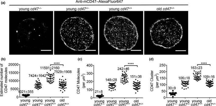
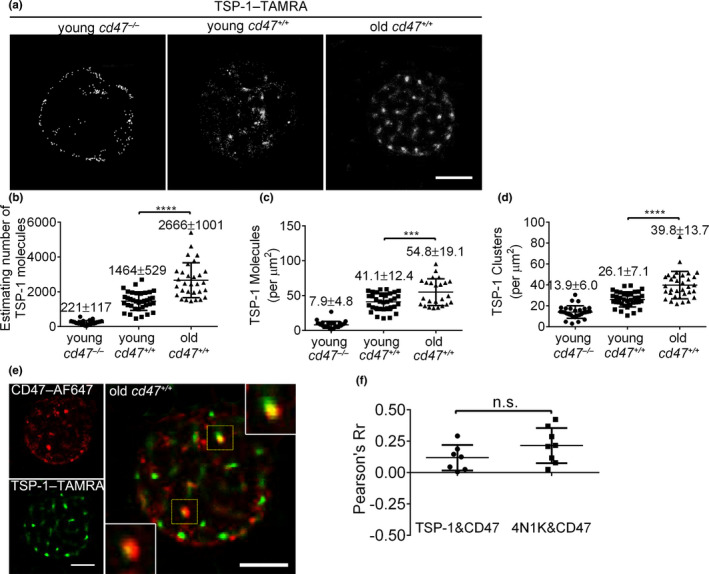
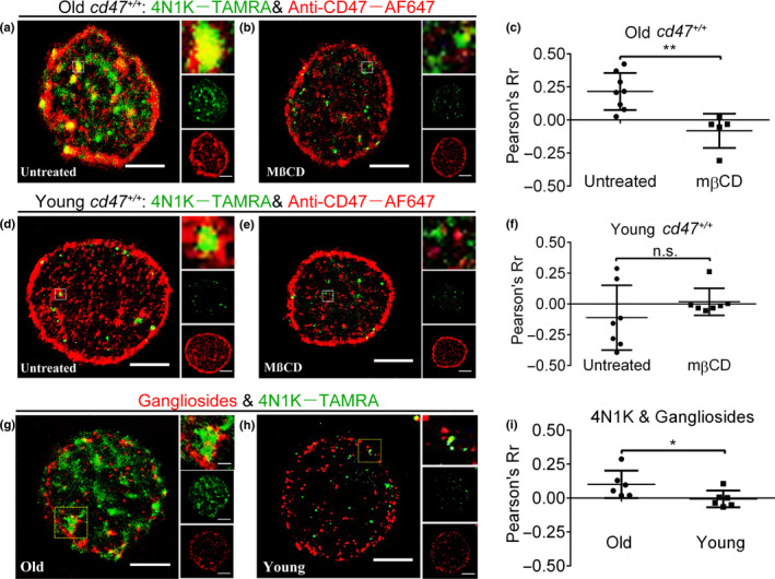
CD47 serves as a ligand for signaling regulatory protein α (SIRPα) and as a receptor for thrombospondin-1 (TSP-1). Although CD47, TSP-1, and SIRPα are thought to be involved in the clearance of aged red blood cells (RBCs), aging-associated changes in the expression and interaction of these molecules on RBCs have been elusive. Using direct stochastic optical reconstruction microscopy (dSTORM)-based imaging and quantitative analysis, we can report that CD47 molecules on young RBCs reside as nanoclusters with little binding to TSP-1, suggesting a minimal role for TSP-1/CD47 signaling in normal RBCs. On aged RBCs, CD47 molecules decreased in number but formed bigger and denser clusters, with increased ability to bind TSP-1. Exposure of aged RBCs to TSP-1 resulted in a further increase in the size of CD47 clusters via a lipid raft-dependent mechanism. Furthermore, CD47 cluster formation was dramatically inhibited on thbs1-/- mouse RBCs and associated with a significantly prolonged RBC lifespan. These results indicate that the strength of CD47 binding to its ligand TSP-1 is predominantly determined by the distribution pattern and not the amount of CD47 molecules on RBCs, and offer direct evidence for the role of TSP-1 in phagocytosis of aged RBCs. This study provides clear nanoscale pictures of aging-associated changes in CD47 distribution and TSP-1/CD47 interaction on the cell surface, and insights into the molecular basis for how these molecules coordinate to remove aged RBCs.

Keywords: CD47; aging; dSTORM; red blood cells; thrombospondin-1.


Conflict of interest statement

The authors declare no competing financial interests.

Figures

Figure 1
Figure 1
Direct STORM imaging and quantitative analysis of CD47 on young vs. old mouse RBCs. (a) Representative dSTORM images of CD47 distribution on cd47 −/−, cd47 +/−, and cd47 +/+ mouse RBCs. Scale bar = 2 μm. (b–d) Quantitative analysis of CD47 on young cd47 −/− mouse RBCs, young cd47 +/− mouse RBCs, young cd47 +/+ mouse RBCs, and old cd47 +/+ mouse RBCs. Data shown are estimated total numbers (b); protein densities (molecules/µm2) (c); and cluster densities (cluster/µm2) (d) of cell surface CD47. Data are combined from 3 independent experiments and presented as mean ± SD (young cd47 −/− mouse RBCs, n = 21; young cd47 +/− mouse RBCs, n = 28; young cd47 +/+ mouse RBCs, n = 30; old cd47 +/+ mouse RBCs, n = 41). ****p < 0.0001 (by two‐tailed unpaired Student's t‐test)
Figure 2
Figure 2
Direct STORM imaging and univariate pair correlation function analysis of CD47 on RBCs from young and old cd47 +/+ mice. (a, b) Left and right panels show RBC dSTORM images (scale bar = 2 μm) and corresponding pair correlation function analysis, respectively, for young (a) and old (b) mouse RBCs. (c–e) Clustering parameters of CD47 clusters on cd47 +/+ RBCs from young or old mice: (c) CD47 cluster radius (nm); (d) density of CD47 proteins in cluster (ψcluster); (e) average numbers of CD47 proteins per cluster (Ncluster). Unpaired t‐test was used; n.s., not significant; *p < 0.05. Data combined from 3 independent experiments are presented as mean ± SD (each symbol represents an individual cell; 20‐25 cells per group were reanalyzed)
Figure 3
Figure 3
Direct STORM imaging and quantitative analysis of 4N1K binding on young vs. old mouse RBCs. (a) Representative dSTORM images of 4N1K binding on young cd47 −/−, young cd47 +/+ and old cd47 +/+ mouse RBCs; scale bar = 2 μm. (b–d) Quantitative analysis of 4N1K binding on young cd47 −/− mouse RBCs, young cd47 +/+ mouse RBCs, and old cd47 +/+ mouse RBCs. Data shown are estimated numbers (b), protein densities (molecules/µm2) (c) and cluster densities (cluster/µm2) (d) of 4N1K molecules on RBCs. Data are combined from three independent experiments and presented as mean ± SD (young cd47 −/− mouse RBCs, n = 25; young cd47 +/+ mouse RBCs, n = 21; old cd47 +/+ mouse RBCs, n = 26). *p < 0.05; **p < 0.01; ***p < 0.001 (by two‐tailed unpaired Student's t‐test)
Figure 4
Figure 4
Two‐color dSTORM imaging and cluster characteristics of CD47 on young vs. old mouse RBCs after 4N1K treatment. (a, b) Representative dSTORM images of CD47 distribution (red) on untreated or 4N1K‐treated young (a) and old (b) cd47 +/+ mouse RBCs. Scale bar = 2 μm. For both untreated and 4N1K‐treated samples, an area of 4 × 4 μm2 was selected, and CD47 distribution characteristics were analyzed with pair correlation function (corresponding g(r) curves of young and old mouse RBCs are shown in c and d, respectively). (c, d) Univariate pair correlation function analysis of CD47 distribution on untreated and 4N1K‐treated young (c) or old (d) cd47 +/+ mouse RBCs. (e–g) Shown are cluster radius (e), ψcluster (f) and Ncluster (g) of CD47 clusters on cd47 +/+ RBCs from young or old mice that were untreated or treated with 4N1K. Data from 3 independent experiments are combined and presented as mean ± SD (untreated young mouse RBCs, n = 41; young mouse RBCs treated with 4N1K, n = 17; untreated old mouse RBCs, n = 24; old mouse RBCs treated with 4N1K, n = 23). n.s. indicates not significant; *p < 0.05; **p < 0.01 (by two‐way ANOVA with Tukey's correction)
Figure 5
Figure 5
Direct STORM imaging and quantitative analysis of TSP‐1 binding on young vs. old mouse RBCs. (a) Representative dSTORM images of TSP‐1 binding on the indicated mouse RBCs. Scale bar = 2 μm. (b–d) Quantitative analysis of TSP‐1 binding on young cd47 −/−, young cd47 +/+, and old cd47 +/+ mouse RBCs. Data shown are estimated numbers (b), protein densities (molecules/μm2) (c), and cluster densities (cluster/μm2) (d) of TSP‐1 molecules. Data are combined from three independent experiments and presented as mean ± SD (young cd47 −/− mouse RBCs, n = 30; young cd47 +/+ mouse RBCs, n = 36; old cd47 +/+ mouse RBCs, n = 30). ***p < 0.001; ****p < 0.0001 (by two‐tailed unpaired Student's t‐test). (e) Two‐color dSTORM images showing co‐localization of TSP‐1–TAMRA with CD47–AF647 on cd47 +/+ old RBCs. High magnification images (insets) of co‐localized TSP‐1 and CD47 clusters are shown (scale bar = 2 μm). (f) Pearson coefficient (Pearson's Rr) showing levels of co‐localization between CD47 with TSP‐1 (0.12 ± 0.09; n = 6) or with 4N1K (0.22 ± 0.14; n = 8) on old cd47 +/+ RBCs
Figure 6
Figure 6
RBC life span and CD47 distribution pattern in old WT and thbs1 −/− mice. Circulating RBCs in 18 months of age WT or thbs1 −/− mice were labeled by biotinylation as described under Materials and Methods. Blood cells were collected at 1, 5, 10, 15, 20, 30, 35, 40, and 45 days postbiotin injection. (a) Flow cytometric profiles showing expression of biotin and CD47 in RBCs at days 1, 5, 20, and 35 post‐biotinylation in WT (upper row) and thbs1 −/− (lower row) were presented. Since newly produced RBCs from bone marrow are biotin, so the changes in the biotin+ RBC population closely reflect the characteristics of aging RBCs. (b) Percentages of biotin+ RBCs in blood circulation of WT (n = 8) and thbs1 −/− (n = 9) mice (mean ± SEM). (c, d) Direct STORM imaging of CD47 on biotin RBCs sorted from WT (c) or thbs1 −/− (d) mice at day 30 post‐biotinylation (bar = 2 µm). Zoomed images (insets) highlight the extent of CD47 molecule aggregation on RBCs
Figure 7
Figure 7
CD47 and 4N1K clustered in lipid rafts on young vs. old cd47 +/+ mouse RBCs. (a–f) RBCs from old (a–c) or young (d–f) cd47 +/+ mice were untreated or treated with MßCD, and analyzed by dual‐color dSTORM images using 4N1K–TAMRA and anti‐CD47–AF647. Shown are representative dSTORM images of untreated (a, d) and MßCD‐treated (b, e) RBCs (scale bar = 2 μm; zoomed images (insets) show co‐localized CD47 and 4N1K clusters; membrane‐bound 4N1K (green) and CD47 (red) distributions are also shown in the side row), and levels of co‐localization (Pearson's Rr) between 4N1K and CD47 on RBCs (c, f; n = 5–8 per group). (g–i) Analysis of 4N1K binding sites and gangliosides (a component of lipid raft) on RBCs. (g, h) Representative dual‐color dSTORM images showing 4N1K binding sites and CT‐B–AF647 labeled gangliosides on RBCs from old (g) or young (h) mice. Zoomed images (insets) show co‐localization between 4N1K and gangliosides. Membrane‐bound 4N1K (green) and gangliosides (red) distributions were also shown in the side row. (i) Levels of co‐localization between gangliosides and 4N1K on old (n = 6) or young (n = 8) mouse RBCs

References

    1. Adams, J. C. , & Lawler, J. (2011). The thrombospondins. Cold Spring Harbor Perspectives in Biology, 3(10), a009712 10.1101/cshperspect.a009712 - DOI - PMC - PubMed
    1. Annibale, P. , Vanni, S. , Scarselli, M. , Rothlisberger, U. , & Radenovic, A. (2011). Quantitative photo activated localization microscopy: unraveling the effects of photoblinking. PLoS One, 6(7), e22678 10.1371/journal.pone.0022678 - DOI - PMC - PubMed
    1. Arias, C. F. , & Arias, C. F. (2017). How do red blood cells know when to die? Royal Society Open Science, 4(4), 160850 10.1098/rsos.160850 - DOI - PMC - PubMed
    1. Barazi, H. O. , Li, Z. , Cashel, J. A. , Krutzsch, H. C. , Annis, D. S. , Mosher, D. F. , & Roberts, D. D. (2002). Regulation of integrin function by CD47 ligands. Differential effects on alpha vbeta 3 and alpha 4beta1 integrin‐mediated adhesion. Journal of Biological Chemistry, 277(45), 42859–42866. 10.1074/jbc.M206849200 - DOI - PubMed
    1. Bauer, P. M. , Bauer, E. M. , Rogers, N. M. , Yao, M. , Feijoo‐Cuaresma, M. , Pilewski, J. M. , … Isenberg, J. S. (2012). Activated CD47 promotes pulmonary arterial hypertension through targeting caveolin‐1. Cardiovascular Research, 93(4), 682–693. 10.1093/cvr/cvr356 - DOI - PMC - PubMed

Publication types