

Ultrafast Dynamics at Lipid-Water Interfaces
- PMID: 32866390
- PMCID: PMC8041150
- DOI: 10.1021/acs.accounts.0c00302
Ultrafast Dynamics at Lipid-Water Interfaces
Abstract
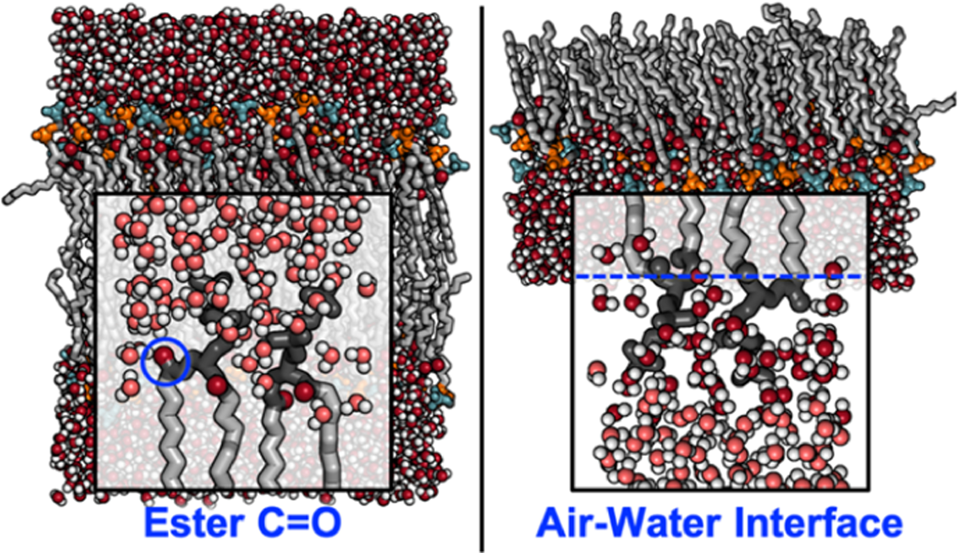
Lipid membranes are more than just barriers between cell compartments; they provide molecular environments with a finely tuned balance between hydrophilic and hydrophobic interactions that enable proteins to dynamically fold and self-assemble to regulate biological function. Characterizing dynamics at the lipid-water interface is essential to understanding molecular complexities from the thermodynamics of liquid-liquid phase separation down to picosecond-scale reorganization of interfacial hydrogen-bond networks.Ultrafast vibrational spectroscopy, including two-dimensional infrared (2D IR) and vibrational sum-frequency generation (VSFG) spectroscopies, is a powerful tool to examine picosecond interfacial dynamics. Two-dimensional IR spectroscopy provides a bond-centered view of dynamics with subpicosecond time resolutions, as vibrational frequencies are highly sensitive to the local environment. Recently, 2D IR spectroscopy has been applied to carbonyl and phosphate vibrations intrinsically located at the lipid-water interface. Interface-specific VSFG spectroscopy probes the water vibrational modes directly, accessing H-bond strength and water organization at lipid headgroup positions. Signals in VSFG arise from the interfacial dipole contributions, directly probing headgroup ordering and water orientation to provide a structural view of the interface.In this Account we discuss novel applications of ultrafast spectroscopy to lipid membranes, a field that has experienced significant growth over the past decade. In particular, ultrafast experiments now offer a molecular perspective on increasingly complex membranes. The powerful combination of ultrafast, interface-selective spectroscopy and simulations opens up new routes to understanding multicomponent membranes and their function. This Account highlights key prevailing views that have emerged from recent experiments: (1) Water dynamics at the lipid-water interface are slow compared to those of bulk water as a result of disrupted H-bond networks near the headgroups. (2) Peptides, ions, osmolytes, and cosolvents perturb interfacial dynamics, indicating that dynamics at the interface are affected by bulk solvent dynamics and vice versa. (3) The interfacial environment is generally dictated by the headgroup structure and orientation, but hydrophobic interactions within the acyl chains also modulate interfacial dynamics. Ultrafast spectroscopy has been essential to characterizing the biophysical chemistry of the lipid-water interface; however, challenges remain in interpreting congested spectra as well as designing appropriate model systems to capture the complexity of a membrane environment.
Conflict of interest statement
The authors declare no competing financial interest.
Figures


References
-
- Valentine ML; Cardenas AE; Elber R; Baiz CR Physiological Calcium Concentrations Slow Dynamics at the Lipid-Water Interface. Biophys. J 2018, 115, 1541–1551. - PMC - PubMed
-
This investigation used isotope-edited ultrafast two-dimensional infrared spectroscopy to probe the lipid–water interface of lipid bilayers with and without Ca2+ in solution. Results indicated a dependence of interfacial dynamics on the Ca2+ concentration in anionic lipid species.
-
- Valentine ML; Cardenas AE; Elber R; Baiz CR Calcium-Lipid Interactions Observed with Isotope-Edited Infrared Spectroscopy. Biophys. J 2020, 118, 2694–2702. - PMC - PubMed
-
This work used isotope-edited ultrafast two-dimensional infrared spectroscopy to investigate the head- group-specific effect of Ca2+ ions on the lipid–water interface, revealing water reorganization at the lipid–water interface of anionic lipids in the presence of Ca2+ that was not observed for zwitterionic lipid species.
-
- Flanagan JC; Baiz CR Site-Specific Peptide Probes Detect Buried Water in a Lipid Membrane. Biophys. J 2019, 116, 1692–1700. - PMC - PubMed
-
Here, isotope-labeled transmembrane peptides were used to probe water penetration within the alkyl tail region of model lipid bilayers with ultrafast two-dimensional infrared spectroscopy. This study found increased hydration ~1 nm into the alkyl region, suggesting the membrane environment is perturbed by the presence of transmembrane peptides.
-
- Flanagan JC; Cardenas AE; Baiz CR Ultrafast Spectroscopy of Lipid–Water Interfaces: Transmembrane Crowding Drives H-Bond Dynamics. J. Phys. Chem. Lett 2020, 11, 4093–4098. - PubMed
-
The effect of the transmembrane peptide content on dynamics at the lipid–water interface of model bilayers was probed by ultrafast two-dimensional infrared spectroscopy. Interfacial dynamics were found to depend nonmonotonically on peptide insertion and are hypothesized to be driven by observed changes in local water structure at the interface.
-
- Singer SJ; Nicolson GL The Fluid Mosaic Model of the Structure of Cell Membranes. Science 1972, 175, 720–731. - PubMed
Publication types
MeSH terms
Substances
Grants and funding
LinkOut - more resources
Full Text Sources
Miscellaneous
