

Age-Related Changes and Sex-Related Differences in Brain Iron Metabolism
- PMID: 32867052
- PMCID: PMC7551829
- DOI: 10.3390/nu12092601
Age-Related Changes and Sex-Related Differences in Brain Iron Metabolism
Abstract
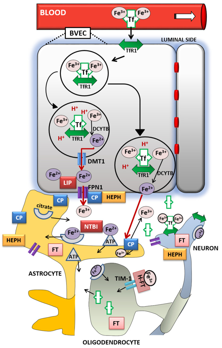
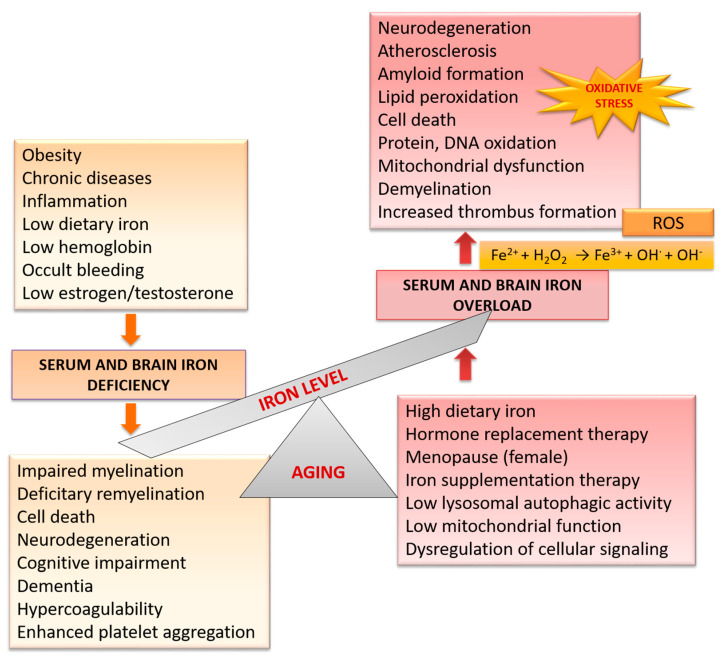
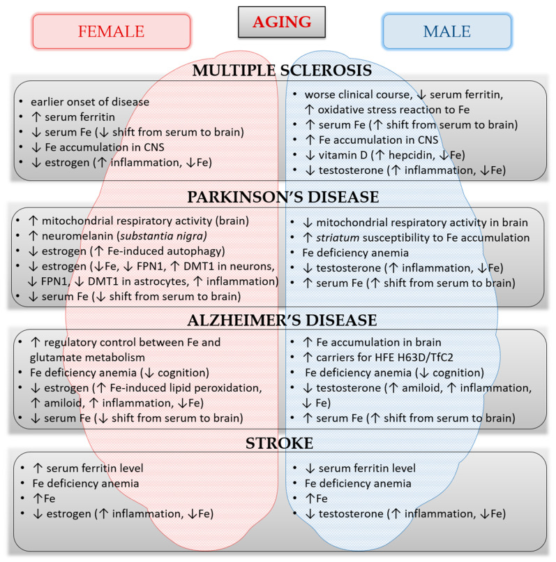
Iron is an essential element that participates in numerous cellular processes. Any disruption of iron homeostasis leads to either iron deficiency or iron overload, which can be detrimental for humans' health, especially in elderly. Each of these changes contributes to the faster development of many neurological disorders or stimulates progression of already present diseases. Age-related cellular and molecular alterations in iron metabolism can also lead to iron dyshomeostasis and deposition. Iron deposits can contribute to the development of inflammation, abnormal protein aggregation, and degeneration in the central nervous system (CNS), leading to the progressive decline in cognitive processes, contributing to pathophysiology of stroke and dysfunctions of body metabolism. Besides, since iron plays an important role in both neuroprotection and neurodegeneration, dietary iron homeostasis should be considered with caution. Recently, there has been increased interest in sex-related differences in iron metabolism and iron homeostasis. These differences have not yet been fully elucidated. In this review we will discuss the latest discoveries in iron metabolism, age-related changes, along with the sex differences in iron content in serum and brain, within the healthy aging population and in neurological disorders such as multiple sclerosis, Parkinson's disease, Alzheimer's disease, and stroke.
Keywords: Alzheimer’s disease; Parkinson’s disease; aging; iron metabolism; multiple sclerosis; sex differences; stroke.
Conflict of interest statement
The authors declare no conflict of interest.
Figures
References
Publication types
MeSH terms
Substances
Grants and funding
LinkOut - more resources
Full Text Sources
Medical
