

Relationship between Visual Perception and Microstructural Change of the Superior Longitudinal Fasciculus in Patients with Brain Injury in the Right Hemisphere: A Preliminary Diffusion Tensor Tractography Study
- PMID: 32867118
- PMCID: PMC7555244
- DOI: 10.3390/diagnostics10090641
Relationship between Visual Perception and Microstructural Change of the Superior Longitudinal Fasciculus in Patients with Brain Injury in the Right Hemisphere: A Preliminary Diffusion Tensor Tractography Study
Abstract
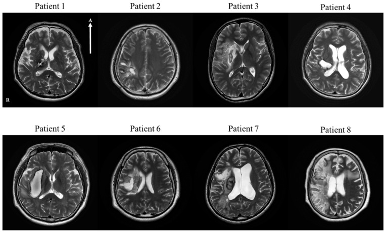
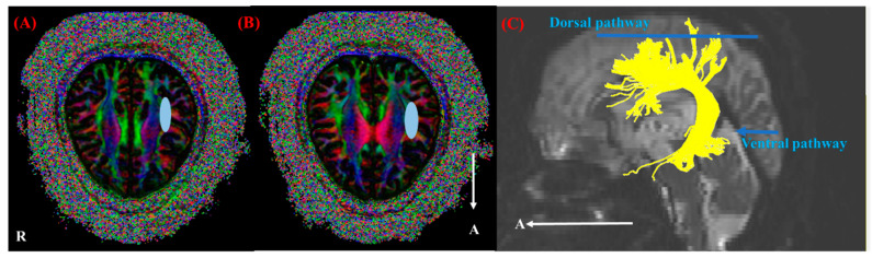
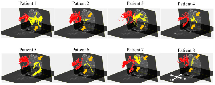
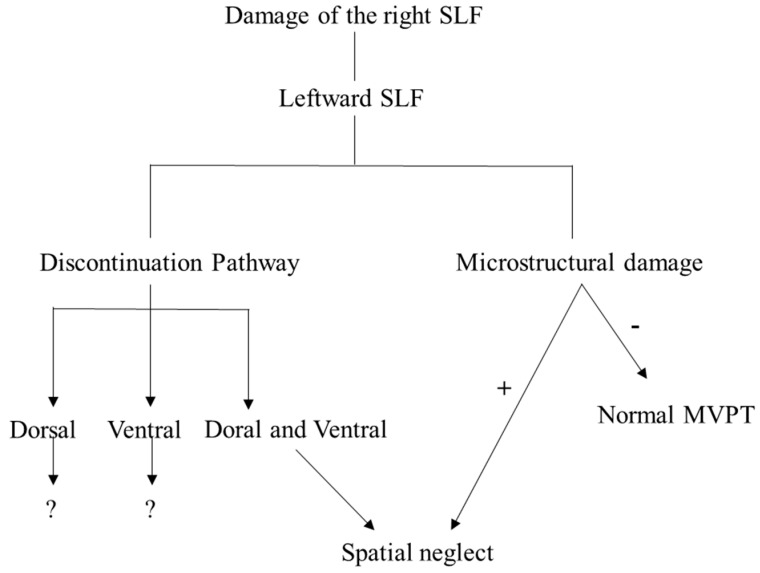
Right hemisphere brain damage often results in visual-spatial deficits. Because various microstructural changes of the superior longitudinal fasciculus (SLF) after a stroke in the right hemisphere affect visual perception, including neglect, the present study investigates the relationship between both microstructural change and lateralization of SLF and visual perception, using diffusion tensor imaging (DTI) in patients with lesions in the right hemisphere. Eight patients with strokes (five patients with intracranial hemorrhage, and three patients with infarction; mean age of 52.5 years) and 16 mean-age-matched healthy control subjects were involved in this study. The visual perception of all eight patients was assessed with the motor-free visual perception test (MVPT), and their SLFs were reconstructed using DTI. The results showed that there was a significant difference between the DTI parameters of the patients and the control subjects. Moreover, patients with microstructural damage to the right SLF showed impairment of visual perception. In patients with damage to both the dorsal and ventral pathways of the right SLF, spatial neglect was present. However, although a leftward SLF asymmetry was revealed in our patients, this lateralization did not show a relationship with visual perception. In conclusion, the microstructural changes of the right SLF play an important role in visual perception, and both pathways contribute to spatial neglect, but leftward lateralization of the right SFL activity after a stroke does not contribute to general visual perception.
Keywords: diffusion tensor imaging; lateralization; neglect; superior longitudinal fasciculus; visual perception.
Conflict of interest statement
The authors declare no conflict of interest.
Figures
Similar articles
-
Correlation of Hemispatial Neglect with White Matter Tract Integrity: A DTI Study.Brain Neurorehabil. 2022 Mar 24;15(1):e6. doi: 10.12786/bn.2022.15.e6. eCollection 2022 Mar. Brain Neurorehabil. 2022. PMID: 36743846 Free PMC article.
-
Structural connectivity in spatial attention network: reconstruction from left hemispatial neglect.Brain Imaging Behav. 2018 Apr;12(2):309-323. doi: 10.1007/s11682-017-9698-7. Brain Imaging Behav. 2018. PMID: 28290071
-
Common brain networks for distinct deficits in visual neglect. A combined structural and tractography MRI approach.Neuropsychologia. 2018 Jul 1;115:167-178. doi: 10.1016/j.neuropsychologia.2017.10.018. Epub 2017 Oct 18. Neuropsychologia. 2018. PMID: 29054427
-
Chronic spatial working memory deficit associated with the superior longitudinal fasciculus: a study using voxel-based lesion-symptom mapping and intraoperative direct stimulation in right prefrontal glioma surgery.J Neurosurg. 2016 Oct;125(4):1024-1032. doi: 10.3171/2015.10.JNS1591. Epub 2016 Feb 19. J Neurosurg. 2016. PMID: 26894458
-
Superior Longitudinal Fasciculus: A Review of the Anatomical Descriptions With Functional Correlates.Front Neurol. 2022 Apr 27;13:794618. doi: 10.3389/fneur.2022.794618. eCollection 2022. Front Neurol. 2022. PMID: 35572948 Free PMC article. Review.
Cited by
-
Implementing New Technologies to Improve Visual-Spatial Functions in Patients with Impaired Consciousness.Int J Environ Res Public Health. 2022 Mar 5;19(5):3081. doi: 10.3390/ijerph19053081. Int J Environ Res Public Health. 2022. PMID: 35270773 Free PMC article.
-
The Effect of Task-Oriented Training on Upper-Limb Function, Visual Perception, and Activities of Daily Living in Acute Stroke Patients: A Pilot Study.Int J Environ Res Public Health. 2022 Mar 8;19(6):3186. doi: 10.3390/ijerph19063186. Int J Environ Res Public Health. 2022. PMID: 35328874 Free PMC article. Clinical Trial.
-
Abnormal White Matter Microstructure in the Limbic System Is Associated With Tuberous Sclerosis Complex-Associated Neuropsychiatric Disorders.Front Neurol. 2022 Mar 14;13:782479. doi: 10.3389/fneur.2022.782479. eCollection 2022. Front Neurol. 2022. PMID: 35359647 Free PMC article.
-
White Matter Function and Network Abnormalities in Patients with Diabetic Retinopathy.Diabetes Metab Syndr Obes. 2024 Nov 3;17:4149-4166. doi: 10.2147/DMSO.S492099. eCollection 2024. Diabetes Metab Syndr Obes. 2024. PMID: 39512603 Free PMC article.
-
Widespread White Matter Alterations in Patients With Visual Snow Syndrome.Front Neurol. 2021 Sep 21;12:723805. doi: 10.3389/fneur.2021.723805. eCollection 2021. Front Neurol. 2021. PMID: 34621237 Free PMC article.
References
LinkOut - more resources
Full Text Sources
