. 2020 Aug 27;13(9):213.
doi: 10.3390/ph13090213.

Proteomics Reveals the Potential Protective Mechanism of Hydrogen Sulfide on Retinal Ganglion Cells in an Ischemia/Reperfusion Injury Animal Model

Affiliations

Proteomics Reveals the Potential Protective Mechanism of Hydrogen Sulfide on Retinal Ganglion Cells in an Ischemia/Reperfusion Injury Animal Model

Hanhan Liu et al. Pharmaceuticals (Basel). .

Abstract

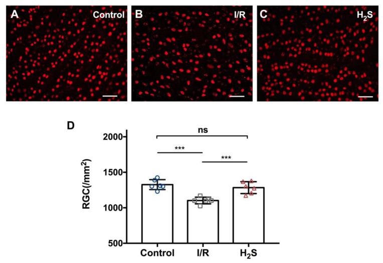
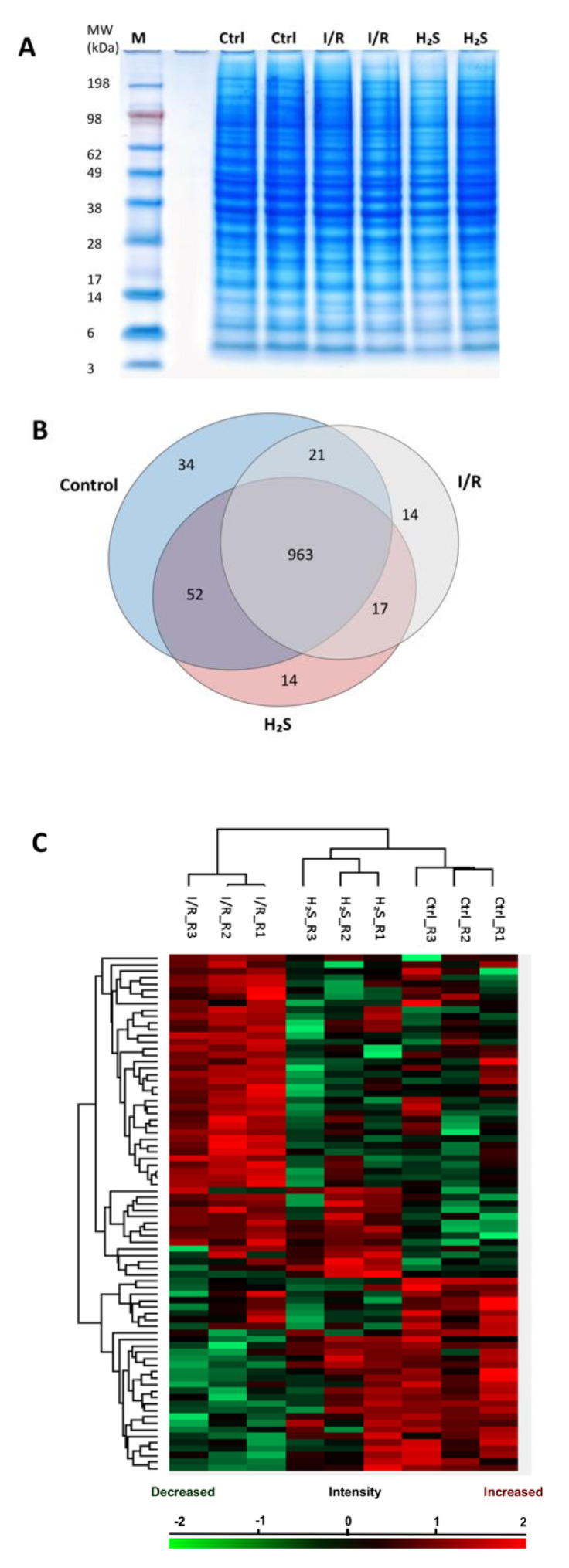
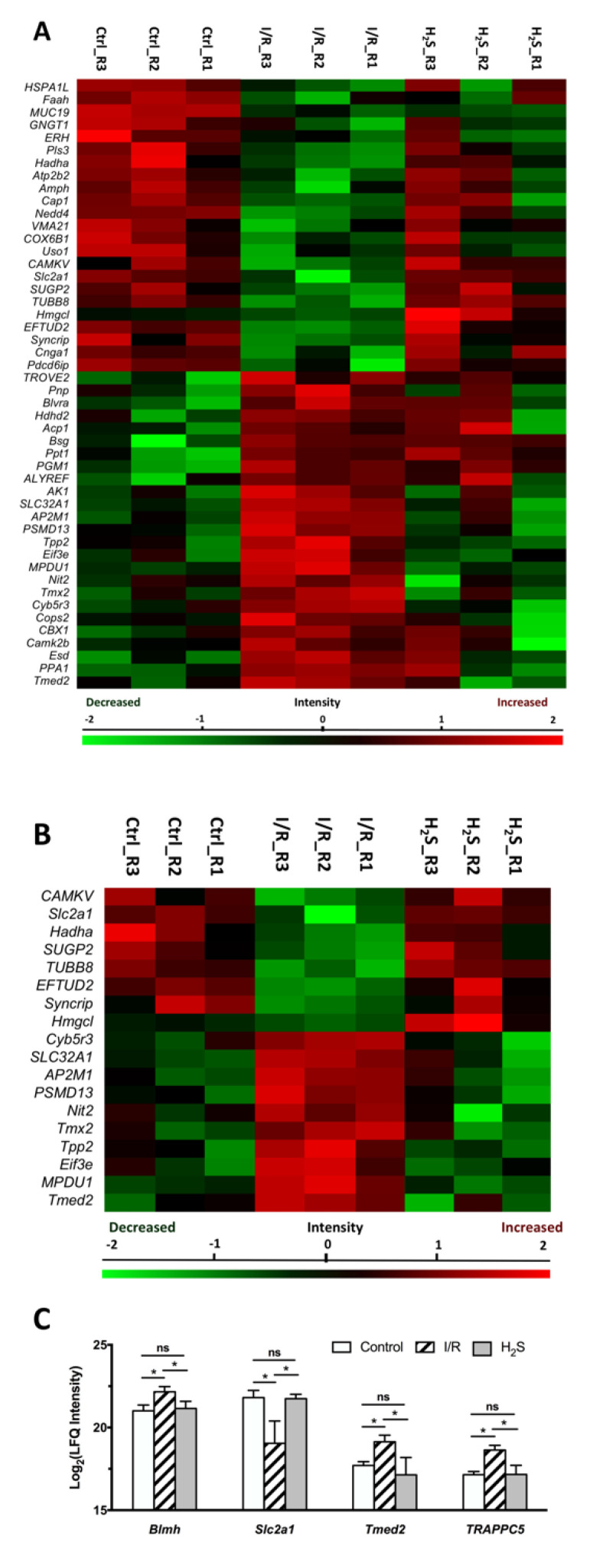
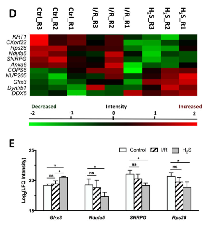
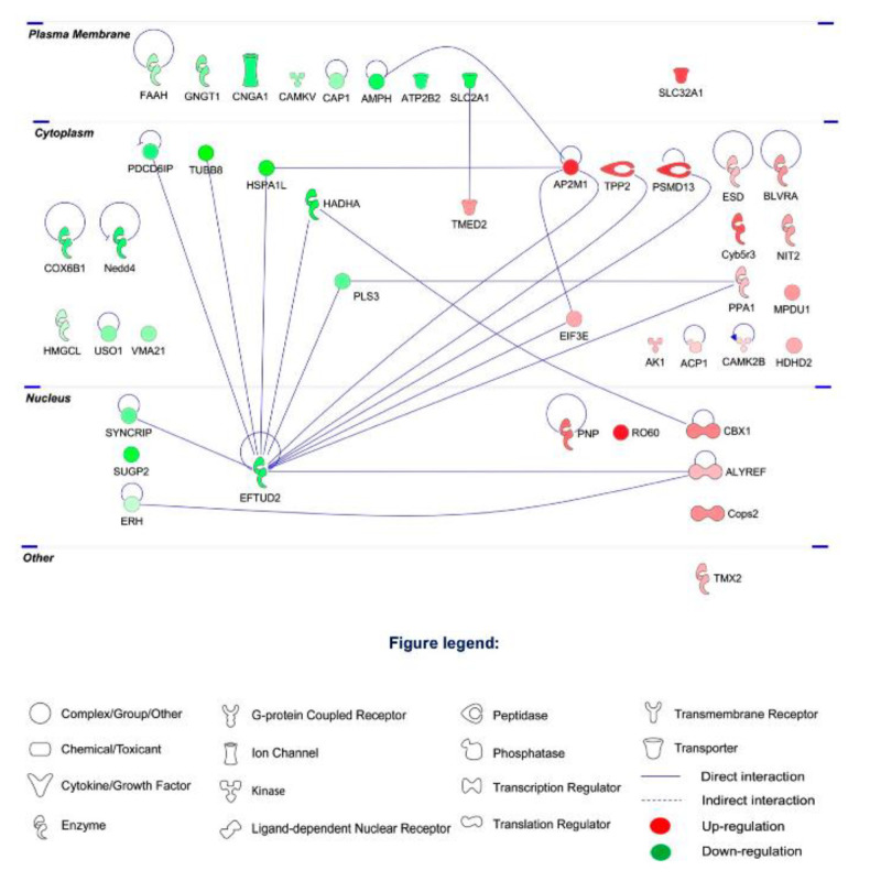
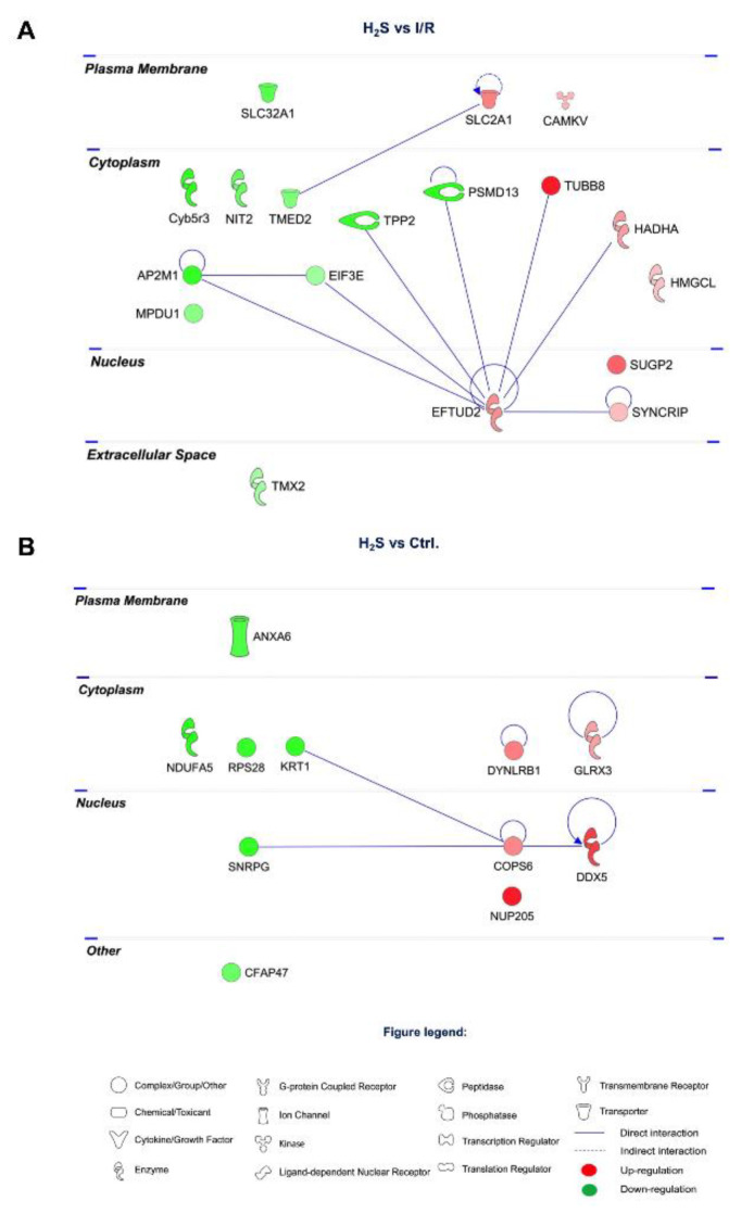
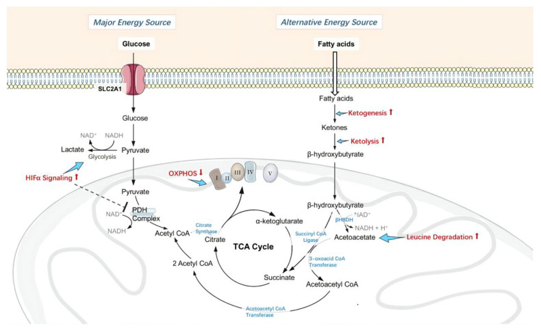
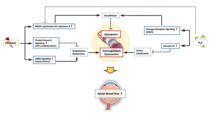
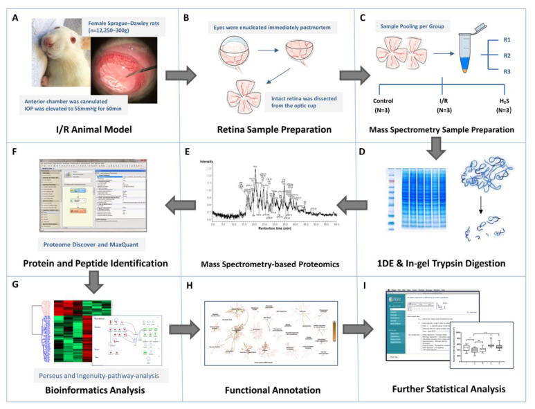
Glaucoma is the leading cause of irreversible blindness and is characterized by progressive retinal ganglion cell (RGC) degeneration. Hydrogen sulfide (H2S) is a potent neurotransmitter and has been proven to protect RGCs against glaucomatous injury in vitro and in vivo. This study is to provide an overall insight of H2S's role in glaucoma pathophysiology. Ischemia-reperfusion injury (I/R) was induced in Sprague-Dawley rats (n = 12) by elevating intraocular pressure to 55 mmHg for 60 min. Six of the animals received intravitreal injection of H2S precursor prior to the procedure and the retina was harvested 24 h later. Contralateral eyes were assigned as control. RGCs were quantified and compared within the groups. Retinal proteins were analyzed via label-free mass spectrometry based quantitative proteomics approach. The pathways of the differentially expressed proteins were identified by ingenuity pathway analysis (IPA). H2S significantly improved RGC survival against I/R in vivo (p < 0.001). In total 1115 proteins were identified, 18 key proteins were significantly differentially expressed due to I/R and restored by H2S. Another 11 proteins were differentially expressed following H2S. IPA revealed a significant H2S-mediated activation of pathways related to mitochondrial function, iron homeostasis and vasodilation. This study provides first evidence of the complex role that H2S plays in protecting RGC against I/R.

Keywords: glaucoma; hydrogen sulfide; label-free mass spectrometry; mitochondrial function; neuronal apoptosis; signalling pathways.


Conflict of interest statement

The authors declare no conflict of interest.

Figures

Figure 1
Figure 1
Pre-treatment with H2S protects RGC against ischemia-reperfusion injury in vivo. (AC) Representative fluorescence microscopy of Brn3a staining of retinal explants 24 h after inducing ischemia/reperfusion injury in vivo. (D) Compared to the contralateral control (1310.6 ± 236.4 RGC/mm2), ischemia-reperfusion injury resulted in significant RGC loss in experimental eyes (1101.7 ± 116.4 RGC/mm2). Pretreatment with GYY4137 showed a significant reduction of RGC loss in vivo (1295.4 ± 136.1 RGC/mm2). Total number of RGC between control and H2S treated group is not significantly different (*** p < 0.0005, n = 6/group, means ± SD, scale bar = 50 μm).
Figure 2
Figure 2
Retinal proteome of Ischemia-reperfusion (I/R) injured and H2S pre-treated retinae. (A) Representative retinal protein profiles of both I/R injured and H2S pre-treated retinae compared to non-operated retinae (designated as CTRL) resolved in PAGE gel stained with colloidal blue. M: marker. (B) Venn diagram depicting overlaps of identified retinal proteins in I/R injured and H2S pre-treated groups compared to the control group. (C) Heat map depicts the hierarchical clustering of the differentially expressed retinal proteins in I/R injured and H2S pre-treated group compared to the control group, H2S pre-treat group demonstrated a more similar protein makeup to the control group (detailed list of proteins can also be found in Supplementary Data S2).
Figure 3
Figure 3
Differential expression profiles of retinal proteins in control, I/R and H2S. The relative protein abundance is represented as heat maps of each protein clusters. (A) A cluster of 48 proteins, the abundance of which were significantly altered due to I/R injury (also see Table 1); (B) a cluster of 18 proteins, the abundance of which were significantly altered due to I/R injury and restored by H2S (Table 2); (D) a cluster of 11 proteins, which were significantly differentially expressed in H2S group (Table 3). Charts showing the different expression profiles of (C) some of the significantly (* p < 0.05) dysregulated proteins in I/R group that were restored due to administration of H2S, (E) some of the significantly differentially expressed retinal proteins in H2S group.
Figure 3
Figure 3
Differential expression profiles of retinal proteins in control, I/R and H2S. The relative protein abundance is represented as heat maps of each protein clusters. (A) A cluster of 48 proteins, the abundance of which were significantly altered due to I/R injury (also see Table 1); (B) a cluster of 18 proteins, the abundance of which were significantly altered due to I/R injury and restored by H2S (Table 2); (D) a cluster of 11 proteins, which were significantly differentially expressed in H2S group (Table 3). Charts showing the different expression profiles of (C) some of the significantly (* p < 0.05) dysregulated proteins in I/R group that were restored due to administration of H2S, (E) some of the significantly differentially expressed retinal proteins in H2S group.
Figure 4
Figure 4
Protein–protein interaction networks of the differentially expressed retinal proteins due to I/R comparing to CTRL group. The major interaction networks of differentially expressed retinal proteins obtained by IPA analysis (QIAGEN Inc., https://www.qiagenbioinformatics.com/products/ingenuity-pathway-analysis) in I/R group.
Figure 5
Figure 5
Protein–protein interaction networks of the differentially expressed retinal proteins modulated by H2S. (A) 18 proteins restored by H2S comparing to I/R group. (B) 11 differentially expressed retinal proteins modulated by H2S comparing to CTRL group. The major interaction networks of restored retinal proteins obtained by IPA analysis in H2S group.
Figure 6
Figure 6
Changes in retinal metabolism, mitochondrial homeostasis and function. Administration of H2S actively inhibited pyruvate dehydrogenase (PDH) complex activity by upregulating HIF1α signaling, therefore limited using glucose as energy source, and suppressed oxidative phosphorylation by inhibiting Complex I activity, consequently increased intracellular oxygen tension under ischemia and limited ROS production during reperfusion. Furthermore, H2S promotes the utilization of ketone bodies as an alternative energy source, which is more energy-efficient than glucose, thus permitted continuous adenosine triphosphate (ATP) production. H2S enhanced the ability of retinal neurons to withstand metabolic stress induced by I/R, therefore less neuronal cell loss.
Figure 7
Figure 7
Changes in retinal vascular function. Due to I/R, PKA (protein kinase A) signaling and NADH-cytochrome b5 reductase 3(Cyb5r3) is upregulated as self-protective mechanism to maintain endothelial function and vasodilation, while eNOS signaling is downregulated as an indication of endothelial dysfunction. Administration of H2S activated estrogen receptor signaling to facilitate vascular relaxation. H2S also reduced the abundance of annexin A6, which plays a central role in artery calcification. Combined together, H2S protected the blood flow regulatory mechanisms and enabled a less constricted vascular environment in retina against I/R injury.
Figure 8
Figure 8
Workflow overview. (A) I/R injury was induced in the left eyes of adult female Sprague-Dawley rats (n = 12), six of which received intravitreal injection of GYY4137, an H2S slow-releasing precursor, shortly before intervention. (B) Animals were executed 24 h after intervention, retinae were harvested immediately postmortem. (CE) Retinae from contralateral eyes were designated as controls. Retinal samples were immediately weighed and lysed by T-PER tissue protein extraction reagent and Bullet Blender Storm. Six retinal protein samples from respective groups were pooled equally into three biological replicates after protein measurements, represented by RI, R2 and R3 (N = 3 replicates per group), and subsequently subjected to PAGE. The protein bands were sliced and digested by trypsin prior to proteomic analysis by LC-ESI-MS/MS. (FH) The emerging datasets were subjected to robust bioinformatics analyses and functional annotations to identify the differential protein expressions and protein interaction networks. (I) To confirm the result of I/R injury and protective effect of H2S in retina, one quarter of each retinal sample (n = 6 per group) were subjected to immunohistochemistry staining against Brn3a for RGC quantification. The averaged RGC density was calculated per mm2. Significance of difference between groups was determined by 1-way ANOVA.

Similar articles

Cited by

References

    1. Flaxman S.R., Bourne R.R.A., Resnikoff S., Ackland P., Braithwaite T., Cicinelli M.V., Das A., Jonas J.B., Keeffe J., Kempen J.H., et al. Global causes of blindness and distance vision impairment 1990–2020: A systematic review and meta-analysis. Lancet Glob. Health. 2017;5:e1221–e1234. doi: 10.1016/S2214-109X(17)30393-5. - DOI - PubMed
    1. Almasieh M., Wilson A.M., Morquette B., Cueva Vargas J.L., Di Polo A. The molecular basis of retinal ganglion cell death in glaucoma. Prog. Retin. Eye Res. 2012;31:152–181. doi: 10.1016/j.preteyeres.2011.11.002. - DOI - PubMed
    1. Keating D.J. Mitochondrial dysfunction, oxidative stress, regulation of exocytosis and their relevance to neurodegenerative diseases. J. Neurochem. 2008;104:298–305. doi: 10.1111/j.1471-4159.2007.04997.x. - DOI - PubMed
    1. Tabassum R., Jeong N.Y., Jung J. Therapeutic importance of hydrogen sulfide in age-associated neurodegenerative diseases. Neural Regen. Res. 2020;15:653–662. doi: 10.4103/1673-5374.266911. - DOI - PMC - PubMed
    1. Snyder S.H., Jaffrey S.R., Zakhary R. Nitric oxide and carbon monoxide: Parallel roles as neural messengers. Brain Res. Brain Res. Rev. 1998;26:167–175. doi: 10.1016/S0165-0173(97)00032-5. - DOI - PubMed

LinkOut - more resources