

Development of Magnetic Torque Stimulation (MTS) Utilizing Rotating Uniform Magnetic Field for Mechanical Activation of Cardiac Cells
- PMID: 32867131
- PMCID: PMC7557977
- DOI: 10.3390/nano10091684
Development of Magnetic Torque Stimulation (MTS) Utilizing Rotating Uniform Magnetic Field for Mechanical Activation of Cardiac Cells
Abstract
Regulation of cell signaling through physical stimulation is an emerging topic in biomedicine.
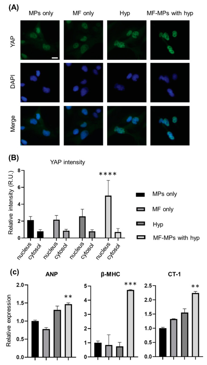
Background: While recent advances in biophysical technologies show capabilities for spatiotemporal stimulation, interfacing those tools with biological systems for intact signal transfer and noncontact stimulation remains challenging. Here, we describe the use of a magnetic torque stimulation (MTS) system combined with engineered magnetic particles to apply forces on the surface of individual cells. MTS utilizes an externally rotating magnetic field to induce a spin on magnetic particles and generate torsional force to stimulate mechanotransduction pathways in two types of human heart cells-cardiomyocytes and cardiac fibroblasts.
Methods: The MTS system operates in a noncontact mode with two magnets separated (60 mm) from each other and generates a torque of up to 15 pN µm across the entire area of a 35-mm cell culture dish. The MTS system can mechanically stimulate both types of human heart cells, inducing maturation and hypertrophy.
Results: Our findings show that application of the MTS system under hypoxic conditions induces not only nuclear localization of mechanoresponsive YAP proteins in human heart cells but also overexpression of hypertrophy markers, including β-myosin heavy chain (βMHC), cardiotrophin-1 (CT-1), microRNA-21 (miR-21), and transforming growth factor beta-1 (TGFβ-1).
Conclusions: These results have important implications for the applicability of the MTS system to diverse in vitro studies that require remote and noninvasive mechanical regulation.
Keywords: cardiac cells; hypoxia; magnetogenetics; mechanotransduction; torsional magnetic stimulation.
Conflict of interest statement
The authors declare no competing financial interest.
Figures


Similar articles
-
Recent Advances in Magnetic-Nanomaterial-Based Mechanotransduction for Cell Fate Regulation.Adv Mater. 2018 Apr;30(17):e1705673. doi: 10.1002/adma.201705673. Epub 2018 Mar 15. Adv Mater. 2018. PMID: 29543348 Review.
-
Molecular Tension Probes for Imaging Forces at the Cell Surface.Acc Chem Res. 2017 Dec 19;50(12):2915-2924. doi: 10.1021/acs.accounts.7b00305. Epub 2017 Nov 21. Acc Chem Res. 2017. PMID: 29160067 Free PMC article. Review.
-
Magnetically actuated tissue engineered scaffold: insights into mechanism of physical stimulation.Nanoscale. 2016 Feb 14;8(6):3386-99. doi: 10.1039/c5nr05500h. Epub 2016 Jan 21. Nanoscale. 2016. PMID: 26790538 Free PMC article.
-
Mechanical stretch via transforming growth factor-β1 activates microRNA-208a to regulate hypertrophy in cultured rat cardiac myocytes.J Formos Med Assoc. 2013 Oct;112(10):635-43. doi: 10.1016/j.jfma.2013.01.002. Epub 2013 Feb 4. J Formos Med Assoc. 2013. PMID: 24120154
-
Magnetic Nanotweezers for Interrogating Biological Processes in Space and Time.Acc Chem Res. 2018 Apr 17;51(4):839-849. doi: 10.1021/acs.accounts.8b00004. Epub 2018 Mar 28. Acc Chem Res. 2018. PMID: 29589897 Free PMC article. Review.
Cited by
-
Biomimetic Cardiac Tissue Models for In Vitro Arrhythmia Studies.Biomimetics (Basel). 2023 Oct 14;8(6):487. doi: 10.3390/biomimetics8060487. Biomimetics (Basel). 2023. PMID: 37887618 Free PMC article. Review.
-
Assessing the combination of magnetic field stimulation, iron oxide nanoparticles, and aligned electrospun fibers for promoting neurite outgrowth from dorsal root ganglia in vitro.Acta Biomater. 2021 Sep 1;131:302-313. doi: 10.1016/j.actbio.2021.06.049. Epub 2021 Jul 13. Acta Biomater. 2021. PMID: 34271170 Free PMC article.
-
Dynamic mechanobiology of cardiac cells and tissues: Current status and future perspective.Biophys Rev (Melville). 2023 Mar;4(1):011314. doi: 10.1063/5.0141269. Epub 2023 Mar 29. Biophys Rev (Melville). 2023. PMID: 37008887 Free PMC article. Review.
-
Magnetogenetics: remote activation of cellular functions triggered by magnetic switches.Nanoscale. 2022 Feb 10;14(6):2091-2118. doi: 10.1039/d1nr06303k. Nanoscale. 2022. PMID: 35103278 Free PMC article. Review.
-
Biomaterials Tailoring at the Nanoscale for Tissue Engineering and Advanced Therapies.Nanomaterials (Basel). 2021 May 6;11(5):1221. doi: 10.3390/nano11051221. Nanomaterials (Basel). 2021. PMID: 34066333 Free PMC article.
References
Grants and funding
LinkOut - more resources
Full Text Sources
Other Literature Sources
