Review
. 2020 Aug 27;10(9):1244.
doi: 10.3390/biom10091244.

Aggrecan, the Primary Weight-Bearing Cartilage Proteoglycan, Has Context-Dependent, Cell-Directive Properties in Embryonic Development and Neurogenesis: Aggrecan Glycan Side Chain Modifications Convey Interactive Biodiversity

Affiliations
Review

Aggrecan, the Primary Weight-Bearing Cartilage Proteoglycan, Has Context-Dependent, Cell-Directive Properties in Embryonic Development and Neurogenesis: Aggrecan Glycan Side Chain Modifications Convey Interactive Biodiversity

Anthony J Hayes et al. Biomolecules. .

Abstract

This review examines aggrecan's roles in developmental embryonic tissues, in tissues undergoing morphogenetic transition and in mature weight-bearing tissues. Aggrecan is a remarkably versatile and capable proteoglycan (PG) with diverse tissue context-dependent functional attributes beyond its established role as a weight-bearing PG. The aggrecan core protein provides a template which can be variably decorated with a number of glycosaminoglycan (GAG) side chains including keratan sulphate (KS), human natural killer trisaccharide (HNK-1) and chondroitin sulphate (CS). These convey unique tissue-specific functional properties in water imbibition, space-filling, matrix stabilisation or embryonic cellular regulation. Aggrecan also interacts with morphogens and growth factors directing tissue morphogenesis, remodelling and metaplasia. HNK-1 aggrecan glycoforms direct neural crest cell migration in embryonic development and is neuroprotective in perineuronal nets in the brain. The ability of the aggrecan core protein to assemble CS and KS chains at high density equips cartilage aggrecan with its well-known water-imbibing and weight-bearing properties. The importance of specific arrangements of GAG chains on aggrecan in all its forms is also a primary morphogenetic functional determinant providing aggrecan with unique tissue context dependent regulatory properties. The versatility displayed by aggrecan in biodiverse contexts is a function of its GAG side chains.

Keywords: HNK-1 trisaccharide; aggrecan; cellular regulation; extracellular matrix; glycosaminoglycan; tissue morphogenesis.


Conflict of interest statement

The authors declare no conflict of interest and have no financial disclosures to make.

Figures
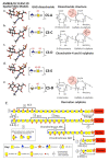
Figure 1
Figure 1
Amber/GLYCAM 3D stick structures of chondroitin sulphate isomers depicting their 3D conformations, disaccharide compositions and Haworth projection disaccharide structures showing sulphation positions (ae). Schematic depiction of the structural organisation of the chondroitin sulphate glycosaminoglycan side chains of aggrecan depicting specific structural features of areas of the chain detected by monoclonal antibodies, putative sulphation patterns, linkage region structure to aggrecan core protein and non-reducing terminal structures (f). These regions on the CS side chain are numbered 1–8. Key: (1) Non-reducing terminal groups present on some cartilage aggrecan CS chains; (2) 3-B-3(−) CS sulphation motif is also present as a non-reducing terminal component on some chains; (3) putative region on CS chain identified by MAb 6C3; (4) putative region on CS chain identified by MAb 4C3; (5) putative region on CS chain identified by MAb 7D4; (6) CS linkage attachment region to Serine residues of the aggrecan core protein; (7) 3-B-3(+) CS sulphation stub epitope generated by exhaustive digestion of the CS chain by chondroitinase ABC and recognised by MAb 3-B-3; and (8) 2-B-6(+) CS sulphation stub epitope generated by exhaustive digestion of the CS chain by chondroitinase ABC and recognised by MAb 2-B-6. Note: Regions 3–5 of the CS chains containing the 6-C-3, 4-C-3 and 7-D-4 reactivity are susceptible to chondroitinase ABC digestion; thus, in graded partial digestions, the 6-C-3 and 4-C-3 reactivity can be selectively removed leaving the 7-D-4 reactive region intact. However, this is also susceptible to chondroitinase ABC, and exhaustive digestion conditions eventually lead to generation of the unsaturated 3-B-3(+) and 2-B-6(+) stub epitopes attached to the linkage region, as shown in this diagram. In (f), the structures shown hypothetical many features such as the sulphation positions on GAGs are variable; the depictions shown are thus generalisations based on literature data.
Figure 2
Figure 2
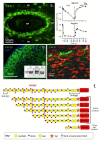
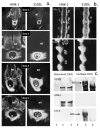
Immunofluorescent localisation of the 3-B-3(−) CS sulphation motifs on aggrecan associated with rudiment cartilage of a rat intervertebral disc (a) and demonstration of the generation of the 3-B-3(−) epitope by digestion of aggrecan and serglycin as model proteoglycans with hyaluronidase-4 (HYAL4) (b). Immunolocalisation of the 3-B-3(−) and 7-D-4 CS sulphation motifs in developmental human foetal knee joint cartilage (14 weeks gestational age) (c,d). The inset of (e) shows foetal aggrecan samples separated by native composite agarose polyacrylamide gel electrophoresis and blotted to nitrocellulose for detection of the 3-B-3(−) and 7-D-4 proteoglycan populations. Two aggrecan populations are discernible. The 3-B-3 (−) CS sulphation epitope has a widespread distribution in the developing rudiment cartilage, whereas the 7-D-4 epitope has a more discrete distribution pattern in small stem cell niches in the cartilage surface. A schematic depicting a typical CS chain and digestion products generated by endoglycolytic cleavage by HYAL4 generating the 3-B-3(−) non-reducing terminal on the cleaved CS chain (f). Exhaustive digestion of CS by chondroitinase (Ch’ase) ABC also depolymerises the CS chain but generates a 3-B-3(+) stub epitope attached to the CS linkage attachment to aggrecan core protein. Inset image (e) modified from [50]. (a,c,d) Images supplied courtesy of Prof B. Caterson, University of Cardiff, UK. As already shown in this manuscript approximately ~1–2 in every seven non-reducing termini of CS chains in cartilage are terminated in the 3-B-3(−) epitope and these vary with age and cartilage type. The 3-B-3(−) epitope is a marker of tissue morphogenesis [36,51,52]. Stem cells are surrounded in proteoglycans decorated with this CS motif [8,9,10]. This motif is also released into synovial fluid in degenerative conditions such as OA [53,54,55,56]. Recently, Farrugia et al. [57] showed that mast cells synthesised HYAL4, a CS hydrolase that could generate the 3-B-3(−) motif in the CS chains of aggrecan and Serglycin in vitro.
Figure 3
Figure 3
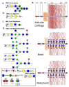
Structure of human natural killer-1 epitope (HNK-1) present on: N-glycans (a); notochordal aggrecan (b); brain phosphacan (c); and myelin Po glycoprotein in nervous tissues (d). Schematic depictions of representative aggrecan structures in articular cartilage, brain perineuronal nets and notochord in embryonic developmental tissues showing their variable relative KS contents and the presence of HNK-1 carbohydrate substitution in brain and notochordal aggrecan (e).
Figure 4
Figure 4
Demonstration of the modulation of growth plate cartilage morphogenesis by aggrecan in wild type (WT) (A) (ah) and nanomelic E7-E12 chick tibia (B) (ar). The ISH images presented demonstrate the expression of: FGFR3 (a); type X collagen (b); and Indian Hedgehog (IHH) (c) in WT (ac); and nanomelic growth plate (df) in E12 (af); E7 (gl); and E8 chick tibia (mr). Images modified from [134] with permission using open access.
Figure 5
Figure 5
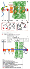
Structural organisation of aggrecan depicting the CS, KS and HS GAG chain distributions (a) and T cell receptor epitopes on the G1 and G3 globular domains (b). The aggrecanopathies showing regions of aggrecan affected by these mutations and the diseases that result (c).
Figure 6
Figure 6
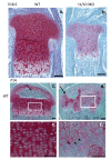
Chondroitin sulphate N-acetylgalactosaminyltransferase-1 and -2 (T1/2) knockout induces dwarfism in mice and altered cartilage structural organisation of the femoral condyle, its ossification centre and growth plate in wild type mice (a,c,e) and T1/2 knockout mice (b,d,f). The boxed area in (c,d) is depicted at higher magnification in (e,f). Safarin O-Fast green stain depicting aggrecan GAG distribution. Arrows depict normal ossification centre in (c) and abnormal structural organisation in T1/2 knockout mice in (d,f). Figure modified from [166]. Figure reproduced under the terms of the Creative Commons Attribution Licence Copyright: 2017 Shimbo et al. [166].
Figure 7
Figure 7
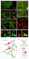
Visualisation of: perineuronal net structures (a,b); vascular features (c); and ganglion cells (d,e) in cerebellum and dorsal root ganglia using MAb 1B5 in confocal images. Immunolocalisation of CS Isomer 1B5 in paraformaldehyde fixed 20-μm cryo-sections of 24-month-old Wistar rat brain and lumbar dorsal root ganglia. Confocal z-stacked images of IB5 CS stub epitope generated by chondroitinase ABC digestion using Alexa 488 secondary antibody for detection and propidium iodide nuclear counterstain, mounted under coverslips using Vectasheld mountant. Images courtesy of Prof B. Caterson, University of Cardiff. Copyright Caterson, Hayes 2012 (af). Immunolocalisation of perineuronal nets surrounding isolated neurons in murine visual cortex using: antibody to parvalbumin (g,i); Wisteria floribunda lectin (h); and in a merged image (j). A schematic model of the perineuronal net structure in the boxed area in (k) showing its constituent lectican proteoglycans (aggrecan, versican, neurocan and brevican) interacting with tenascin hexabrachion and hyaluronan to form an aggregate structure stabilised by link protein. A key is provided to explain items in (k,l). Figure modified from [170] under Open Access under the auspices of a Creative Commons Attribution License.
Figure 8
Figure 8
Demonstration of: Sox 10 (a,e); Seraf (b); and HNK-1 epitope (c,d) expression in migratory neural crest cells (a,c) and in whole mount chick embryos (b,d,e). (a,c,d) In-situ hybridisation images. (b,e) Immunolocalisations with specific antibodies. Seraf (Schwann cell-specific EGF-like repeat autocrine factor) is a unique protein expressed by avian embryo Schwann cell precursor cells [175]. Images reproduced from [176] under the auspices of attribution-non-commercial-no derivatives 4.0 international licence (CC BY-NC-ND 4.0).
Figure 9
Figure 9
Fluorescent immunolocalisation of HNK-1 and aggrecan S103L epitope in 2–5-day-old chick trunk sections associated with the neural tube (NT) and notochord (N) development (a,b) and in Western blots (c) of purified chick notochordal and cartilage aggrecan. Keratan sulphate was also immunolocalised on blots using MAb 5-D-4. Notochordal aggrecan was S103L and HNK-1 positive but did not contain KS and was of a smaller molecular weight; the 400-kDa cartilage aggrecan species was not detected. Cartilage aggrecan did not stain with the HNK-1 antibody. The S103L antibody identifies the sequence 585XXX Glu Ileu Ser Gly Phe Leu Ser Gly Asp Arg615 in the CS attachment domain of aggrecan. Images reproduced from [177,178,179].

Similar articles

Cited by

References

    1. Feng H., Danfelter M., Strömqvist B., Heinegård D. Extracellular Matrix in Disc Degeneration. J. Bone Jt. Surg. Am. Vol. 2006;88:25. doi: 10.2106/jbjs.e.01341. - DOI - PubMed
    1. Hardingham T.E., Fosang A.J., Dudhia J. The structure, function and turnover of aggrecan, the large aggregating proteoglycan from cartilage. Eur. J. Clin. Chem. Clin. Biochem. 1994;32:249–257. - PubMed
    1. Heinegard D. Fell-Muir Lecture: Proteoglycans and More—From Molecules to Biology. Int. J. Exp. Pathol. 2009;90:575–586. doi: 10.1111/j.1365-2613.2009.00695.x. - DOI - PMC - PubMed
    1. Kiani C., Chen L., Wu Y.J., Yee A.J., Yang B.B. Structure and function of aggrecan. Cell Res. 2002;12:19–32. doi: 10.1038/sj.cr.7290106. - DOI - PubMed
    1. Roughley P.J., Lee E.R. Cartilage proteoglycans: Structure and potential functions. Microsc. Res. Tech. 1994;28:385–397. doi: 10.1002/jemt.1070280505. - DOI - PubMed

Publication types

LinkOut - more resources