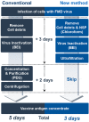
Efficient Removal of Non-Structural Protein Using Chloroform for Foot-and-Mouth Disease Vaccine Production
- PMID: 32867254
- PMCID: PMC7565013
- DOI: 10.3390/vaccines8030483
Efficient Removal of Non-Structural Protein Using Chloroform for Foot-and-Mouth Disease Vaccine Production
Abstract
To differentiate foot-and-mouth disease (FMD)-infected animals from vaccinated livestock, non-structural proteins (NSPs) must be removed during the FMD vaccine manufacturing process. Currently, NSPs cannot be selectively removed from FMD virus (FMDV) culture supernatant. Therefore, polyethylene glycol (PEG) is utilized to partially separate FMDV from NSPs. However, some NSPs remain in the FMD vaccine, which after repeated immunization, may elicit NSP antibodies in some livestock. To address this drawback, chloroform at a concentration of more than 2% (v/v) was found to remove NSP efficiently without damaging the FMDV particles. Contrary to the PEG-treated vaccine that showed positive NSP antibody responses after the third immunization in goats, the chloroform-treated vaccine did not induce NSP antibodies. In addition to this enhanced vaccine purity, this new method using chloroform could maximize antigen recovery and the vaccine production time could be shortened by two days due to omission of the PEG processing phase. To our knowledge, this is the first report to remove NSPs from FMDV culture supernatant by chemical addition. This novel method could revolutionize the conventional processes of FMD vaccine production.
Keywords: chloroform; foot-and-mouth disease virus; inactivated vaccine; non-structural protein; purity.
Conflict of interest statement
The authors declare no conflict of interest.
Figures


Similar articles
-
Application of Heparin Affinity Chromatography to Produce a Differential Vaccine without Eliciting Antibodies against the Nonstructural Proteins of the Serotype O Foot-and-Mouth Disease Viruses.Viruses. 2020 Dec 7;12(12):1405. doi: 10.3390/v12121405. Viruses. 2020. PMID: 33297420 Free PMC article.
-
Factors Involved in Removing the Non-Structural Protein of Foot-and-Mouth Disease Virus by Chloroform and Scale-Up Production of High-Purity Vaccine Antigens.Vaccines (Basel). 2022 Jun 24;10(7):1018. doi: 10.3390/vaccines10071018. Vaccines (Basel). 2022. PMID: 35891182 Free PMC article.
-
A simple and rapid assay to evaluate purity of foot-and-mouth disease vaccine before animal experimentation.Vaccine. 2019 Jun 27;37(29):3825-3831. doi: 10.1016/j.vaccine.2019.05.053. Epub 2019 May 25. Vaccine. 2019. PMID: 31138453
-
Foot-and-mouth disease vaccines: progress and problems.Expert Rev Vaccines. 2016 Jun;15(6):783-9. doi: 10.1586/14760584.2016.1140042. Epub 2016 Jan 22. Expert Rev Vaccines. 2016. PMID: 26760264 Review.
-
Developments in diagnostic techniques for differentiating infection from vaccination in foot-and-mouth disease.Vet J. 2004 Jan;167(1):9-22. doi: 10.1016/s1090-0233(03)00087-x. Vet J. 2004. PMID: 14623146 Review.
Cited by
-
Application of Heparin Affinity Chromatography to Produce a Differential Vaccine without Eliciting Antibodies against the Nonstructural Proteins of the Serotype O Foot-and-Mouth Disease Viruses.Viruses. 2020 Dec 7;12(12):1405. doi: 10.3390/v12121405. Viruses. 2020. PMID: 33297420 Free PMC article.
-
Detection of clinical and subclinical Foot and Mouth Disease Virus in Cattle in Al-Najaf Province.Arch Razi Inst. 2022 Jun 30;77(3):1185-1189. doi: 10.22092/ARI.2022.357621.2074. eCollection 2022 Jun. Arch Razi Inst. 2022. PMID: 36618320 Free PMC article.
-
Factors Involved in Removing the Non-Structural Protein of Foot-and-Mouth Disease Virus by Chloroform and Scale-Up Production of High-Purity Vaccine Antigens.Vaccines (Basel). 2022 Jun 24;10(7):1018. doi: 10.3390/vaccines10071018. Vaccines (Basel). 2022. PMID: 35891182 Free PMC article.
-
A review of foot-and-mouth disease in Ethiopia: epidemiological aspects, economic implications, and control strategies.Virol J. 2023 Dec 15;20(1):299. doi: 10.1186/s12985-023-02263-0. Virol J. 2023. PMID: 38102688 Free PMC article. Review.
-
Post-vaccination Monitoring to Assess Foot-and-Mouth Disease Immunity at Population Level in Korea.Front Vet Sci. 2021 Aug 4;8:673820. doi: 10.3389/fvets.2021.673820. eCollection 2021. Front Vet Sci. 2021. PMID: 34422940 Free PMC article.
References
-
- Murphy F.A., Gibbs E.P.J., Horzinek M.C., Studdert M.J. Veterinary Virology. Elsevier; Amsterdam, The Netherlands: 1999.
-
- International Committee, Biological Standards Commission, International Office of Epizootics . Manual of Diagnostic Tests and Vaccines for Terrestrial Animals: Mammals, Birds, and Bees. Renouf Publishing Co. Ltd.; Ottawa, ON, Canada: 2018. [(accessed on 26 August 2020)]. Available online: https://www.oie.int/fileadmin/Home/eng/Health_standards/tahm/3.01.08_FMD....
Grants and funding
LinkOut - more resources
Full Text Sources

