Review
. 2020 Aug 27;21(17):6213.
doi: 10.3390/ijms21176213.

GM2 Gangliosidoses: Clinical Features, Pathophysiological Aspects, and Current Therapies

Affiliations
Review

GM2 Gangliosidoses: Clinical Features, Pathophysiological Aspects, and Current Therapies

Andrés Felipe Leal et al. Int J Mol Sci. .

Abstract

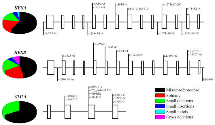
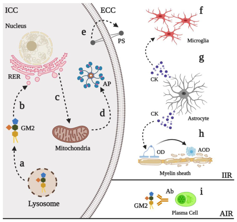
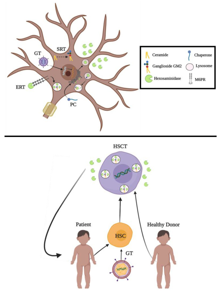
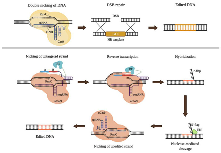
GM2 gangliosidoses are a group of pathologies characterized by GM2 ganglioside accumulation into the lysosome due to mutations on the genes encoding for the β-hexosaminidases subunits or the GM2 activator protein. Three GM2 gangliosidoses have been described: Tay-Sachs disease, Sandhoff disease, and the AB variant. Central nervous system dysfunction is the main characteristic of GM2 gangliosidoses patients that include neurodevelopment alterations, neuroinflammation, and neuronal apoptosis. Currently, there is not approved therapy for GM2 gangliosidoses, but different therapeutic strategies have been studied including hematopoietic stem cell transplantation, enzyme replacement therapy, substrate reduction therapy, pharmacological chaperones, and gene therapy. The blood-brain barrier represents a challenge for the development of therapeutic agents for these disorders. In this sense, alternative routes of administration (e.g., intrathecal or intracerebroventricular) have been evaluated, as well as the design of fusion peptides that allow the protein transport from the brain capillaries to the central nervous system. In this review, we outline the current knowledge about clinical and physiopathological findings of GM2 gangliosidoses, as well as the ongoing proposals to overcome some limitations of the traditional alternatives by using novel strategies such as molecular Trojan horses or advanced tools of genome editing.

Keywords: GM2 gangliosidoses; Sandhoff disease; Tay–Sachs disease; lysosomal storage disorders; therapeutic alternatives; β-Hexosaminidases.


Conflict of interest statement

The authors declare no conflict of interest. The funders had no role in the design of the study; in the collection, analyses, or interpretation of data; in the writing of the manuscript, or in the decision to publish the review.

Figures

Figure 1
Figure 1
M6PR-dependent transport of β-Hexosaminidase A and ganglioside degradation. α and β subunits of Hex are synthesized in the rough endoplasmic reticulum (RER) and transported to the Cis-Golgi network. In this compartment, Hex is subject to N-glycosylations and phosphorylations from Cis-Golgi network to the Trans-Golgi network [33]. Monomers are dimerized in the Trans-Golgi network and coupled to mannose-6 phosphate receptors (M6PR) [33,37]. New vesicles are sorted to both early endosomes (EE) and to the secretory pathway, where can be uptake by neighbor cells through M6PR [37,45]. Hexosaminidases are dissociated from the M6PR in the EE; which allows the M6PR recycling to the Trans-Golgi network by both clathrin-dependent and independent mechanisms [37]. On the other side, gangliosides are placed in caveolae-rich microdomains (CvRM), and in the turnover of the plasma membrane undergo caveolae-mediated endocytosis (CME) [1,38]. New caveosomes containing gangliosides (CCV-GM) reach the EE and further fusion events result in a late endosome, which can be fused with the lysosome to give rise to the endo-lysosome (EL, pH: 4.5) [37]. Gangliosides degradation starts with the hydrolysis of the galactose of the GM1 ganglioside to generate GM2 ganglioside which are harbored on intralysosomal vesicles (ILV) [1]. GM2 interacts with HexA through a GM2AP-mediated mechanism to removes the N-acetylgalactosamine resides [38]. Additional reactions implied in the ganglioside degradation to glucosylceramide (GluCer) are shown. The enzymes of each reaction are as follow: 1 and 4: β-Galactosidase/GM2AP, 2: β-Hexosaminidase A/GM2AP, and 3: Neuraminidase. LacCer: Lactoceramide. ECC: Extracellular compartment. ICC: Intracellular compartment.
Figure 2
Figure 2
Structure of HexA and HexB. HexA (PDB 2gjx) isolated from human placenta, while HexB (PDB 1o7a) was recombinantly expressed in insect cells. α- and β-subunits are colored in light blue and green, respectively. N-glycans and active sites are colored in red and orange, respectively. The residues present in the active site of each subunit are also shown.
Figure 3
Figure 3
Common mutations on HEXA, HEXB, and GM2A genes. The figure shows some of the most common mutations identified on HEXA, HEXB, and GM2A, as well as their distribution throughout the gene. Mutations can be found either on exons (boxes), introns, and the 5′ and 3′UTRs. 14 exons and 13 introns are represented to HEXA and HEXB, whereas 4 exons and 3 introns are shown for GM2A. This figure was made according to the reviewed in [49,50,51,52,53,54,55].
Figure 4
Figure 4
Physiopathological events in GM2 gangliosidoses. Innate (IIR) and adaptative (AIR) immune response have been described in GM2 gangliosidoses. Upon the impaired of lysosomal degradation of the GM2 ganglioside, this can be released into the cytoplasm (a) where its sialic acid can interact with the rough endoplasmic reticulum (RER) (b). Sustained stress into RER induces the activation of proapoptotic proteins such as CHOP (c) that promotes mitochondrial-mediated apoptosis (d) In early apoptosis states, the phosphatidylserine (PS) is externalized (e), which promotes the recruitment of several proinflammatory cells such as microglia (f). Astrogliosis has also been reported in the GM2 gangliosidoses (g). The release of several astrocyte-derived cytokines (CK), such as CCL2 and CXCL10, increases the recruitment of microglia and the apoptosis in myelinating oligodendrocytes (AOD), which induces neuron demyelination (h). Finally, auto antibodies (Ab) against GM2 ganglioside seem to contribute to the physiopathology of the GM2 gangliosidoses (i), although the precise mechanism of its release has not described. ECC: Extracellular compartment, ICC: Intracellular compartment, AP: Apoptosome, OD: Oligodendrocyte.
Figure 5
Figure 5
Therapeutic alternatives for GM2 gangliosidoses. The figure shows current proposals for in vivo (upper) and ex vivo (lower) approaches. Extracellular Hex represents exocytosis of the enzyme upon its translation, which supports the cross-correction hypothesis. GT: Gene Therapy. SRT: Substrate Reduction Therapy. PC: Pharmacological Chaperones. ERT: Enzyme Replacement Therapy. HSCT: Hematopoietic Stem Cell Transplantation. HSC: Hematopoietic Stem Cell.
Figure 6
Figure 6
Approaches for genome editing using CRISPR/Cas9. The upper panel shows the classical strategy of knock-in using a ribonucleoprotein complex (sgRNA-guide Cas9) to guide to Cas9 to the target DNA and cut the double-strand (DSB). After the DSB, repair machinery is activated. In the presence of a donor sequence (HR template), homologous recombination is favored. To promote the recombination event of a gene of interest (GOI), the HR template must be flanked by homologous recombination arms which are complementary to the 5′ and 3′-ends of the sequence into the gene that will be subject of edition. Typically, between 100-150bp and 400-800bp are suitable for small (<50 bp) and large (>100) insertions, respectively [165,166]. In the lower panel, Prime Editing (PE) is represented. PE uses a nickase Cas9 (nCas9-H840A) fused to reverse transcriptase (RT) and a guide RNA (pegRNA) which is engineered with a sequence in the 5′-end.20 nucleotides guide to nCas9 to the target DNA and a sequence in the 3-end with a primer-binding site (A) as well as an RT template (B) that could be between 7 to 12 nucleotides [167]. Upon reverse transcription, newly synthesized strand hybridizes to the unedited strand (US) forming a mismatch and a 5′-flap strand which is removed by exonucleases (EN) such as EXO1 [168]. The mismatch is resolved with the introduction of a new nCas9 coupled to a simple sgRNA which guide to nCas9 to the edited strand (ES), about 50 bp from the pegRNA-mediated nick, to cut the US and use the sequence of the ES as a template for repair de simple cut [167]. In both cases upper and lower panels, newly edited DNA is successfully obtained with different efficiencies.

References

    1. Sandhoff K., Harzer K. Gangliosides and Gangliosidoses: Principles of Molecular and Metabolic Pathogenesis. J. Neurosci. 2013;33:10195–10208. doi: 10.1523/JNEUROSCI.0822-13.2013. - DOI - PMC - PubMed
    1. Schnaar R.L. The Biology of Gangliosides. Adv. Carbohydr. Chem. Biochem. 2019;76:113–148. - PubMed
    1. Ledeen R., Wu G. Gangliosides of the Nervous System. Methods Mol. Biol. 2018;1804:19–55. - PubMed
    1. Cachon-Gonzalez M.B., Zaccariotto E., Cox T.M. Genetics and Therapies for GM2 Gangliosidosis. Curr. Gene Ther. 2018;18:68–89. doi: 10.2174/1566523218666180404162622. - DOI - PMC - PubMed
    1. Virgolini M.J., Feliziani C., Cambiasso M.J., Lopez P.H., Bollo M. Neurite Atrophy and Apoptosis Mediated by PERK Signaling after Accumulation of GM2-Ganglioside. Biochim. Biophys. Acta Mol. Cell Res. 2019;1866:225–239. doi: 10.1016/j.bbamcr.2018.10.014. - DOI - PubMed

MeSH terms

LinkOut - more resources