. 2020 Aug 27;21(17):6216.
doi: 10.3390/ijms21176216.

A Novel Peptide Antibiotic, Pro10-1D, Designed from Insect Defensin Shows Antibacterial and Anti-Inflammatory Activities in Sepsis Models

Affiliations

A Novel Peptide Antibiotic, Pro10-1D, Designed from Insect Defensin Shows Antibacterial and Anti-Inflammatory Activities in Sepsis Models

Manigandan Krishnan et al. Int J Mol Sci. .

Abstract

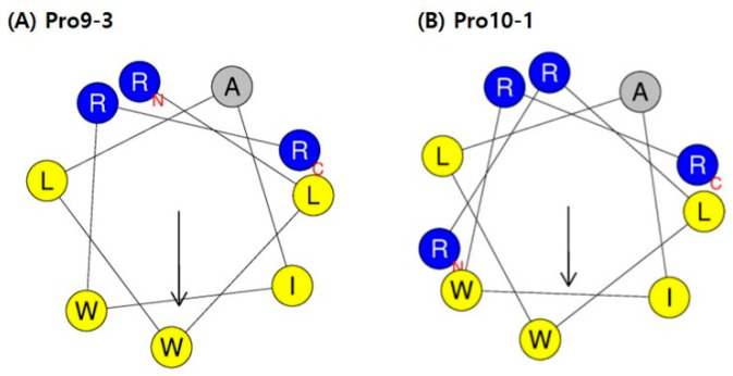
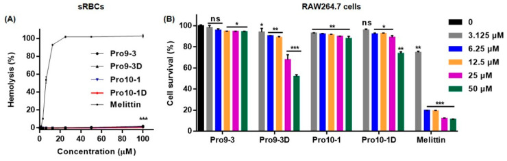
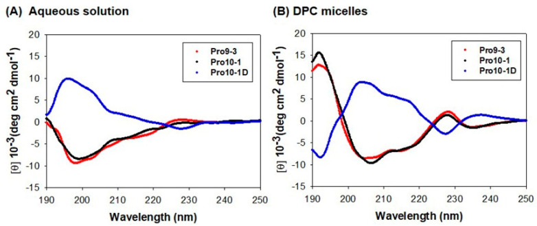
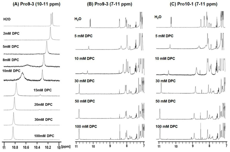
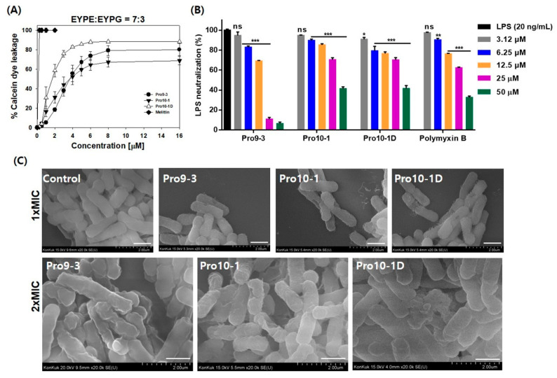
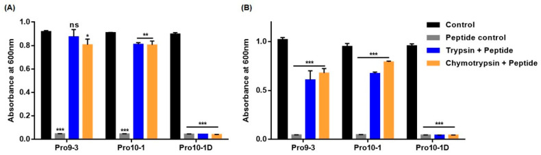
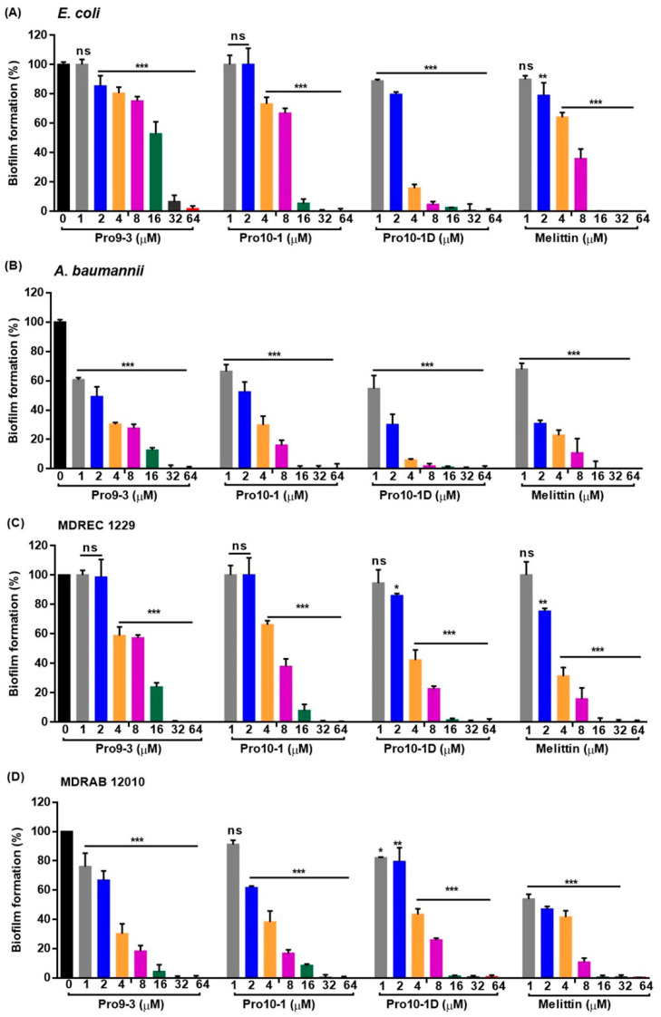
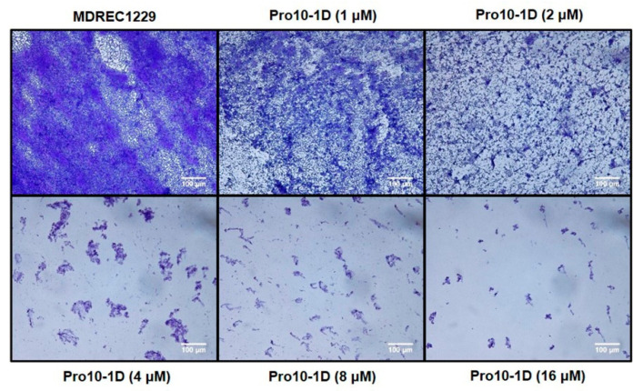
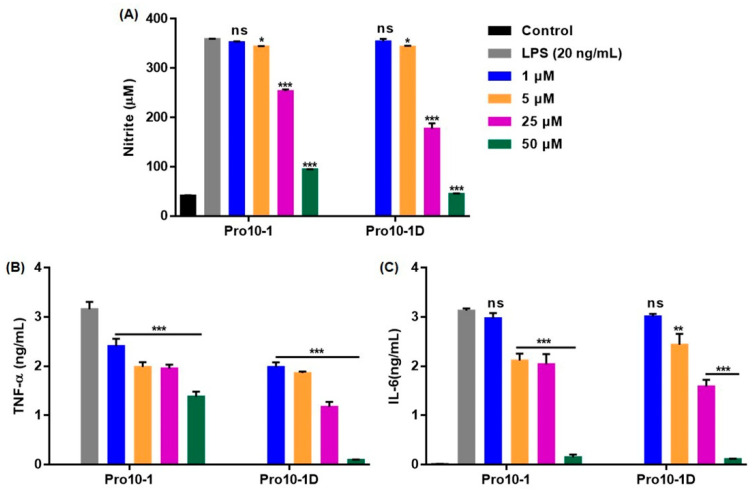
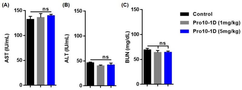
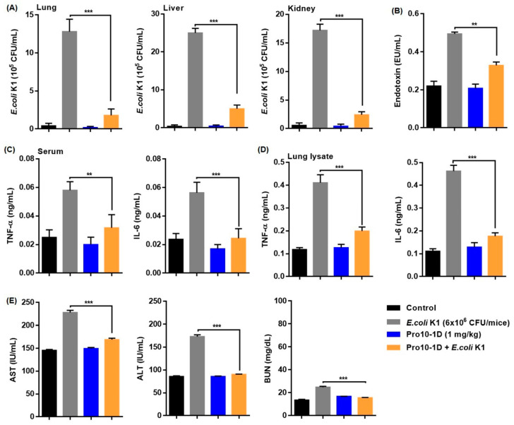
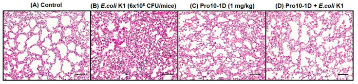
Owing to the challenges faced by conventional therapeutics, novel peptide antibiotics against multidrug-resistant (MDR) gram-negative bacteria need to be urgently developed. We had previously designed Pro9-3 and Pro9-3D from the defensin of beetle Protaetia brevitarsis; they showed high antimicrobial activity with cytotoxicity. Here, we aimed to develop peptide antibiotics with bacterial cell selectivity and potent antibacterial activity against gram-negative bacteria. We designed 10-meric peptides with increased cationicity by adding Arg to the N-terminus of Pro9-3 (Pro10-1) and its D-enantiomeric alteration (Pro10-1D). Among all tested peptides, the newly designed Pro10-1D showed the strongest antibacterial activity against Escherichia coli, Acinetobacter baumannii, and MDR strains with resistance against protease digestion. Pro10-1D can act as a novel potent peptide antibiotic owing to its outstanding inhibitory activities against bacterial film formation with high bacterial cell selectivity. Dye leakage and scanning electron microscopy revealed that Pro10-1D targets the bacterial membrane. Pro10-1D inhibited inflammation via Toll Like Receptor 4 (TLR4)/Nuclear factor-κB (NF-κB) signaling pathways in lipopolysaccharide (LPS)-stimulated RAW264.7 cells. Furthermore, Pro10-1D ameliorated multiple-organ damage and attenuated systemic infection-associated inflammation in an E. coli K1-induced sepsis mouse model. Overall, our results suggest that Pro10-1D can potentially serve as a novel peptide antibiotic for the treatment of gram-negative sepsis.

Keywords: antimicrobial peptide; biofilm; gram-negative infection; peptide antibiotics; sepsis.


Conflict of interest statement

The authors declare no conflict of interest.

Figures

Figure 1
Figure 1
Helical-wheel diagrams of (A) Pro9-3 and (B) Pro10-1 using HeliQuest online tool [32]: Positively charged amino acid residues are shown in blue, negatively charged residues are in red, and hydrophobic residues are in yellow at the bottom of the wheel. Alanine (A) is shown in gray. The arrows indicate the helical hydrophobic moment.
Figure 2
Figure 2
Cytotoxic effects of Pro9-3 and its analogs: (A) dose–response curves for sheep red blood cell (sRBC) hemolysis induced by peptides and (B) dose-dependent (0–50 μM) cytotoxicity of the peptides against mouse macrophage RAW264.7 cells for 24 h. Melittin was used as a control. The values are expressed as the mean ± SEM of three independent experiments and are statistically significant at * p < 0.05, ** p < 0.01, and *** p < 0.001; ns, not significant (by one-way ANOVA, Tukey’s post hoc test).
Figure 3
Figure 3
Circular dichroism spectra of Pro9-3 and its analogs at 100 μM in (A) aqueous solution and (B) 50 mM dodecylphosphocholine (DPC) micelles acquired with 10 scans: Double negative maxima at 205 and 220 nm are characteristic of α-helical structures.
Figure 4
Figure 4
Chemical shift perturbations in the NH region of the 1H NMR spectra at 298 K in 9:1 (v/v) H2O/D2O induced by dodecylphosphocholine (DPC): Sets of 1D 1H NMR spectra of a 1 mM solution of peptides were acquired with 128 scans. One-dimensional 1H NMR spectra of Pro9-3 were recorded with 9 different concentrations of DPC from 10 to 11 ppm (A), and 1D 1H NMR spectra of Pro9-3 (B) and Pro10-1 (C) from 7 to 11 ppm were recorded for six different concentrations of DPC. Changes in chemical shifts and line broadenings in the spectra of the peptides upon addition of DPC indicate conformational changes and interactions with DPC.
Figure 5
Figure 5
Antibacterial mechanism of peptides: (A) Dose–response curves of calcein leakage from egg yolk L-α-phosphatidylethanolamine (EYPE)/egg yolk L-α-phosphatidylglycerol (EYPG) (7:3, w/w) large unilamellar vesicles (LUVs) (induced by the peptides) were constructed. The concentration-dependent dye release indicates the membrane-permeabilizing ability of the peptides. Melittin was used as a control. (B) Lipopolysaccharide (LPS)-neutralizing capacity of the peptides and polymyxin B (positive control): the percentage of inhibition signifies the LPS-neutralizing abilities of peptides. (C) Scanning electron micrographs of E. coli (KCTC 1682) treated with peptides at 1× and 2 × of the minimum inhibitory concentrations (MICs) for 4 h: ultrastructural changes to the outer membrane of E. coli indicate the membrane damage caused by the peptides. Scale bar, 100 μm. Data are expressed as the mean ± SEM of three independent experiments and are statistically significant at * p < 0.05, ** p < 0.01, and *** p < 0.001; ns, not significant (by one-way ANOVA, Tukey’s post hoc test).
Figure 6
Figure 6
Inhibition of antimicrobial activity of peptides by trypsin and chymotrypsin assessed using the broth microdilution method: peptides at their respective minimum inhibitory concentrations were preincubated with trypsin and chymotrypsin for 6 h, and the treated peptides were examined for their antimicrobial activity against (A) E. coli and (B) A. baumannii for 16 h. Inhibited bacterial growth signifies the proteolytic stability of peptides. Data are shown as the mean ± SEM of three independent experiments and are statistically significant at * p < 0.05, ** p < 0.01, and *** p < 0.001; ns, not significant (by one-way ANOVA, Tukey’s post hoc test).
Figure 7
Figure 7
Inhibitory effects of Pro9-3, Pro10-1, and Pro10-1D on bacterial biofilm formation: Biofilm degradation properties of peptides were measured against (A) E. coli, (B) A. baumannii, (C) MDREC 1229, and (D) MDRAB 12010 in a dose-dependent manner. Melittin was used as a control. Data are shown as the mean ± SEM (n = 3) and are statistically significant at * p < 0.05, ** p < 0.01, and *** p < 0.001; ns, not significant by one-way ANOVA, Tukey’s post hoc test.
Figure 8
Figure 8
Light microscopic images representing the antibiofilm effect of Pro10-1D against MDREC 1229: MDREC 1229 cells were allowed to adhere the polystyrene surface of the culture plate and were then exposed to Pro10-1D (0–16 μM) for 16 h. Strains were cultured in Mueller–Hinton (MH) media, and biofilm formation was evaluated by crystal violet staining. Pro10-1D-exposed biofilms appeared thinner and scantier than untreated MDREC 1229; these effects were dose-dependent. Scale bar, 100 μm.
Figure 9
Figure 9
Pro10-1D modulates lipopolysaccharide (LPS)-induced inflammatory response in RAW264.7 cells. (A) Graphical data signify the dose-dependent inhibition of nitrite production and (B,C) expression of inflammatory cytokines (TNF-α and IL-6) in RAW264.7 cells incubated with peptides (0–50 μM) and then challenged with LPS (20 ng/mL) for 16 h. Data are expressed as the mean ± SEM of three independent experiments and are statistically significant, based on one-way ANOVA (Tukey’s post hoc test, * p < 0.05, ** p < 0.01, and *** p < 0.001; ns, not significant).
Figure 10
Figure 10
Pro10-1D is a target of toll-like receptor 4 (TLR4) and downregulates MAPK and NF-κB translocation in LPS-stimulated RAW264.7 cells. (A) Immunoblot analysis revealed the suppression of LPS-mediated TLR4, total/phospho-JNK, total/phospho-ERK, and cytosolic and nuclear translocation of NF-κB in RAW264.7 cells treated with peptides (20 μM) and LPS (50 ng/mL). β-actin was used as an internal control. (B) Graphical data represent the densitometry analysis of respective proteins, quantified by ImageJ. The values are expressed as the mean ± SEM of three independent experiments and are statistically significant, based on one-way ANOVA (Tukey’s post hoc test, ** p < 0.01, and *** p < 0.001; ns, not significant).
Figure 11
Figure 11
Pro10-1D exerts nontoxic effects in vivo. The representative graph indicates (A) aspartate aminotransferase (AST), (B) alanine aminotransferase (ALT), and (C) blood urea nitrogen (BUN) levels of mice treated with Pro10-1D (1 mg/kg and 5 mg/Kg) for 24 h. Control mice only received phosphate-buffered saline. Data are presented as the mean ± SEM (5 mice/group). Statistical analysis was performed by one-way ANOVA (Tukey’s post hoc test; ns, not significant).
Figure 12
Figure 12
Pro10-1D treatment alleviates septic shock in E. coli K1-infected mice: (A) Inhibition of bacterial growth by Pro10-1D in the lung, liver, and kidney lysates of the septic mice. Mice were pretreated with 1 mg/kg of Pro10-1D (1 h) and E. coli K1 (6 × 10⁶ Colony-Forming Unit (CFU)/mice, 16 h). (B) Pro10-1D reduces circulating endotoxin (LPS) levels in serum of septic mice. Effect of Pro10-1D on TNF-α and IL-6 levels in the (C) serum, and (D) lung lysates of septic mice. (E) Pro10-1D normalizes the serum levels of (E) aspartate aminotransferase (AST), alanine aminotransferase (ALT), and blood urea nitrogen (BUN) in septic mice infected with E. coli K1. The graphs indicate the mean ± SEM (5 mice/group). ** p < 0.01; *** p < 0.001; ns, nonsignificant (by one-way ANOVA, Tukey’s post hoc test).
Figure 13
Figure 13
Pro10-1D treatment ameliorates lung microanatomical changes in E. coli K1-infected mice. Representative hematoxylin and eosin staining of lung tissue of (A) the control shows normal lung anatomy, while that of (B) E. coli K1-infected mice (6 × 10⁶ CFU/mice, Intra-peritoneal injection (I.P.)) displays severe lung damage associated with neutrophil infiltration. (C) No sign of lung damage can be observed in the Pro10-1D-treated control mice (1 mg/Kg, I.P.), while (D) Pro10-1D pretreatment distinctly ameliorated the aberrant changes induced by E. coli K1 infection (×20 magnification, scale bar, 100 μm).

Similar articles

Cited by

References

    1. Van Der Poll T., Van De Veerdonk F.L., Scicluna B., Netea M.M. The immunopathology of sepsis and potential therapeutic targets. Nat. Rev. Immunol. 2017;17:407–420. doi: 10.1038/nri.2017.36. - DOI - PubMed
    1. Rudd K.E., Kissoon N., Limmathurotsakul D., Bory S., Mutahunga B., Seymour C.W., Angus D.C., West T.E. The global burden of sepsis: Barriers and potential solutions. Crit. Care. 2018;22:1–11. doi: 10.1186/s13054-018-2157-z. - DOI - PMC - PubMed
    1. Oh S.-Y., Cho S., Kim G.H., Jang E.J., Choi S., Lee H., Ryu H.G. Incidence and Outcomes of Sepsis in Korea. Crit. Care Med. 2019;47:e993–e998. doi: 10.1097/CCM.0000000000004041. - DOI - PubMed
    1. Delcour A.H. Outer membrane permeability and antibiotic resistance. Biochim. Biophys. Acta (BBA) Proteins Proteom. 2009;1794:808–816. doi: 10.1016/j.bbapap.2008.11.005. - DOI - PMC - PubMed
    1. Tucureanu M.M., Rebleanu D., Constantinescu C.A., Deleanu M., Voicu G., Butoi E., Calin M., Manduteanu I. Lipopolysaccharide-induced inflammation in monocytes/macrophages is blocked by liposomal delivery of Gi-protein inhibitor. Int. J. Nanomed. 2017;13:63–76. doi: 10.2147/IJN.S150918. - DOI - PMC - PubMed

MeSH terms

LinkOut - more resources