

Peripheral Mechanobiology of Touch-Studies on Vertebrate Cutaneous Sensory Corpuscles
- PMID: 32867400
- PMCID: PMC7504094
- DOI: 10.3390/ijms21176221
Peripheral Mechanobiology of Touch-Studies on Vertebrate Cutaneous Sensory Corpuscles
Abstract
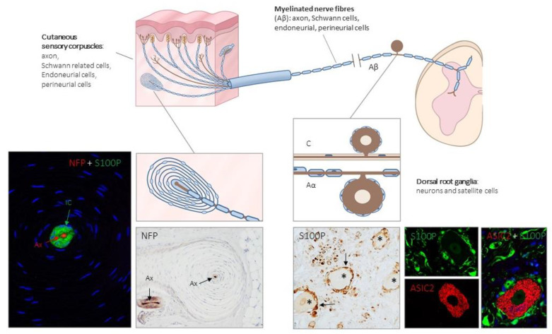
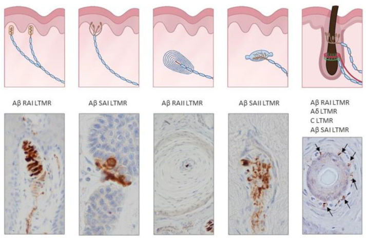
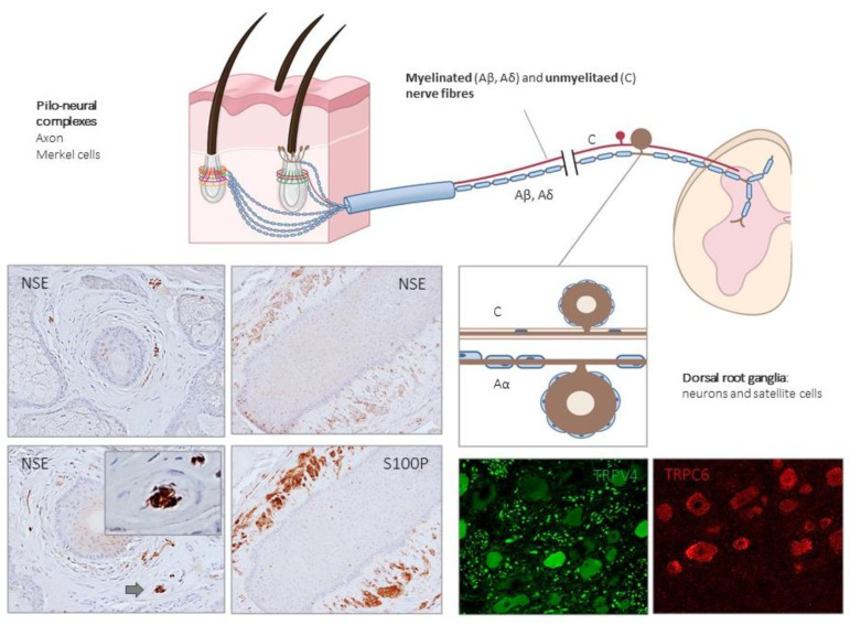
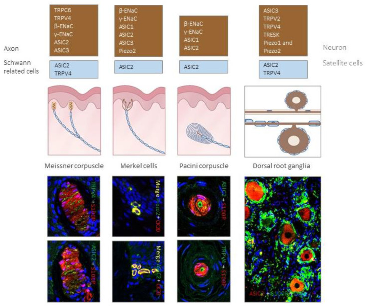
The vertebrate skin contains sensory corpuscles that are receptors for different qualities of mechanosensitivity like light brush, touch, pressure, stretch or vibration. These specialized sensory organs are linked anatomically and functionally to mechanosensory neurons, which function as low-threshold mechanoreceptors connected to peripheral skin through Aβ nerve fibers. Furthermore, low-threshold mechanoreceptors associated with Aδ and C nerve fibers have been identified in hairy skin. The process of mechanotransduction requires the conversion of a mechanical stimulus into electrical signals (action potentials) through the activation of mechanosensible ion channels present both in the axon and the periaxonal cells of sensory corpuscles (i.e., Schwann-, endoneurial- and perineurial-related cells). Most of those putative ion channels belong to the degenerin/epithelial sodium channel (especially the family of acid-sensing ion channels), the transient receptor potential channel superfamilies, and the Piezo family. This review updates the current data about the occurrence and distribution of putative mechanosensitive ion channels in cutaneous mechanoreceptors including primary sensory neurons and sensory corpuscles.
Keywords: Piezo2; acid-sensing ion channels; low-threshold mechanoreceptors; mechanoproteins; sensory corpuscles; skin; transient receptor potential channels.
Conflict of interest statement
The authors declare no conflict of interest.
Figures
Similar articles
-
Mechanosensory neurons, cutaneous mechanoreceptors, and putative mechanoproteins.Microsc Res Tech. 2012 Aug;75(8):1033-43. doi: 10.1002/jemt.22028. Epub 2012 Mar 28. Microsc Res Tech. 2012. PMID: 22461425 Review.
-
Merkel cells and Meissner's corpuscles in human digital skin display Piezo2 immunoreactivity.J Anat. 2017 Dec;231(6):978-989. doi: 10.1111/joa.12688. Epub 2017 Sep 14. J Anat. 2017. PMID: 28905996 Free PMC article.
-
Piezo2 voltage-block regulates mechanical pain sensitivity.Brain. 2024 Oct 3;147(10):3487-3500. doi: 10.1093/brain/awae227. Brain. 2024. PMID: 38984717 Free PMC article.
-
Immunohistochemical detection of PIEZO1 and PIEZO2 in human digital Meissner´s corpuscles.Ann Anat. 2024 Feb;252:152200. doi: 10.1016/j.aanat.2023.152200. Epub 2023 Dec 17. Ann Anat. 2024. PMID: 38109982
-
Transduction and encoding sensory information by skin mechanoreceptors.Pflugers Arch. 2015 Jan;467(1):109-19. doi: 10.1007/s00424-014-1651-7. Epub 2014 Nov 23. Pflugers Arch. 2015. PMID: 25416542 Review.
Cited by
-
Somatosensory innervation of healthy human oral tissues.J Comp Neurol. 2021 Aug 1;529(11):3046-3061. doi: 10.1002/cne.25148. Epub 2021 Apr 29. J Comp Neurol. 2021. PMID: 33786834 Free PMC article.
-
Piezo 1 and Piezo 2 in the Chemosensory Organs of Zebrafish (Danio rerio).Int J Mol Sci. 2024 Jul 5;25(13):7404. doi: 10.3390/ijms25137404. Int J Mol Sci. 2024. PMID: 39000511 Free PMC article.
-
Sensory innervation of the human palmar aponeurosis in healthy individuals and patients with palmar fibromatosis.J Anat. 2022 May;240(5):972-984. doi: 10.1111/joa.13609. Epub 2021 Dec 8. J Anat. 2022. PMID: 34881452 Free PMC article.
-
Contributions and future potential of animal models for geroscience research on sensory systems.Geroscience. 2025 Feb;47(1):61-83. doi: 10.1007/s11357-024-01327-5. Epub 2024 Sep 23. Geroscience. 2025. PMID: 39312151 Free PMC article. Review.
-
The Human Cutaneous Sensory Corpuscles: An Update.J Clin Med. 2021 Jan 10;10(2):227. doi: 10.3390/jcm10020227. J Clin Med. 2021. PMID: 33435193 Free PMC article. Review.
References
-
- Rice F., Albrecht P. The Senses: A Comprehensive Reference. Volume 6. Elsevier BV; Amsterdam, The Netherlands: 2008. Cutaneous Mechanisms of Tactile Perception: Morphological and Chemical Organization of the Innervation to the Skin; pp. 1–31.
Publication types
MeSH terms
Substances
LinkOut - more resources
Full Text Sources
