Review
. 2020 Aug 27;13(17):3795.
doi: 10.3390/ma13173795.

Mesoporous Silica Nanoparticles in Bioimaging

Affiliations
Review

Mesoporous Silica Nanoparticles in Bioimaging

Daohe Yuan et al. Materials (Basel). .

Abstract

A biomedical contrast agent serves to enhance the visualisation of a specific (potentially targeted) physiological region. In recent years, mesoporous silica nanoparticles (MSNs) have developed as a flexible imaging platform of tuneable size/morphology, abundant surface chemistry, biocompatibility and otherwise useful physiochemical properties. This review discusses MSN structural types and synthetic strategies, as well as methods for surface functionalisation. Recent applications in biomedical imaging are then discussed, with a specific emphasis on magnetic resonance and optical modes together with utility in multimodal imaging.

Keywords: bioimaging; imaging modality; magnetic resonance imaging; mesoporous silica nanoparticles; multi-modality imaging; nanoparticles; optical imaging.


Conflict of interest statement

The authors declare no conflict of interest.

Figures

Figure 1
Figure 1
A schematic illustration of the synthesis of MSNs. Initially, surfactant micelles are formed which then self-assemble forming a lyotropic liquid-crystal phase. Condensation of the silica source around the surfactant template and subsequent surfactant template removal produces MCM-41. Here, tetraethyl orthosilicate (TEOS) is used as the silica source and cetyltrimethylammonium bromide (CTAB) as the surfactant template.
Figure 2
Figure 2
A schematic detailing the three synthetic routes for fabrication of hollow mesoporous silica nanoparticles (HMSNs), namely “soft-templating”, “hard-templating” and “self-templating”. All three routes start from the formation of the core template. Route (A) is a two-step process, resulting in the formation of HMSNs after the removal of both the mesopore and core templates. Route (B) illustrates the formation of HMSNs by a hard-templating method after the removal of both the mesopore and core templates. Route (C) shows the process of self-templating fabrication of HMSNs. The final hollow mesoporous silica nanoparticles are obtained by a complete etching of self-templating solid silica spheres (sSiO2). Common etching agents include strong acids such as HCl and HF, an alkaline medium such as ammonia and NaOH, NaBH4 or hot water [49].
Figure 3
Figure 3
A schematic illustration of the synthesis of magnetic mesoporous silica nanoparticles by different methods. (i) Synthesis using an oil-in-water technique. Hydrophobic-ligand-capped iron oxide nanoparticles dispersed in a nonpolar organic solvent are added to an aqueous solution containing cetyltrimethylammonium bromide (CTAB) and TEOS, allowing the formation of core–shell magnetic mesoporous silica nanoparticles (M-MSNs). Adapted with permission from reference [51]. Copyright (2006) American Chemistry Society. (ii) Synthesis using a water-in-oil method. The synthesis starts with micellular solution of an amphiphilic surfactant (such as Igepal CO-520), and core–shell iron oxide nanoparticle@nSiO2 (IONP@nSiO2) with a reverse-microemulsion system formed on the addition of ammonia. Finally, the core–shell M-MSNs are produced by the introduction of an additional mesoporous silica shell (mSiO2). (iii) Synthesis using a hard-templating strategy. This method starts with the introduction of mesoporous silica structures on the surface of native iron oxide nanoparticles by adding the silica source and the pore directing agent (e.g., TEOS and cetyltrimethylammonium salt). The desired M-MSNs are produced after calcination to remove the embedded surfactant template. Adapted with permission from reference [54]. Copyright (2009) The Royal Society of Chemistry.
Figure 4
Figure 4
The immobilisation of chemical functionalities by both co-condensation and post synthetic functionalisation. Path (A) shows the direct synthetic co-condensation functionalisation by organo-substituted trialkoxysilanes on the surface of silica mesopores. Path (B) indicates the co-condensation process for periodic mesoporous organosilicas (PMOs), where bridged organosilanes (R’O)3Si-R-Si(OR’)3 and tetraalkoxysilanes are within the three-dimensional network structure of the silica matrix. Path (C) illustrates the post synthetic grafting of terminal trialkoxyorgnanosilanes.
Figure 5
Figure 5
A figure detailing how the position of paramagnetic chelates within MSNs affects the relaxivity (r2) values. (A) General schematic of Dy-MSNs. (B) The different available locations for the tethered Dy-chelate. S, L and P refer to the synthetic procedure i.e., short-delay, long-delay and post-grafting, respectively. (C) Associated relaxivity values where the highest relaxivity can be observed for Dy-MSNs-L with r2 = 143.5 ± 8.2 mM−1 s−1 at 11.7 T. All three locations show substantially improved r2 in comparison to native molecular Dy-DOTA. (D) MRI phantoms characterising the response of the system. Adapted with permission from reference [80]. Copyright (2018) The Royal Society of Chemistry.
Figure 6
Figure 6
A schematic representation of high-intensity focused ultrasound (HIFU)-responsive magnetic resonance imaging (MRI)-active MSNs. In this work, the particles were initially amine Figure 2. (green) and the pore channels capped with poly(ethylene glycol) (PEG) (brown). Finally, the effect of HIFU stimulation on the release of Gd(DTPA)2− can be observed. Reprinted with permission from reference [94]. Copyright (2019) American Chemical Society.
Figure 7
Figure 7
A core/shell hybrid based on a platinum core and MSN shell. Tethering with Gd-DTPA provides MRI activity with photothermal therapy mediated by the Pt core. APTES refers to (3-aminopropyl)triethoxysilane. Reprinted with permission from reference [111]. Copyright (2017) The Royal Society of Chemistry.
Figure 8
Figure 8
Schematic illustration of two types of MSNs as applied in optical imaging where a luminescent moiety is incorporated into the MSNs substructure (Section 4.1) or is confined within an MSN-wrapped luminescent core (Section 4.2).
Figure 9
Figure 9
HCFA-MSNs. (i) A schematic illustration of hypoxia-activatable and cytoplasmic protein-powered fluorescence cascade amplifying (HCFA) MSNs. The HCFA-MSNs are composed of the β-cyclodextrin polymer (β-CDP) gatekeeper, loaded with both squarylium (SQ) dyes and black hole quencher 2 (BHQ2). (ii) In vivo fluorescence images of nude mice after the injection of a commercial small molecular fluorescent probe, the HCFA-MSNs, the control group or convalescent group of the HCFA-MSNs (from right to left), respectively. The most intense fluorescent signal can be observed the HCFA-MSNs. (iii) TEM image of the as-synthesised MSNs. Adapted with permission from Ref. [124]. Copyright (2020) American Chemical Society.
Figure 10
Figure 10
Chitosan-gated fluorescent mesoporous silica nanoparticles (FMSNs). (i) Schematic illustration of the design of chitosan-gated MSNs for monitoring drug release in vitro. The as-synthesised nanoparticles are composed of naphthalimide-dye-functionalised chitosan (Cs-Nac) as the gatekeeper, an MSN nanoparticulate scaffold and Rhodamine 6G (R6) as the model cargo agent. On glutathione (GSH) addition two fluorescent emissions corresponding to the release of R6 and cleaved Cs-Nac are observed. (ii) A TEM image of as-synthesised MSNs. (iii) Fluorescence images of MRC–5 cells with different GSH concentration. (A) The as-synthesised MSNs with low GSH concentration. (B) MSNs in MRC-5 without any other treatment. (C) MSNs with high GSH concentration. Adapted with permission from reference [126]. Copyright (2020) American Chemical Society.
Figure 11
Figure 11
Schematic illustration of persistent luminescent nanocrystal-incorporated MSNs for afterglow luminescence imaging and photodynamic therapy in vivo. The as-synthesised nanoparticles are composed of the mesoporous silica-templated zinc gallogermanate (mZGGOs) nanocomposite core, an outer PEGylated coating and the loading of silicon phthalocyanine photosensitiser (Si-Pc). This design exhibited rechargeable X-ray-excited persistent luminescence (XEPL) properties for bioimaging and the ability of tumour suppression. Reprinted with permission from reference [139]. Copyright (2020) Wiley-VCH.
Figure 12
Figure 12
(A) Schematic illustration of DOX-loaded zinc gallogermanate (ZGGO)@mSiO2@GFLG-SS-pHLIP composed of persistent luminescent nanoparticle (PLNP)/MSN core/shell structure, glutathione/cathepsin B dual-responsive polypeptide coating (pHLIP-SS-GFLG) and anticancer drug (DOX). This nanosystem is designed to achieve drug delivery under glutathione or/and cathepsin B conditions. (B) TEM image of ZGGO@mSiO2. (C) Confocal laser scanning microscopy images of stained A549 cells, ZGGO@mSiO2@GFLG-SS-pHLIP, and merged (from left to right). (D) The drug release profile of DOX-loaded ZGGO@mSiO2@GFLG-SS-pHLIP in PBS buffer solution with or without stimulus (glutathione and cathepsin B). Adapted with permission from reference [147]. Copyright (2020) American Chemical Society.
Figure 13
Figure 13
A schematic representation for the design of 18F-radiolabelled MSNs. Cyclooctyne functionalised MSNs (DBCO-MSNs) were first incubated with RAW cells, with the incubated samples then injected into nude mice bearing a tumour or atherosclerosis plaques. One to eight days later, 18F-labelled azide was injected into the mice to visualise the tumour/atherosclerotic sites in vivo. Reprinted with permission from reference [152]. Copyright (2019) Elsevier.
Figure 14
Figure 14
Schematic illustration of mesoporous silica-coated upconversion nanoparticles composed of a gadolinium doped UCNP core (noted as NP), mesoporous silica shell (labelled SiO2) and Aβ oligomer-selective cyanine dye coating (noted as F-SLOH). The gadolinium-doped UCNP core provides T1 MRI contrast and the selective binding of Aβ oligomers to the F-SLOH dye facilitates target activated NIR fluorescence (denoted near infrared imaging, NIRI). UCL refers to upconversion luminescence. Reprinted with permission from reference [171]. Copyright (2020) Wiley-VCH.
Figure 15
Figure 15
A schematic representing a multi-modal Janus nanoparticulate system composed of a SPION/MSN core/shell face and Au nanoparticle face. T2 MRI contrast capabilities arise from the encapsulated SPION with Au nanoparticles providing the possibility for CT imaging. As shown in this schematic, the system is functionalised with a fluorescent dye (Alexa Fluor® 647) for optical imaging in addition to cRDG for active tumour targeting. Reprinted with permission from reference [178]. Copyright (2018) American Chemical Society.

References

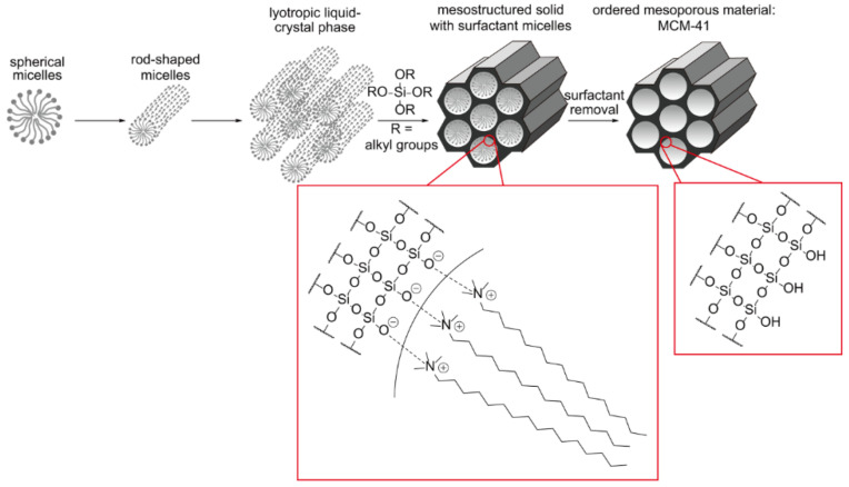
    1. Kresge C.T., Leonowicz M.E., Roth W.J., Vartuli J.C., Beck J.S. Ordered mesoporous molecular sieves synthesized by a liquid-crystal template mechanism. Nature. 1992;359:710–712. doi: 10.1038/359710a0. - DOI
    1. Beck J.S., Vartuli J.C., Roth W.J., Leonowicz M.E., Kresge C.T., Schmitt K.D., Chu C.T.W., Olson D.H., Sheppard E.W., McCullen S.B., et al. A new family of mesoporous molecular sieves prepared with liquid crystal templates. J. Am. Chem. Soc. 1992;114:10834–10843. doi: 10.1021/ja00053a020. - DOI
    1. Tan S.Y., Teh C., Ang C.Y., Li M., Li P., Korzh V., Zhao Y. Responsive mesoporous silica nanoparticles for sensing of hydrogen peroxide and simultaneous treatment toward heart failure. Nanoscale. 2017;9:2253–2261. doi: 10.1039/C6NR08869D. - DOI - PubMed
    1. Liao Y.-T., Lee C.-H., Chen S.-T., Lai J.-Y., Wu K.C.W. Gelatin-functionalized mesoporous silica nanoparticles with sustained release properties for intracameral pharmacotherapy of glaucoma. J. Mater. Chem. B. 2017;5:7008–7013. doi: 10.1039/C7TB01217A. - DOI - PubMed
    1. Hou L., Zheng Y., Wang Y., Hu Y., Shi J., Liu Q., Zhang H., Zhang Z. Self-Regulated Carboxyphenylboronic Acid-Modified Mesoporous Silica Nanoparticles with “Touch Switch” Releasing Property for Insulin Delivery. ACS Appl. Mater. Interfaces. 2018;10:21927–21938. doi: 10.1021/acsami.8b06998. - DOI - PubMed