

Basal cell carcinoma and squamous cell carcinoma in a single tumor in the anterior auricular area
- PMID: 32867417
- PMCID: PMC7463126
- DOI: 10.7181/acfs.2020.00262
Basal cell carcinoma and squamous cell carcinoma in a single tumor in the anterior auricular area
Abstract
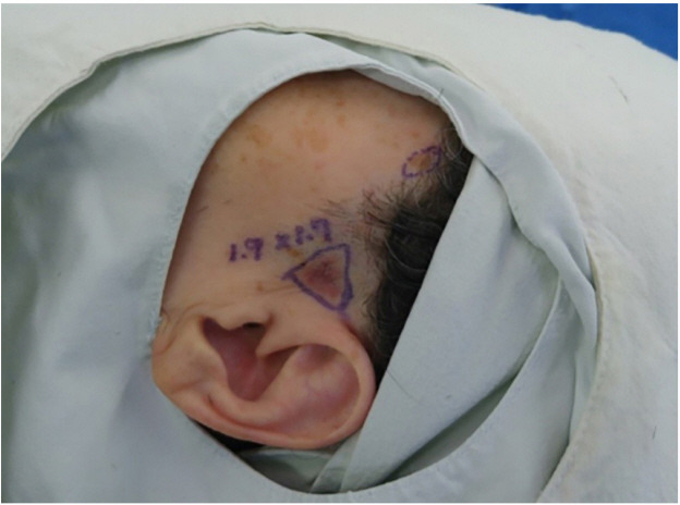
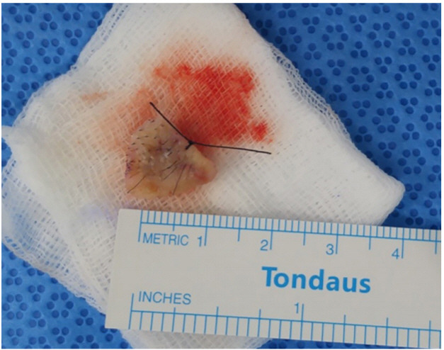
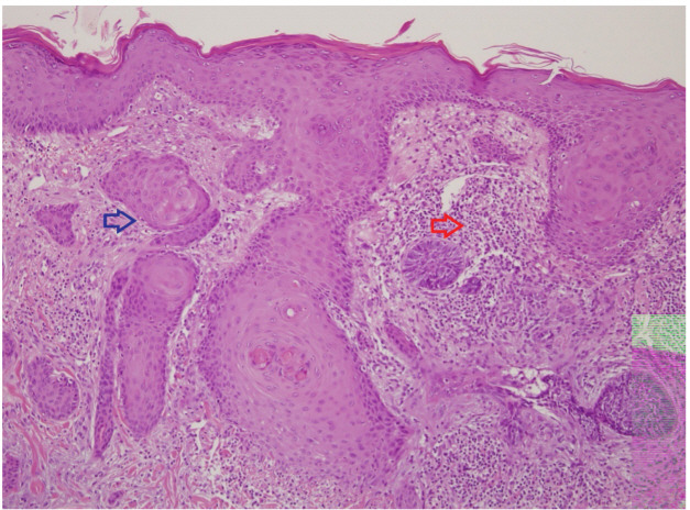
The concurrence of basal cell carcinoma (BCC) and squamous cell carcinoma (SCC) in a single tumor is rarely encountered. We report a case of BCC and SCC in a single tumor in the anterior auricular area. A 70-year-old woman had been diagnosed with BCC by a punch biopsy performed at a dermatology clinic. We performed wide excision of the tumor with an ulcer in the anterior auricular area. Analysis of the biopsy specimen revealed the presence of both BCC and SCC in the tumor. This case illustrates that it is necessary to establish a precise diagnosis and formulate appropriate surgical and treatment plans considering the possibility that two carcinomas may coexist, although the possibility is low in patients with skin cancer.
Keywords: Basal cell carcinoma; Co-existence; Squamous cell carcinoma.
Conflict of interest statement
No potential conflict of interest relevant to this article was reported.
Figures
References
-
- Pierard GE, Fazaa B, Henry F, Kamoun MR, Pierard-Franchimont C. Collision of primary malignant neoplasms on the skin: the connection between malignant melanoma and basal cell carcinoma. Dermatology. 1997;194:378–9. - PubMed
-
- Inoshita T, Laurain AR, Youngberg GA, Musil G. Metastasis of bronchogenic carcinoma to the skin involved by melanoma. Arch Pathol Lab Med. 1984;108:595–8. - PubMed
-
- Boyd AS, Rapini RP. Cutaneous collision tumors: an analysis of 69 cases and review of the literature. Am J Dermatopathol. 1994;16:253–7. - PubMed
-
- Hirakawa E, Miki H, Kobayashi S, Nomura Y, Ohmori M. Collision tumor of cutaneous malignant melanoma and basal cell carcinoma. Pathol Res Pract. 1998;194:649–53. - PubMed
-
- Burkhalter A, White WL. Malignant melanoma in situ colonizing basal cell carcinoma: a simulator of invasive melanoma. Am J Dermatopathol. 1997;19:303–7. - PubMed
Publication types
LinkOut - more resources
Full Text Sources
Research Materials
