. 2020 Sep 1;18(1):332.
doi: 10.1186/s12967-020-02499-2.

MiR-199a-3p/5p participated in TGF-β and EGF induced EMT by targeting DUSP5/MAP3K11 in pterygium

Affiliations

MiR-199a-3p/5p participated in TGF-β and EGF induced EMT by targeting DUSP5/MAP3K11 in pterygium

Siying He et al. J Transl Med. .

Abstract

Background: Recently, it has been reported that miRNA is involved in pterygium, however the exact underlying mechanism in pterygium is unrevealed and require further investigation.

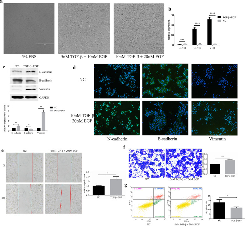
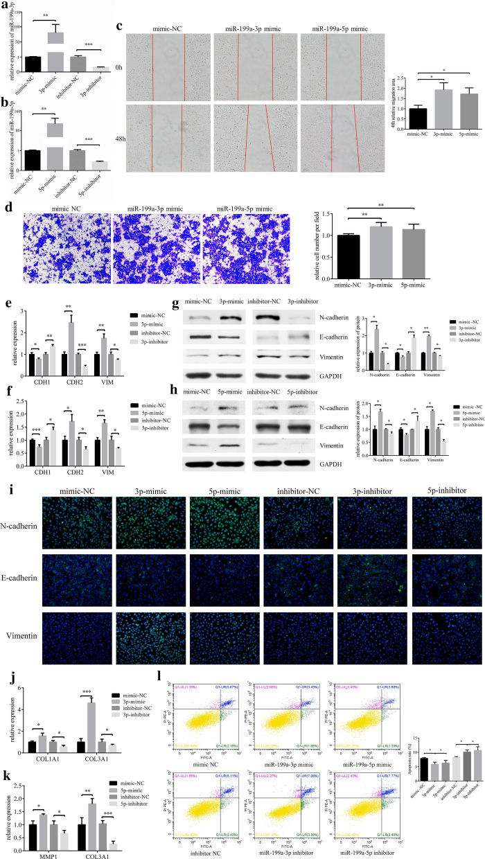
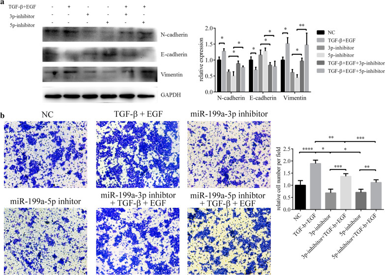
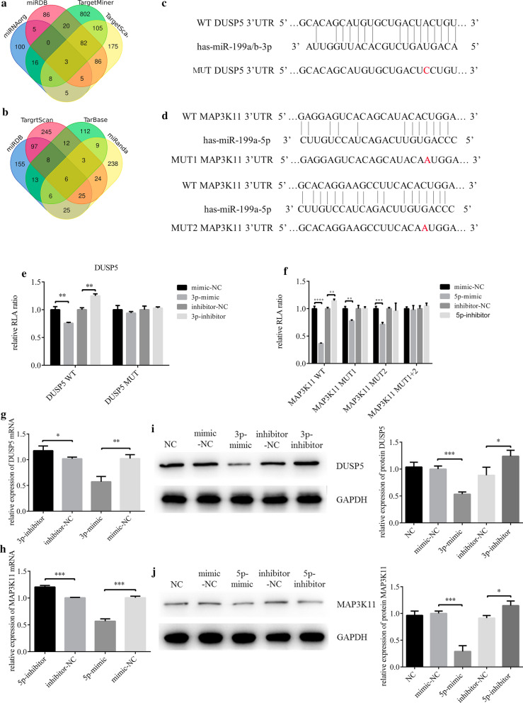
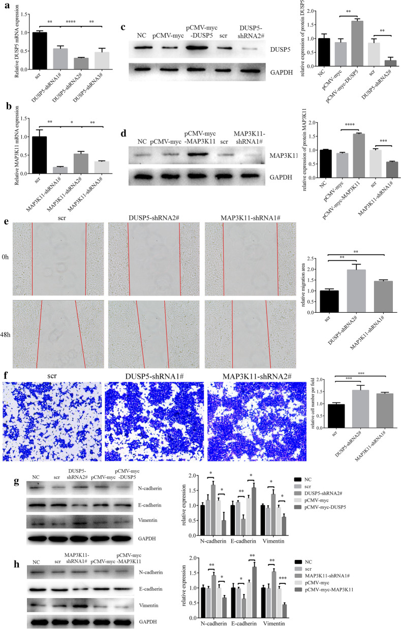
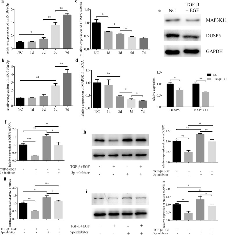
Methods: The differential expression of miRNA in pterygium was profiled using microarray and validated with quantitative real-time polymerase chain reaction (qRT-PCR). Human conjunctival epithelial cells (HCEs) were cultured and treated with transforming growth factor β (TGF-β) and epidermal growth factor (EGF) and transfected with miR-199a-3p/5p mimic and inhibitor. Markers of epithelial-mesenchymal transition (EMT) in HCEs were detected using western blot and immunohistochemistry. Cell migration ability was determined using wound healing and transwell assay, while apoptosis was determined by flow cytometry. The target genes of miR-199a were confirmed by the dual-luciferase reporter assay.

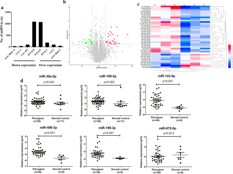
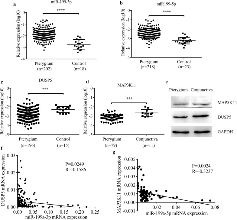
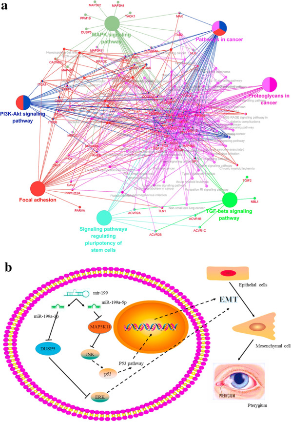
Results: TGF-β and EGF could induced EMT in HCEs and increase miR-199a-3p/5p but suppress target genes, DUSP5 and MAP3K11. With the occurrence of EMT, cell migration ability was enhanced, and apoptosis was impeded. Promoting miR-199a-3p/5p expression could induce EMT in HCEs without TGF-β and EGF, while suppressing miR-199a-3p/5p could inhibit EMT in TGF-β and EGF induced HCEs. In a word, TGF-β and EGF induced EMT could be regulated with miR-199a-3p/5p-DUSP5/MAP3K11 axes. The validated results in tissues showed that, compared with control conjunctival tissues, miR-199a-3p/5p were more overexpressed in pterygium, while DUSP5/MAP3K11 were lower expressed. In addition, bioinformatics analysis indicated the miR-199a-3p/5p-DUSP5/MAP3K11 was belong to MAPK signalling pathway.

Conclusions: TGF-β and EGF induce EMT of HCEs through miR-199a-3p/5p-DUSP5/MAP3K11 axes, which explains the pathogenesis of EMT in pterygium and may provide new targets for pterygium prevention and therapy.

Keywords: DUSP5; EMT; MAP3K11; Pterygium; miR-199a.


Conflict of interest statement

The authors declare that they have no competing interests.

Figures

Fig. 1
Fig. 1
Differential expressed miRNAs in pterygium. a The log scales of most miRNAs expression in pterygium tissues were found from -1 to 1. X-axis indicates the log scale of miRNA expression in control conjunctiva and pterygium tissues. Negative scale indicates downregulation, and positive scale indicates overexpression. b The volcano plot of the microarray with the criteria of ∣lg (FC)∣ > 1 and p value < 0.05. The green nodes presented the downregulated miRNAs and the red nodes presented the upregulated miRNAs. c The heat map showed the expression levels of miRNAs which were indicated on a colour gradation scale from low expression (blue) to high expression (red) in pterygium (P1, P2, P3) and control conjunctiva (C1, C2, C3) groups. d The relative expression of six validated miRNAs in pterygium. MiR-30a-5p (p = 0.043) and miR-199a-5p (p = 0.002) were up-regulated in pterygium samples (n = 55) compared with control conjunctiva tissues (n = 11), and miR-143-5p (p < 0.0001), miR-486-3p (p < 0.0001), miR-199a-3p (p = 0.001) were up-regulated in pterygium samples (n = 40) compared with control conjunctiva tissues (n = 6). The verification result of miR-675-5p (p = 0.613) was inconsistent with the chip
Fig. 2
Fig. 2
TGF-β and EGF could induced EMT in HCEs. a A combination of TGF-β 10 nM and EGF (20 nM) could induced HCEs to show an EMT phenotype. bd The changes in EMT markers were detected with qRT-PCR, western blot and immunofluorescence (*p < 0.05, **p < 0.01, ***p < 0.001, ****p < 0.0001). e, f Cell migration in HCEs induced by TGF-β (10 nM) and EGF (20 nM). Cell migration was determined by the wound healing assay and transwell assay (*p < 0.05, ***p < 0.001). g Apoptosis in HCEs induced by TGF-β (10 nM) and EGF (20 nM). The apoptosis cells were decreased compared with the HCEs without inducement (*p < 0.05)
Fig. 3
Fig. 3
MiR-199a-3p and miR-199a-5p promotes migration of EMT in HCEs. a, b HCEs were transfected with miR-199a-3p/miR-199a-5p mimics/mimics NC, inhibitors/inhibitor NC. The expression of miR-199a-3p and miR-199a-5p was detected by qRT-PCR (**p < 0.01, ***p < 0.001). HCEs were transfected with miR-199a-3p mimic, miR-199a-5p mimic and mimic NC. c Cell migration was determined by the wound healing assay (*p < 0.05). d Cell migration was determined by the transwell assay (**p < 0.01). ei HCEs were transfected with miR-199a-3p/miR-199a-5p mimic, mimic NC, miR-199a-3p/miR-199a-5p inhibitor and inhibitor NC. The EMT markers (E-cadherin (CDH1), N-cadherin (CDH2) and Vimentin (VIM)) were detected by qRT-PCR, western blot and immunofluorescence (*p < 0.05, **p < 0.01, ***p < 0.001). j After HCEs were transfected with miR-199a-3p mimic, mimic NC, miR-199a-3p inhibitor and inhibitor NC, COL1A1 and COL3A1 were upregulated only by miR-199a-3p, detected by qRT-PCR (*p < 0.05, ***p < 0.001). k After HCEs were transfected with miR-199a-5p mimic, mimic NC, miR-199a-5p inhibitor and inhibitor NC, MMP1 and COL3A1 were upregulated only by miR-199a-5p, detected by qRT-PCR (*p < 0.05, **p < 0.01, ***p < 0.001). l HCEs were transfected with miR-199a-3p/miR-199a-5p mimics/mimics NC, inhibitors/inhibitor NC. Transfection of the miR-199a-3p mimic and miR-199a-5p mimic decreased the apoptosis cells, while miR-199a-3p inhibitor and miR-199a-5p inhibitor increased the apoptosis cells (*p < 0.05, **p < 0.01)
Fig. 4
Fig. 4
MiR-199a inhibitor hindered TGF-β and EGF induced EMT in HCEs. a The changes of EMT biomarkers detected with western blot (*p < 0.05, **p < 0.01). b The migration ability of HCEs measured by transwell assay (*p < 0.05, **p < 0.01, ***p < 0.001, ****p < 0.0001)
Fig. 5
Fig. 5
MiR-199a-3p and miR-199a-5p target DUSP5 and MAP3K11 respectively. a, b Venn diagram of miR-199a-3p and miR-199a-5p target genes prediction results from 4 databases. c, d Putative miR-199a-3p and miR-199a-5p binding sites in the 3′UTR of DUSP5 and MAP3K11, and the designed mutation sites. e, f A luciferase reporter plasmid containing wild type or mutant type were co-transfected with relevant mimic, mimic NC, inhibitor and inhibitor NC into HCEs. Luciferase activity was normalized to Renilla luciferase activity. Of note, MAP3K11 3′UTR had 2 binding sites withmiR-199a-5p, so 3 kinds of mutants were construct, including mutant at the first site, mutant at the second site and mutant at both sites (**p < 0.01, ***p < 0.001, ****p < 0.0001). gj After HCEs were transfected with miR-199a-3p/miR-199a-5p mimic, mimic NC, miR-199a-3p/miR-199a-5p inhibitor and inhibitor NC, the variations of DUSP5 and MAP3K11 expression were analyzed with qRT-PCR and western blot (*p < 0.05, **p < 0.01, ***p < 0.001)
Fig. 6
Fig. 6
Downregulating DUSP5 or MAP3K11could promote HCEs migration and EMT. a, b The shRNAs which had the best knockdown efficiency were selected with qRT-PCR. c, d The effect of pCMV-myc-DUSP5, pCMV-myc-MAP3K11, DUSP5-shRNA2# and MAP3K11-shRNA#1. HCEs were transfected with pCMV-myc and scr as control. e Cell migration was determined by the wound healing assay (**p < 0.01). f Cell migration was determined by the transwell assay (**p < 0.01). g, h The changes in EMT markers, after HCEs were transfected with pCMV-myc-DUSP5, pCMV-myc-MAP3K11, pCMV-myc, DUSP5-shRNA2#, MAP3K11-shRNA#1 and shRNA control. (*p < 0.05, **p < 0.01, ***p < 0.001)
Fig. 7
Fig. 7
The effects of TGF-β and EGF on expressions of DUSP5 and MAP3K11 could be regulated by miR-199a-3p and miR-199a-5p. ad HCEs were induced with TGF-β (10 nM) and EGF (20 nM) for 7 days. The expressions of miR-199a-3p, miR-199a-5p, DUSP5 and MAP3K11 were detected by qRT-PCR analysis (*p < 0.05, **p < 0.01). e The protein expression of DUSP5 and MAP3K11 were analyzed with western blot (*p < 0.05, **p < 0.01). f, g. The mRNA changes of DUSP5 and MAP3K11 before and after induction and transfection (*p < 0.05, **p < 0.01, ***p < 0.001). h, i The protein changes of DUSP5 and MAP3K11 before and after induction and transfection (*p < 0.05, **p < 0.01)
Fig. 8
Fig. 8
Inhibition on miR-199a-3p and miR-199a-5p alleviates the EMT promoted by DUSP5 and MAP3K11 in induced HCEs. HCEs were induced with TGF-β (10 nM) and EGF (20 nM) for 7 days, and subsequently transfected with pCMV-myc-DUSP5 (or pCMV-myc-MAP3K11) and pCMV-myc, miR-199a-3p (miR-199a-5p) inhibitor/inhibitor NC and miR-199a-3p (miR-199a-5p) mimic/mimic NC. Un-transfected HCEs with or without inducement were also included. a, b The migration ability of HCE was measured by transwell assay (**p < 0.01, ***p < 0.001). c, d The EMT progression of HCE was measured as 3 EMT markers by western blot (*p < 0.05, **p < 0.01, ***p < 0.001)
Fig. 9
Fig. 9
MiR-199a-3p, miR-199a-5p, DUSP5 and MAP3K11 expressions in pterygium. a, b Both miR-199a-3p (****p < 0.0001) and miR-199a-5p (****p < 0.0001) were upregulated in pterygium samples (n = 234) compared with control conjunctiva tissues (n = 29). c, d The relative expression of DUSP5 mRNA was downregulated in pterygium samples (n = 196) compared with control conjunctiva tissues (n = 15) (***p < 0.001). The relative expression of MAP3K11 mRNA was downregulated in pterygium samples (n = 79) compared with control conjunctiva tissues (n = 11) (***p < 0.001). e DUSP5 and MAP3K11 expressions were analyzed by western blot in pterygium samples and control conjunctiva tissues. f, g The relationship between DUSP5 mRNA and miR-199a-3p expressions, MAP3K11 mRNA and miR-199a-5p expressions were detected by Spearman’s correlation analysis, and both of them significantly showed a negative correlation (R = -0.1586, P = 0.0249; R = -0.3237, p = 0.0024)
Fig. 10
Fig. 10
MiR-199a-3p and miR-199a-5p might affect MAPK signaling pathway by regulating DUSP5 and MAP3K11. a KEGG pathway network for miR-199a-3p and miR-199a-5p. Genes that enrich for selected terms are displayed as small nodes connecting to the larger pathway nodes. Both of DUSP5 and MAP3K11 were related to MAPK signaling pathway. b Based on KEGG pathway analysis, miR-199a-3p and miR-199a-5p had an effect on EMT process through MAPK by targeting DUSP5 or MAP3K11

Similar articles

Cited by

References

    1. Lucas RM, McMichael AJ, Armstrong BK, Smith WT. Estimating the global disease burden due to ultraviolet radiation exposure. Int J Epidemiol. 2008;37:654–667. - PubMed
    1. Lan W, Chen S, Tong L. MicroRNA-215 regulates fibroblast function: insights from a human fibrotic disease. Cell Cycle. 2015;14:1973–1984. - PMC - PubMed
    1. Todani A, Melki SA. Pterygium: current concepts in pathogenesis and treatment. Int Ophthalmol Clin. 2009;49:21–30. - PubMed
    1. Kocamis O, Bilgec M. Evaluation of the recurrence rate for pterygium treated with conjunctival autograft. Graefes Arch Clin Exp Ophthalmol. 2014;252:817–820. - PubMed
    1. Di Girolamo N. Signalling pathways activated by ultraviolet radiation: role in ocular and cutaneous health. Curr Pharm Des. 2010;16:1358–1375. - PubMed

Publication types

MeSH terms

LinkOut - more resources