. 2020 Aug 31;18(1):138.
doi: 10.1186/s12964-020-00560-7.

TRPP2 and STIM1 form a microdomain to regulate store-operated Ca2+ entry and blood vessel tone

Affiliations

TRPP2 and STIM1 form a microdomain to regulate store-operated Ca2+ entry and blood vessel tone

Jizheng Guo et al. Cell Commun Signal. .

Abstract

Background: Polycystin-2 (TRPP2) is a Ca2+ permeable nonselective cationic channel essential for maintaining physiological function in live cells. Stromal interaction molecule 1 (STIM1) is an important Ca2+ sensor in store-operated Ca2+ entry (SOCE). Both TRPP2 and STIM1 are expressed in endoplasmic reticular membrane and participate in Ca2+ signaling, suggesting a physical interaction and functional synergism.

Methods: We performed co-localization, co-immunoprecipitation, and fluorescence resonance energy transfer assay to identify the interactions of TRPP2 and STIM1 in transfected HEK293 cells and native vascular smooth muscle cells (VSMCs). The function of the TRPP2-STIM1 complex in thapsigargin (TG) or adenosine triphosphate (ATP)-induced SOCE was explored using specific small interfering RNA (siRNA). Further, we created TRPP2 conditional knockout (CKO) mouse to investigate the functional role of TRPP2 in agonist-induced vessel contraction.

Results: TRPP2 and STIM1 form a complex in transfected HEK293 cells and native VSMCs. Genetic manipulations with TRPP2 siRNA, dominant negative TRPP2 or STIM1 siRNA significantly suppressed ATP and TG-induced intracellular Ca2+ release and SOCE in HEK293 cells. Inositol triphosphate receptor inhibitor 2-aminoethyl diphenylborinate (2APB) abolished ATP-induced Ca2+ release and SOCE in HEK293 cells. In addition, TRPP2 and STIM1 knockdown significantly inhibited ATP- and TG-induced STIM1 puncta formation and SOCE in VSMCs. Importantly, knockdown of TRPP2 and STIM1 or conditional knockout TRPP2 markedly suppressed agonist-induced mouse aorta contraction.

Conclusions: Our data indicate that TRPP2 and STIM1 are physically associated and form a functional complex to regulate agonist-induced intracellular Ca2+ mobilization, SOCE and blood vessel tone. Video abstract.

Keywords: Blood vessel tone; Polycystin-2; Store-operated Ca2+ entry; Stromal interaction molecule 1; Vascular smooth muscle cells.


Conflict of interest statement

The authors declare that they have no competing interests.

Figures

Fig. 1
Fig. 1
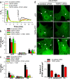
Co-localization and co-immunoprecipitation of TRPP2 and STIM1 in transfected HEK293 cells. a Upper penal: GFP-tagged TRPP2 (TRPP2-GFP) co-localized with endoplasmic reticulum maker (ER-DsRed); Lower penal: TRPP2-GFP co-localized with mCherry-tagged STIM1 (mCherry-STIM1). b Representative images showing co-immunoprecipitation followed by immunoblots [left, immunoblot with anti-GFP; right, immunoblot with anti-mCherry]. GFP or mCherry antibody pulled down the proteins from TRPP2-GFP and mCherry-STIM1 co-expressing HEK293 cells. The experiment was repeated 4 times
Fig. 2
Fig. 2
FRET efficiency of TRPP2-STIM1 interaction in TRPP2 and STIM1 co-expressing HEK293 cells. a Representative images showing GFP, mCherry and FRET efficiency channels. The cells expressed TRPP2-mCherry and STIM1-GFP (upper), mCherry-TRPP2 and STIM1-GFP (middle) or GFP-mCherry (lower) proteins respectively. b Summarized data showing FRET efficiency in transfected HEK293 cells (four groups: mCherry and GFP, mCherry-GFP, TRPP2-mCherry and STIM1-GFP, mCherry-TRPP2 and STIM1-GFP). Values are shown as the mean ± SEM (n = 9–20 cells). *P < 0.05 for GFP and mCherry co-expression vs. GFP-mCherry expression; #P < 0.05 for TRPP2-mCherry and STIM1-GFP co-expression vs. mCherry-TRPP2 and STIM1-GFP co-expression. c Schematic diagram of full-length TRPP2 and its truncated derivatives: TRPP2 without N1 (2-111aa, ∆N1) and TRPP2 without N2 (112-221aa, ∆N2). d Representative images of co-immunoprecipitation experiments in HEK293 cells co-expressed with STIM1 plus GFP-tagged full-length TRPP2 (WT) or ∆N1 or ∆N2. IP, GFP antibody; IB, anti-EGFP antibody. The GFP-only vector was used as a negative control. e Summarized data showing the relative binding strength of STIM1 with TRPP2 (WT), or ∆N1 or ∆N2. The optical density of ∆N1 or ∆N2 blot was normalized to that of WT blot (= 100%) and expressed as the relative binding strength. Values are shown as the mean ± SEM (n = 3). *P < 0.05 for STIM1 and ∆N1 or ∆N2 co-expression vs. STIM1 and TRPP2 co-expression
Fig. 3
Fig. 3
Effect of TRPP2-STIM1 interaction on store-operated Ca2+ entry (SOCE) in TRPP2 and STIM1 co-expressing HEK293 cells. a-d Summary of data showing changes in Ca2+ release (a, c) and SOCE (b, d) in HEK293 cells transfected with TRPP2 siRNA, STIM1 siRNA, STIM1, TRPP2 and/or dominant negative TRPP2 (D511V), and treated by ATP (10 μmol/L), 2APB (100 μmol/L) + ATP (10 μmol/L) and thapsigargin (TG, 2.5 μmol/L) for 8 min in Ca2+-free solution. SOCE was evoked by extracellular Ca2+ (1 mmol/L) application. Values are shown as the mean ± SEM (n = 3–5). *P < 0.05 compared with scrambled siRNA in each treatment. e Summarized data showing ATP (10 μmol/L)-induced SOCE in HEK293 cells co-expressed with STIM1 and GFP-tagged full-length TRPP2 (GFP-TRPP2) or TRPP2 without N1 (GFP-TRPP2-∆N1, deletion of 2-111aa in TRPP2) or TRPP2 without N2 (GFP-TRPP2-∆N2, deletion of 112-221aa in TRPP2). Values are shown as mean ± SEM (n = 5). *P < 0.05 for STIM1 and GFP-TRPP2 or GFP-TRPP2-∆N2 co-expression vs. STIM1 and GFP-TRPP2-∆N1 co-expression
Fig. 4
Fig. 4
Co-immunoprecipitation and in situ proximity ligation assay (PLA) of TRPP2 and STIM1 in mouse aortic smooth muscle cells. (a, b) Immunoblots showing that anti-TRPP2 and anti-STIM1 recognized TRPP2 and STIM1 proteins, and co-immunoprecipitation followed by immunoblots (a, immunoblot with anti-TRPP2; b, immunoblot with anti-STIM1). Proteins from the mouse aortic smooth muscle cells were immunoprecipitated with indicated antibody (+) or no antibody (−). (c) PLA analysis was used to detect the interaction between TRPP2 and STIM1. Representative images were displayed in the presence of anti-STIM1 antibody alone ((a)-(c)), or in the presence of anti-TRPP2 and anti-STIM1 antibodies ((d)-(f)). ((c), (f)) were merged with bright view. Red doted fluorescence showing positive signal. Nuclei were marked by DAPI staining (blue color). Scale bar represents 5 μm. The experiment was repeated 4 times
Fig. 5
Fig. 5
Role of TRPP2 in store-operated Ca2+ entry (SOCE) and STIM1 puncta formation in mouse aortic smooth muscle cells. a Representative traces showing ATP (10 μmol/L)-induced Ca2+ release in Ca2+-free solution and SOCE in the mouse aortic smooth muscle cells transfected with TRPP2, STIM1, both TRPP2 and STIM1, or scrambled siRNAs. b Summary of data showing changes in intracellular Ca2+ concentration increase in response to extracellular ATP (10 μmol/L) or thapsigargin (TG, 2.5 μmol/L) application in the mouse aortic smooth muscle cells treated with or without 2APB (100 μmol/L) for 10 min in Ca2+-free solution. c Summary of data showing changes in intracellular Ca2+ concentration increase in response to extracellular Ca2+ (1 mmol/L) application in the mouse aortic smooth muscle cells treated by ATP (10 μmol/L), 2APB (100 μmol/L) + ATP (10 μmol/L) and thapsigargin (TG, 2.5 μmol/L) for 10 min in Ca2+-free solution. Values are shown as mean ± SEM (n = 3–6 experiments). *P < 0.05 for scrambled siRNA vs. TRPP2 or STIM1 or TRPP2 + STIM1 siRNA transfection in each treatment. d Representative images showing STIM1 puncta formation in the mouse aortic smooth muscle cells transfected with TRPP2 siRNA or scrambled siRNA and treated by ATP (10 μmol/L), 2APB (100 μmol/L) + ATP (10 μmol/L) and thapsigargin (TG, 2.5 μmol/L) for 10 min in Ca2+-free solution. Scale bar represents 10 μm. The experiment was repeated 4 times
Fig. 6
Fig. 6
Role of TRPP2 and STIM1 in Ca2+ release and store-operated Ca2+ entry (SOCE)-induced mouse aorta contraction. a Representative traces showing phenylephrine (Phe, 10 μmol/L)-induced contraction in Ca2+-free solution and extracellular Ca2+ (2.5 mmol/L) re-addition-induced contraction in mice aortae. b-e Summarized data showing Phe-induced contraction in Ca2+-free solution (b, d) and extracellular Ca2+ re-addition (c, e)-induced contractions in mice aortae, which were transfected with TRPP2 siRNA (b, c), SITM1 siRNA (d, e) or scrambled siRNA. f-i Summarized data showing endothelin 1 (ET-1, 100 nmom/L)-induced contraction in Ca2+-free solution (f, h) and extracellular Ca2+ re-addition (g, i)-induced contractions in mice aortae transfected with TRPP2 siRNA (f, g), SITM1 siRNA (h, i) or scrambled siRNA. Values are shown as mean ± SEM (n = 3–4 mice). *P < 0.05 for scrambled siRNA vs. TRPP2 siRNA or STIM1 siRNA transfection. j Representative traces showing thapsigargin (TG, 2.5 μmol/L)-induced contraction in Ca2+-free solution and extracellular Ca2+ (2.5 mmol/L) re-addition-induced contraction in mice aortae. k-n Summarized data showing TG-induced contraction in Ca2+-free solution (k, m) and extracellular Ca2+ re-addition (l, n)-induced contraction in mice aortae, which were transfected with TRPP2 siRNA (j-l), SITM1 siRNA (m-n) or scrambled siRNAs. Values are shown as the mean ± SEM (n = 3–4 mice). *P < 0.05 for TRPP2 siRNA or STIM1 siRNA vs. scrambled siRNA
Fig. 7
Fig. 7
Role of TRPP2 and STIM1 in agonist-induced mouse aorta contraction. Phenylephrine (10 μmol/L, a, b, e, f) and endothelin 1 (100 nmol/L, c, d, g, h) concentration-dependently induced the contraction of the mice aortae transfected with TRPP2 (a-d), STIM1 (e-h) or scrambled siRNA. b, d, f, h The mice aortae were pretreated by heparin (1 mg/ml) using a reversible permeabilization loading procedure. Values are shown as mean ± SEM (n = 4–6 mice). *P < 0.05 for scrambled siRNA vs. TRPP2 or STIM1 siRNA transfection
Fig. 8
Fig. 8
Generation and verification of the TRPP2 conditional knockout (CKO) mice. a Schematic description of gene-targeting map of PKD2 gene. Targeting construct is referring to the floxed allele. Flp-recombined is obtained from deletion of the Neomycin cassette. Cre-recombined is the mutant allele yielded from the deletion with Cre-recombinase. b Genomic PCR analysis data from mice tail DNA samples showing the presence of LoxP+/+ sites and Cre-transgens. c Immunoblots data showing TRPP2 expressing in aortic smooth muscle cells from TRPP2 CKO or Cre control mice. d, e Agonist-induced mouse aorta contraction. Phenylephrine (10 μmol/L, d) and endothelin 1 (100 nmol/L, e) concentration-dependently induced the contraction of TRPP2 CKO or Cre control mice aortae. Values are shown as mean ± SEM (n = 4–8 mice). *P < 0.05 for Cre control vs. TRPP2 CKO mouse

Similar articles

Cited by

References

    1. Hofherr A, Kottgen M. TRPP channels and polycystins. Adv Exp Med Biol. 2011;704:287–313. - PubMed
    1. Grantham JJ, Mulamalla S, Swenson-Fields KI. Why kidneys fail in autosomal dominant polycystic kidney disease. Nat Rev Nephrol. 2011;7:556–566. - PubMed
    1. Chen XZ, Li Q, Wu Y, Liang G, Lara CJ, Cantiello HF. Submembraneous microtubule cytoskeleton: interaction of TRPP2 with the cell cytoskeleton. FEBS J. 2008;275:4675–4683. - PubMed
    1. Du J, Wong WY, Sun L, Huang Y, Yao X. Protein kinase G inhibits flow-induced Ca2+ entry into collecting duct cells. J Am Soc Nephrol. 2012;23:1172–1180. - PMC - PubMed
    1. Bai CX, Giamarchi A, Rodat-Despoix L, Padilla F, Downs T, Tsiokas L, Delmas P. Formation of a new receptor-operated channel by heteromeric assembly of TRPP2 and TRPC1 subunits. EMBO Rep. 2008;9:472–479. - PMC - PubMed

Publication types

LinkOut - more resources