. 2020 Aug 31;17(1):258.
doi: 10.1186/s12974-020-01925-y.

Benzo(a)pyrene exposure induced neuronal loss, plaque deposition, and cognitive decline in APP/PS1 mice

Affiliations

Benzo(a)pyrene exposure induced neuronal loss, plaque deposition, and cognitive decline in APP/PS1 mice

Dan Liu et al. J Neuroinflammation. .

Abstract

Background: Exposure to benzo(a)pyrene (BaP) was associated with cognitive impairments and some Alzheimer's disease (AD)-like pathological changes. However, it is largely unknown whether BaP exposure participates in the disease progression of AD.

Objectives: To investigate the effect of BaP exposure on AD progression and its underlying mechanisms.

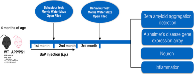
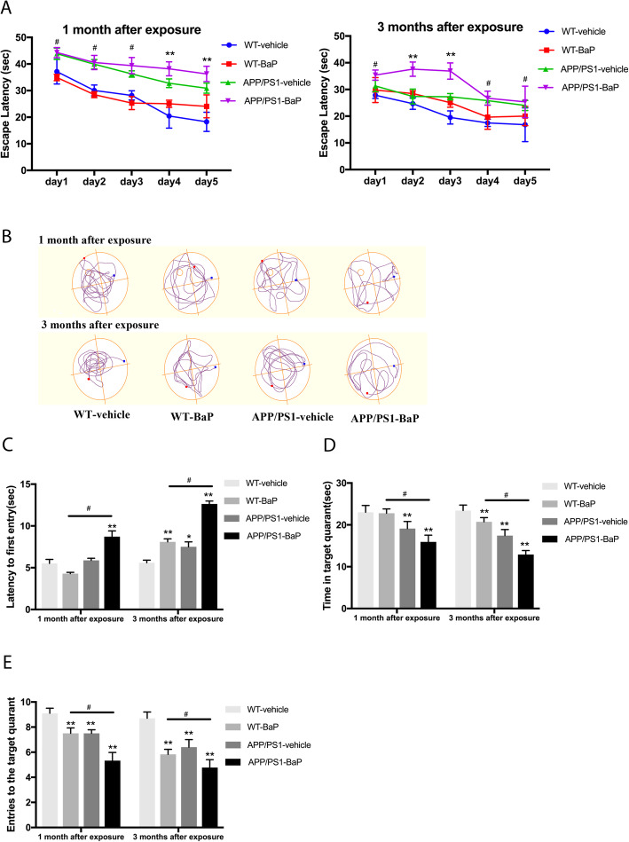
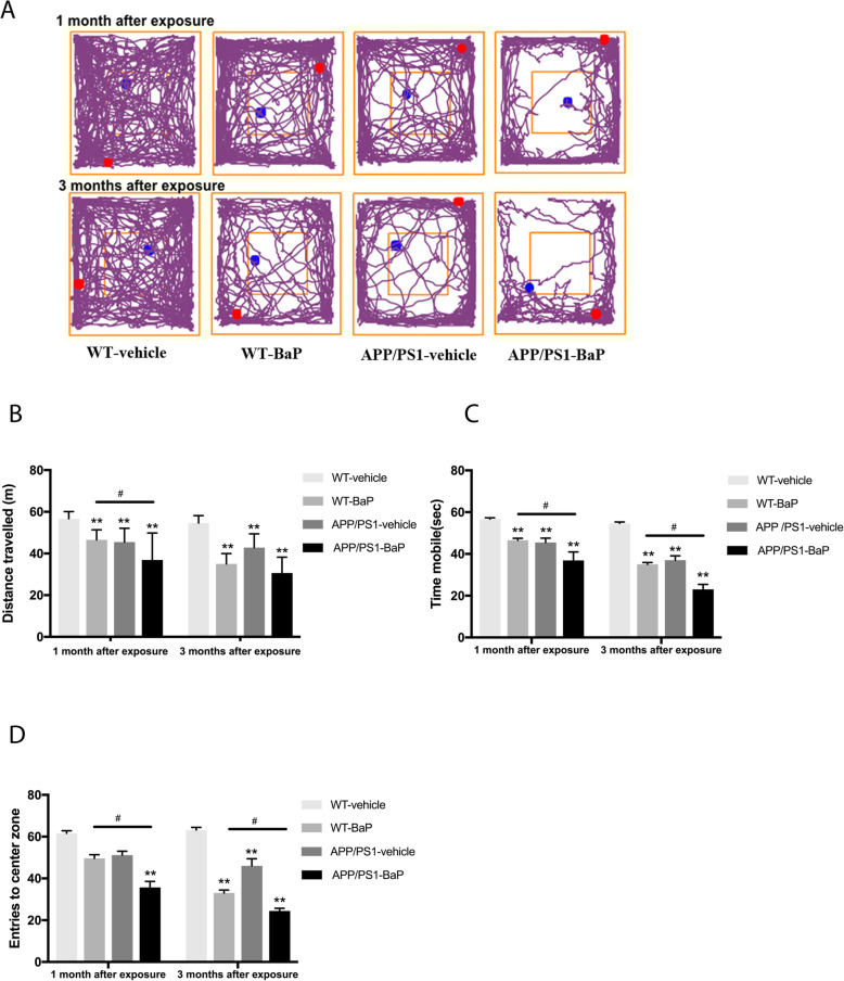
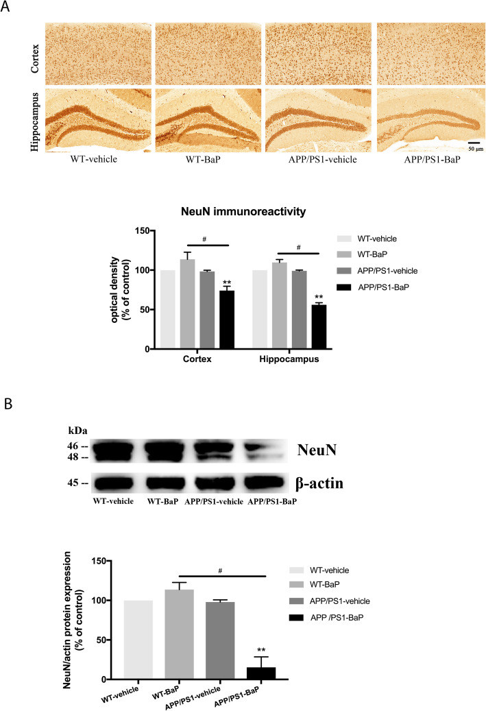
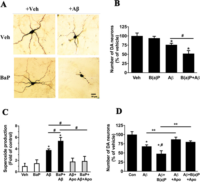
Methods: BaP or vehicle was administered to 4-month-old APPswe/PS1dE9 transgenic (APP/PS1) mice and wildtype (WT) mice for 2 months. Learning and memory ability and exploratory behaviors were evaluated 1 month after the initiation/termination of BaP exposure. AD-like pathological and biochemical alterations were examined 1 month after 2-month BaP exposure. Levels of soluble beta-amyloid (Aβ) oligomers and the number of Aβ plaques in the cortex and the hippocampus were quantified. Gene expression profiling was used to evaluate alternation of genes/pathways associated with AD onset and progression. Immunohistochemistry and Western blot were used to demonstrate neuronal loss and neuroinflammation in the cortex and the hippocampus. Treatment of primary neuron-glia cultures with aged Aβ (a mixture of monomers, oligomers, and fibrils) and/or BaP was used to investigate mechanisms by which BaP enhanced Aβ-induced neurodegeneration.

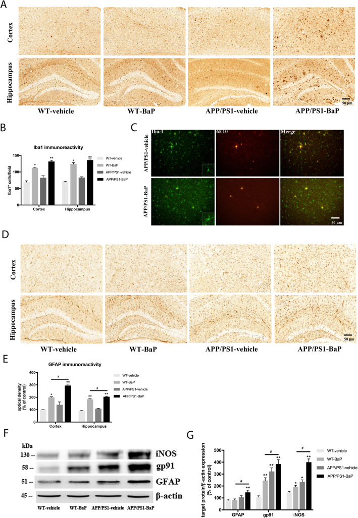
Results: BaP exposure induced progressive decline in spatial learning/memory and exploratory behaviors in APP/PS1 mice and WT mice, and APP/PS1 mice showed severer behavioral deficits than WT mice. Moreover, BaP exposure promoted neuronal loss, Aβ burden and Aβ plaque formation in APP/PS1 mice, but not in WT mice. Gene expression profiling showed most robust alteration in genes and pathways related to inflammation and immunoregulatory process, Aβ secretion and degradation, and synaptic formation in WT and APP/PS1 mice after BaP exposure. Consistently, the cortex and the hippocampus of WT and APP/PS1 mice displayed activation of microglia and astroglia and upregulation of inducible nitric oxide synthase (iNOS), glial fibrillary acidic protein (GFAP), and NADPH oxidase (three widely used neuroinflammatory markers) after BaP exposure. Furthermore, BaP exposure aggravated neurodegeneration induced by aged Aβ peptide in primary neuron-glia cultures through enhancing NADPH oxidase-derived oxidative stress.

Conclusion: Our study showed that chronic exposure to environmental pollutant BaP induced, accelerated, and exacerbated the progression of AD, in which elevated neuroinflammation and NADPH oxidase-derived oxidative insults were key pathogenic events.

Keywords: Alzheimer’s disease; Amyloid; Benzo(a)pyrene; Cognition; Neuroinflammation.


Conflict of interest statement

The authors declare that they have no known competing financial interests or personal relationships that could have appeared to influence the work reported in this paper.

Figures

Fig. 1
Fig. 1
Scheme of experimental design for in vivo studies. BaP (1 mg/kg/bw/day) or corn oil was administered into 4-month-old WT mice and APP/PS1 transgenic mice (WT-vehicle, WT-BaP, APP/PS1-vehicle, APP/PS1-BaP) for 2 consecutive months. Morris water maze and open field behavior tests were performed one month after the initiation/termination of BaP exposure. Then, the mice were sacrificed for gene expression profiling and histological/biochemical analyses 1 month after 2-month BaP exposure
Fig. 2
Fig. 2
BaP exposure accelerated progressive spatial and working memory decline in the Morris water maze test. a Escape latency during 5 days training performed immediately after BaP exposure for 1 month or 1 month after 2-month BaP exposure. b Representative path maps of each group in the probe trail. c Latency to the first entry to the target quadrant in the probe trail. d Time spent in the target quadrant in the probe trail. e Entries to the platform quadrant in the probe trail. N = 10/group. Data are mean ± SEM, *P < 0.05 and **P < 0.01 compared with WT-Vehicle control; #P < 0.05 compared with WT-BaP group
Fig. 3
Fig. 3
BaP exposure exacerbated progressive exploratory and anxiety impairments. a Representative path maps of each group in the open filed test. b Total distance traveled during the test. c Total time mobile during the test. d Entries to the center zone of the open filed. N = 10/group. Data are mean ± SEM, **P < 0.01 compared with WT-Vehicle control; #P < 0.05 compared with WT-BaP group
Fig. 4
Fig. 4
BaP exposure induced neuronal loss only in APP/PS1 mice. a Representative images of immunohistochemical staining of NeuN in the cortex and the hippocampus. The quantification results of NeuN immunoreactivity are expressed as a percentage of the corresponding WT-Vehicle control. b Levels of NeuN protein in the cortex and the hippocampus were detected by Western blot. β-actin was used to monitor loading errors. Data are expressed as a percentage of the WT-Vehicle control. Data are mean ± SEM of 3–4 mice in each group. Significance was determined by two-way ANOVA followed by LSD multiple comparisons post-hoc test. **P < 0.01 compared with WT-Vehicle control, #P < 0.05 compared with WT-BaP group
Fig. 5
Fig. 5
BaP exposure exacerbated Aβ burden and plaque formation in APP/PS1 mice. a Representative images of immunohistochemical staining of human Aβ using 6E10 antibody in the cortex and the hippocampus of APP/PS1 mice. Total number of Aβ plaques was counted. b Thioflavin-T staining showed Aβ plaques in the cortex and the hippocampus of APP/PS1 mice. Number of Aβ plaques was counted. c Levels of RIPA soluble Aβ species and APP were detected by Western blot using 6E10 antibody for human Aβ/APP. β-actin was used to monitor loading errors. The ratio of densitometry values of Aβ monomer (measured in the image with long exposure time), oligomers, and APP (measured in the image with short exposure time) was normalized to β-actin. Data are expressed as a percentage of APP/PS1-Vehicle control and are mean ± SEM of 3–4 mice in each treatment group. Significance was determined by t test. **P < 0.01 compared with APP/PS1-Vehicle. Arrow indicates Aβ fibrils stuck in the loading well of the SDS-PAGE gel and then transferred to the PVDF membrane
Fig. 6
Fig. 6
Glial activation and neuroinflammation were induced after BaP exposure. a, b Microglial activation was indicated by immunostaining with Iba-1 antibody (a) and counting of Iba-1-positive cells (b) in the cortex and the hippocampus. c Confocal double-staining of microglia and Aβ plaques by anti-Iba1 and anti-Aβ antibodies in the hippocampus. d Astroglial activation was detected with anti-GFAP antibody. e The GFAP immunoreactivity was measured and expressed as a percentage of the WT-Vehicle. f, g Levels of iNOS, gp91, and GFAP (three widely used neuroinflammatory markers) were detected by Western blot. β-Actin was used to monitor loading errors. Data are expressed as percentage of WT-Vehicle and are mean ± SEM of 3–4 mice in each treatment group. Significance was determined by two-way ANOVA followed by LSD multiple comparisons post-hoc test. *P < 0.05, **P < 0.01 compared with WT-Vehicle control, #P < 0.05 compared with WT-BaP group
Fig. 7
Fig. 7
Inhibition of NADPH oxidase prevented neuronal loss induced by aged Aβ alone or in combination with BaP. a, b In primary neuron-glia cultures, loss of TH-positive neurons was examined by immunostaining (a) and cell counting (b) at 7 days after treatment with 5 μg/ml (1.108 μM) aged Aβ (a mixture of monomers, oligomers, and fibrils) and/or BaP (0.1 μM). c Extracellular superoxide production in primary neuron-glia cultures treated with Aβ and/or BaP with or without pre-treatment for 30 min with NADPH oxidase inhibitor apocynin (Apo, 0.25 mM) was measure at 24 h after Aβ/BaP treatment by SOD-inhibitable reduction of WST-1. d Neuroprotective effects of pre-treatment with Apo (0.25 mM) for 30 min against neurotoxic effects of Aβ and BaP determined by immunostaining and cell counting at 7 days after Aβ/BaP treatment. Data are expressed as fold or percent of vehicle-treated control and are mean ± SEM of 3–4 experiments performed in quadruplicate. Significance was determined by one-way ANOVA (b) or two-way ANOVA (c and d) followed by LSD multiple comparisons post-hoc test. *P < 0.05 compared with vehicle-treated control, #P < 0.05 compared with Aβ-treated cultures. **P < 0.05

References

    1. Mayeux R, Stern Y. Epidemiology of Alzheimer disease. Cold Spring Harb Perspect Med. 2012;2:137. - PMC - PubMed
    1. Scheltens P, Blennow K, Breteler MM, de Strooper B, Frisoni GB, Salloway S, et al. Alzheimer’s disease. Lancet. 2016;388:505–517. - PubMed
    1. Chin-Chan M, Navarro-Yepes J, Quintanilla-Vega B. Environmental pollutants as risk factors for neurodegenerative disorders: Alzheimer and Parkinson diseases. Front Cell Neurosci. 2015;9:124. - PMC - PubMed
    1. Wallin C, Sholts SB, Osterlund N, Luo J, Jarvet J, Roos PM, et al. Alzheimer's disease and cigarette smoke components: effects of nicotine, PAHs, and cd(II), Cr(III), Pb(II), Pb(IV) ions on amyloid-beta peptide aggregation. Sci Rep. 2017;7:14423. - PMC - PubMed
    1. Calderon-Garciduenas L, Reed W, Maronpot RR, Henriquez-Roldan C, Delgado-Chavez R, Calderon-Garciduenas A, et al. Brain inflammation and Alzheimer's-like pathology in individuals exposed to severe air pollution. Toxicol Pathol. 2004;32:650–658. - PubMed

MeSH terms