. 2020 Sep 1;21(1):64.
doi: 10.1186/s40360-020-00442-1.

Boswellic acid sensitizes gastric cancer cells to Cisplatin-induced apoptosis via p53-mediated pathway

Affiliations

Boswellic acid sensitizes gastric cancer cells to Cisplatin-induced apoptosis via p53-mediated pathway

Shadia Al-Bahlani et al. BMC Pharmacol Toxicol. .

Abstract

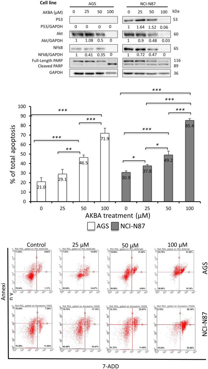
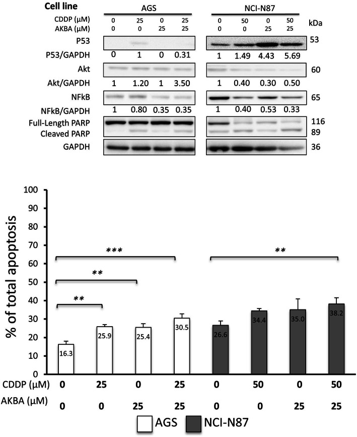
Background: Cisplatin (CDDP) is an effective anticancer drug for Gastric cancer (GC) that induces apoptosis by altering pro- (p53) and anti-apoptotic (Akt and NFkB) proteins; however, chemoresistance remains a big challenge. Additional compounds with promising anticancer effects such as AKBA (Acetyl-keto-beta boswellic acid) may overcome the resistance. However, its role in CDDP-induced apoptosis in GC has not been studied. This study aimed to examine the effectiveness of AKBA on p53-mediated, CDDP-induced apoptosis in GC cells. AGS and NCI-N87 cells were treated with different concentrations (0, 25, 50, 100 μM) of CDDP and/or AKBA.

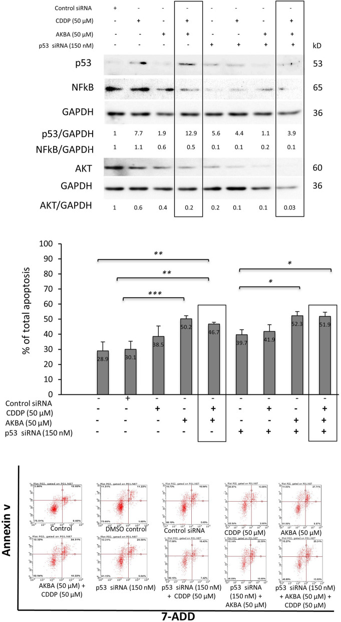
Methods: P53, Akt and NFkB proteins and apoptosis were assessed by Western blot and flow cytometry. The role of p53 was determined by inhibiting its function via the siRNA approach.

Results: The results revealed that CDDP and AKBA significantly increased p53 content in both cells, while Akt and NFkB were significantly decreased. Both compounds significantly induced apoptosis in a dose-dependent manner. AKBA sensitized GC cells to CDDP-induced apoptosis by altering the protein expression. P53 downregulation affected Akt and NFkB proteins with a slight increase in apoptosis induction in the combination treated groups.

Conclusions: Altogether, our findings suggest that AKBA enhances GC cell sensitivity to CDDP-induced apoptosis via the p53 pathway.

Keywords: AKBA; Apoptosis and gastric cancer; CDDP; P53.


Conflict of interest statement

The authors declare no competing Interests.

Figures

Fig. 1
Fig. 1
CDDP had differential effect on proteins expression and induced apoptosis in GC cells. a Western blot shows the effect of CDDP on protein expression in AGS and NCI - N87 cell lines. The expression of tumor suppressor protein 53 increased with CDDP concentration, whilst Akt and NFkB expressions reduced, notice the cleaved PARP which indicates apoptosis b Graphical presentation of apoptosis percentages in AGS and NCI-N87 cell lines treated with different concentrations of CDDP showing significant increase in apoptosis in comparison to control concentration (*P < 0.05, **P < 0.01, ***P < 0.001). The response of AGS cells is more evident than NCI-N87. c Flow cytometry graphs showing the effect of different doses of CDDP on AGS and NCI-N87 cells, each graph shows alive cells, early and late apoptotic cells, and dead cells
Fig. 2
Fig. 2
AKBA exhibited similar effect as CDDP on proteins expression and apoptosis in GC cells except for p53. a Western blot shows the effect of AKBA on protein expression in AGS and NCI - N87 cell lines. The expression followed a similar pattern to the cells treated with CDDP b Graphical presentation of apoptosis percentage in AGS and NCI-N87 cell lines treated with different concentrations of AKBA showing significant increase in apoptosis in comparison to control concentration (*P < 0.05, **P < 0.01, ***P < 0.001). The concentration of 100 μM was found to be highly toxic and the apoptosis percentages reached 71.9 and 85.4% in AGS and NCI-N87 cell lines respectively. c Flow cytometry graphs showing the effect of different doses of AKBA on AGS and NCI-N87 cells. Each graph shows alive cells, early and late apoptotic cells, and dead cells
Fig. 3
Fig. 3
AKBA sensitized GC cells to CDDP-induced apoptosis via altering proteins expression. a Western blot shows the effect of combined CDDP and AKBA on protein expression in AGS and NCI - N87 cell lines. b Graphical presentation of apoptosis percentage in AGS and NCI-N87 cell lines treated with different concentrations of CDDP and AKBA showing significant increase in apoptosis in comparison to control concentration (*P < 0.05, **P < 0.01, ***P < 0.001)
Fig. 4
Fig. 4
P53-dependent pathway mediated CDDP- and AKBA-induced apoptosis in GC a Western blot shows the effect of siRNA, CDDP and AKBA on protein expression in NCI - N87 cell line. P53 siRNA was successful in reducing the increase in p53 expression post treatment of CDDP and AKBA up to approximately 70%. The expression of Akt and NFkB proteins was further reduced in the combined treatment when p53 protein was down-regulated b Graphical presentation of apoptosis percentage in NCI-N87 cell line treated with different treatments showing varying degrees in apoptosis in comparison to control concentration. Apoptosis induction showed a slight trend of increase with no significant difference between treated groups with and without p53 down regulation. (*P < 0.05, **P < 0.01, ***P < 0.001). (c) Flow cytometry graphs showing the effect of different treatments on NCI-N87 cells. Each graph shows alive cells, early and late apoptotic cells, and dead cells

References

    1. Bray F, Ferlay J, Soerjomataram I, Siegel R, Torre L, Jemal A. Global cancer statistics 2018: GLOBOCAN estimates of incidence and mortality worldwide for 36 cancers in 185 countries. CA Cancer J Clin. 2018;68(6):394–424. doi: 10.3322/caac.21492. - DOI - PubMed
    1. WHO. Cancer. Who.int. http://www.who.int/mediacentre/factsheets/fs297/en/. Published 2017.
    1. Abbas A, Aster J, Kumar V, Perkins J. Robbins Basic Pathology. 9th ed: Elsevier Saunders; 2013. p. 570–3.
    1. Al-Lawati N, Al-Bahrani B, Al-Raisi S, Al-Lawati J. Twenty-year trends of Cancer incidence in Omanis, 1996-2015. Oman Med J. 2019;34(4):361–387. doi: 10.5001/omj.2019.55. - DOI - PMC - PubMed
    1. Clark M, Finkel R. Lippincott's Illustrated Reviews: Pharmacology. 5. Baltimore: Lippincott Williams & Wilkins; 2012. pp. 507–508.

Publication types

MeSH terms