. 2020 Nov 6;295(45):15366-15375.
doi: 10.1074/jbc.RA120.015009. Epub 2020 Aug 31.

Capping protein is dispensable for polarized actin network growth and actin-based motility

Affiliations

Capping protein is dispensable for polarized actin network growth and actin-based motility

Majdouline Abou-Ghali et al. J Biol Chem. .

Abstract

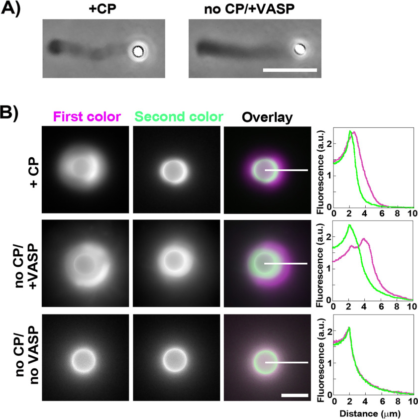
Heterodimeric capping protein (CP) binds the rapidly growing barbed ends of actin filaments and prevents the addition (or loss) of subunits. Capping activity is generally considered to be essential for actin-based motility induced by Arp2/3 complex nucleation. By stopping barbed end growth, CP favors nucleation of daughter filaments at the functionalized surface where the Arp2/3 complex is activated, thus creating polarized network growth, which is necessary for movement. However, here using an in vitro assay where Arp2/3 complex-based actin polymerization is induced on bead surfaces in the absence of CP, we produce robust polarized actin growth and motility. This is achieved either by adding the actin polymerase Ena/VASP or by boosting Arp2/3 complex activity at the surface. Another actin polymerase, the formin FMNL2, cannot substitute for CP, showing that polymerase activity alone is not enough to override the need for CP. Interfering with the polymerase activity of Ena/VASP, its surface recruitment or its bundling activity all reduce Ena/VASP's ability to maintain polarized network growth in the absence of CP. Taken together, our findings show that CP is dispensable for polarized actin growth and motility in situations where surface-directed polymerization is favored by whatever means over the growth of barbed ends in the network.

Keywords: Arp2/3 complex; Ena/VASP proteins; actin; actin polymerization; biophysics; capping protein; cell motility; cytoskeleton; in vitro systems.


Conflict of interest statement

Conflict of interest—The authors declare that they have no conflicts of interest with the contents of this article.

Figures

Figure 1.
Figure 1.
VASP can replace CP in bead motility and polarized actin network growth. A, 1-μm diameter beads form actin comets and move in the presence of 25 nm CP or without CP but with 50 nm added VASP. Reaction time is ∼8 min. B, the polarity of actin assembly on 4.5-μm beads is assessed with a two-color assay in the presence or absence of 25 nm CP, and in the absence of CP with 50 nm added VASP. The first color is magenta (actin Alexa 594) and the second is green (actin Alexa 488). Separate channels and overlay images are shown (colocalization is white in the magenta/green overlays). Line scans are taken as indicated by white lines, and fluorescence intensity (arbitrary units) is plotted versus distance from the bead center. Superposition of magenta and green curves indicates unpolarized growth, and separation of magenta and green curves indicates polarized growth. For all panels, the concentration of the Arp2/3 complex is 50 nm. Phase-contrast and epifluorescence microscopy. All scale bars, 5 μm.
Figure 2.
Figure 2.
Interplay of Arp2/3 complex activity and VASP for polarized actin growth in the absence of CP. The extent of polarized actin growth on 4.5-μm beads is assessed using the two-color approach. A, separate channels and overlay images are shown (first color magenta, representing actin Alexa 594, second color green, representing actin Alexa 488). Line scans are measured as indicated by white lines, fluorescence intensity (arbitrary units) is plotted versus distance from the bead center, and separation of magenta and green curve maxima is taken as a segregation event, indicative of polarized growth. Top panels: actin growth in the presence of excess (150 nm) Arp2/3 complex in the absence of CP and VASP. Bottom panels: beads are coated with a tetrameric form of pVCA (streptavidin-pVCA or S-pVCA), which is a more effective activator of the Arp2/3 complex than GST-pVCA. The polymerization mix contains no CP and no VASP, and 50 nm Arp2/3 complex. B, actin growth in the presence of varying amounts of the Arp2/3 complex and VASP in the absence of CP. Overlay images are shown. % color segregation and number of beads analyzed is indicated on an image of each condition; images were chosen to represent the majority case for each condition. The phase space where segregation occurs more than 50% of the time is depicted by the red boundary. C, images of actin and Arp2/3 complex in 25 nm CP and no CP/37 nm VASP conditions at about 10 min reaction time. The Arp2/3 complex concentration is 50 nm. A medial plane is shown. Quantification of total fluorescent intensity in the actin and Arp2/3 complex channels (n ≥ 7). n.s. indicates a nonsignificant difference. A and B, epifluorescence microscopy. C, spinning disc microscopy. All scale bars, 5 μm.
Figure 3.
Figure 3.
How the actin network grows with other elongators and over time. A, separate channels and overlay images are shown (first color magenta, representing actin Alexa 594, second color green, representing actin Alexa 488). Line scans are measured as indicated by white lines, fluorescence intensity (arbitrary units) is plotted versus distance from the bead center, and separation of magenta and green curve maxima is taken as a segregation event, indicative of polarized growth. Top panels: actin growth in the presence of 100 nm chimeric human/Dictyostelium VASP dimer (VASP-2M). Color segregation occurs on 80% of the beads (n = 88). Bottom panels: actin growth in the presence of 50 nm FMNL2-8P, which gave 0% color segregation (n = 44). Epifluorescence microscopy was used. Scale bar, 5 μm. B, evolution over time of the total fluorescence of the actin network (medial plane, spinning disc images) in no CP/37 nm VASP conditions (open symbols) and in 25 nm CP conditions (closed symbols). Linear fits are shown. The Arp2/3 complex concentration was 50 nm (n ≥ 8) for beads in each condition. S.D. are large because of low signal to noise in spinning disc slices, but differences between no CP/+VASP and +CP conditions are significant except for actin fluorescence at 1 min.

Similar articles

Cited by

References

    1. Loisel T. P., Boujemaa R., Pantaloni D., and Carlier M. F. (1999) Reconstitution of actin-based motility of Listeria and Shigella using pure proteins. Nature 401, 613–616 10.1038/44183 - DOI - PubMed
    1. Cameron L. A., Footer M. J., Van Oudenaarden A., and Theriot J. A. (1999) Motility of ActA protein-coated microspheres driven by actin polymerization. Proc. Natl. Acad. Sci. U.S.A. 96, 4908–4913 10.1073/pnas.96.9.4908 - DOI - PMC - PubMed
    1. Bernheim-Groswasser A., Wiesner S., Golsteyn R. M., Carlier M.-F., and Sykes C. (2002) The dynamics of actin-based motility depend on surface parameters. Nature 417, 308–311 10.1038/417308a - DOI - PubMed
    1. Achard V., Martiel J.-L., Michelot A., Guérin C., Reymann A.-C., Blanchoin L., and Boujemaa-Paterski R. (2010) A “primer”-based mechanism underlies branched actin filament network formation and motility. Curr. Biol. 20, 423–428 10.1016/j.cub.2009.12.056 - DOI - PubMed
    1. Reymann A.-C., Boujemaa-Paterski R., Martiel J.-L., Guérin C., Cao W., Chin H. F., De La Cruz E. M., Théry M., and Blanchoin L. (2012) Actin network architecture can determine myosin motor activity. Science 336, 1310–1314 10.1126/science.1221708 - DOI - PMC - PubMed

Publication types

LinkOut - more resources