. 2020 Sep 22;117(38):23802-23806.
doi: 10.1073/pnas.2006109117. Epub 2020 Aug 31.

Helper bacteria halt and disarm mushroom pathogens by linearizing structurally diverse cyclolipopeptides

Affiliations

Helper bacteria halt and disarm mushroom pathogens by linearizing structurally diverse cyclolipopeptides

Ron Hermenau et al. Proc Natl Acad Sci U S A. .

Abstract

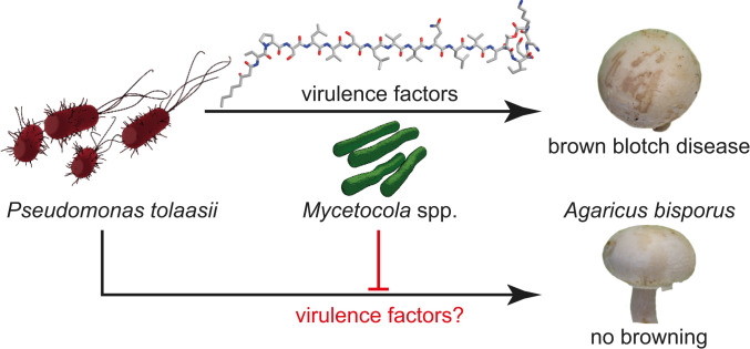
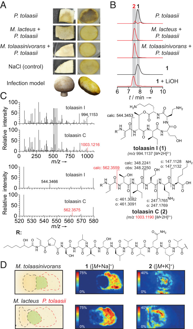
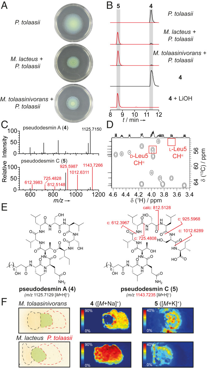
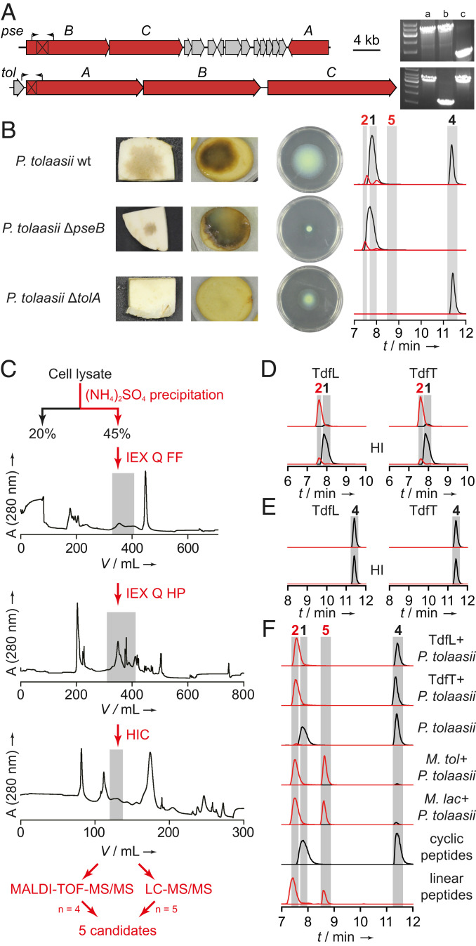
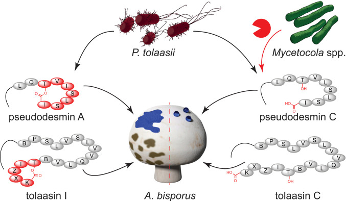
The bacterial pathogen Pseudomonas tolaasii severely damages white button mushrooms by secretion of the pore-forming toxin tolaasin, the main virulence factor of brown blotch disease. Yet, fungus-associated helper bacteria of the genus Mycetocola (Mycetocola tolaasinivorans and Mycetocola lacteus) may protect their host by an unknown detoxification mechanism. By a combination of metabolic profiling, imaging mass spectrometry, structure elucidation, and bioassays, we found that the helper bacteria inactivate tolaasin by linearizing the lipocyclopeptide. Furthermore, we found that Mycetocola spp. impair the dissemination of the pathogen by cleavage of the lactone ring of pseudodesmin. The role of pseudodesmin as a major swarming factor was corroborated by identification and inactivation of the corresponding biosynthetic gene cluster. Activity-guided fractionation of the Mycetocola proteome, matrix-assisted laser desorption/ionization (MALDI) analyses, and heterologous enzyme production identified the lactonase responsible for toxin cleavage. We revealed an antivirulence strategy in the context of a tripartite interaction that has high ecological and agricultural relevance.

Keywords: Mycetocola; antivirulence; brown blotch disease; cyclic lipopeptides; tolaasin.


Conflict of interest statement

The authors declare no competing interest.

Figures

Fig. 1.
Fig. 1.
Tripartite interaction of mushroom pathogen, helper bacteria, and fungal host. P. tolaasii produces virulence factors that cause bacterial brown blotch in A. bisporus. Mycetocola spp. can prevent decay of the mushroom cap.
Fig. 2.
Fig. 2.
Helper bacteria inactivate the toxin produced by the mushroom pathogen. (A) In vivo infection assays with mushroom and potato tuber slices. (B) Extracted-ion chromatogram (EIC) traces of 1 (black, 994.1137 [M + 2H]2+) and 2 (red, 1003.1190 [M + 2H]2+) obtained from high-performance liquid chromatography (HPLC) analysis of culture extracts, isolated 1, and 1 hydrolyzed with 5 eq LiOH, 1 h, RT. (C) Comparison of MS/MS fragmentation patterns of 1 and 2 with highlighted key fragments. Gray bar indicates magnified area. c: calculated; o: observed masses. (D) MALDI imaging of solid cultures of P. tolaasii with M. lacteus, and M. tolaasinivorans, respectively, showing spatial distribution of 1 and 2. Black: colony of M. tolaasinivorans and M. lacteus, respectively; red: colony of P. tolaasii; green: mixed colonies.
Fig. 3.
Fig. 3.
Cleavage of 4 impairs dissemination of the pathogen. (A) Swarming assays of P. tolaasii and respective cocultures on soft Kings B agar. (B) EIC traces of 4 (black, 1125.7129 [M + H]+) and 5 (red, 1143.7235 [M + H]+) obtained from HPLC analysis of culture extracts, isolated 4, and 4 hydrolyzed with 5 eq LiOH, 1 h, RT. (C) MS/MS analysis of 4 and 5 with key fragments highlighted in red. (D) Heteronuclear single quantum coherence correlations of the CHα region of 4 with red boxes indicating expected signals for d-Leu CHα and l-Leu CHα in position 5 based on literature data. (E) Structures of 4 and 5 with key fragments highlighted in red. c: calculated masses of fragments. (F) MALDI imaging of cultures of P. tolaasii with M. lacteus or M. tolaasinivorans, showing spatial distribution of 4 and 5. Black: colony of helper bacteria; red: colony of pathogen; green: mixed colonies.
Fig. 4.
Fig. 4.
Phenotypes of P. tolaasii knockout mutants, and identification of hydrolytic principle. (A) Gene clusters coding for pseudodesmin (pse) and tolaasin (tol) biosynthesis. Black boxes mark deleted areas, arrows indicate primer binding sites used for knockout verification. Agarose gel shows obtained amplicons from (a) P. tolaasii wild type; (b) P. tolaasii ΔtolA; (c) P. tolaasii ΔpseB. (B) Pathogenicity and swarming ability of P. tolaasii strains deficient in the production of tolaasin and pseudodesmin; EIC traces of 1, 2, 4, and 5. (C) Exemplary flow chart of bioactivity-guided fractionation approach. IEX: anion-exchange chromatography; Q: quarternary ammonium anion exchanger; FF: sepharose fast flow; HP: high performance; HIC: hydrophobic interaction chromatography. Gray boxes indicate active fractions that were further fractionated. (D) EIC traces of 1 (black, 994.1137 [M + 2H]2+) and 2 (red, 1003.1190 [M + 2H]2+) obtained from HPLC analyses of in vitro assays of TdfL and TdfT using 1 as a substrate. HI: heat-inactivated. (E) EIC traces of 4 (black, 1125.7129 [M + H]+) and 5 (red, 1143.7235 [M + H]+) obtained from HPLC analysis of in vitro assays of TdfL and TdfT using 4 as a substrate. (F) EIC traces of 1, 2, 4, and 5. In all LC traces: black: cyclic peptides 1 and 4; red: linearized peptides 2 and 5.
Fig. 5.
Fig. 5.
P. tolaasii produces pseudodesmins as swarming agents and tolaasins as toxins against A. bisporus. In the presence of protective helper bacteria (Mycetocola spp.) the virulence factors are cleaved by enzymes, such as TdfL or TdfT. One letter code used to represent amino acids in the peptides. B: dehydrobutyrine; X: 2,4-diaminobutanoic acid; Z: homoserine.

References

    1. Scherlach K. et al. ., Biosynthesis and mass spectrometric imaging of tolaasin, the virulence factor of brown blotch mushroom disease. ChemBioChem 14, 2439–2443 (2013). - PubMed
    1. Thongkongkaew T. et al. ., Two types of threonine-tagged lipopeptides synergize in host colonization by pathogenic Burkholderia species. ACS Chem. Biol. 13, 1370–1379 (2018). - PubMed
    1. Graupner K. et al. ., Imaging mass spectrometry and genome mining reveal highly antifungal virulence factor of mushroom soft rot pathogen. Angew. Chem. Int. Ed. Engl. 51, 13173–13177 (2012). - PubMed
    1. Fischer D. et al. ., Disruption of membrane integrity by the bacterium-derived antifungal jagaricin. Antimicrob. Agents Chemother. 63, e00707–e00719 (2019). - PMC - PubMed
    1. Nutkins J. C. et al. ., Structure determination of tolaasin, an extracellular lipodepsipeptide produced by the mushroom pathogen, Pseudomonas tolaasii Paine. J. Am. Chem. Soc. 113, 2621–2627 (1991).

Publication types

Supplementary concepts