Review
. 2020 Sep 30;39(3):244-258.
doi: 10.23876/j.krcp.20.082.

Mitochondrial dysfunction in kidney injury, inflammation, and disease: Potential therapeutic approaches

Affiliations
Review

Mitochondrial dysfunction in kidney injury, inflammation, and disease: Potential therapeutic approaches

Divya Bhatia et al. Kidney Res Clin Pract. .

Abstract

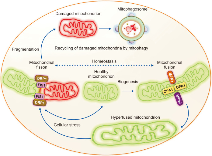
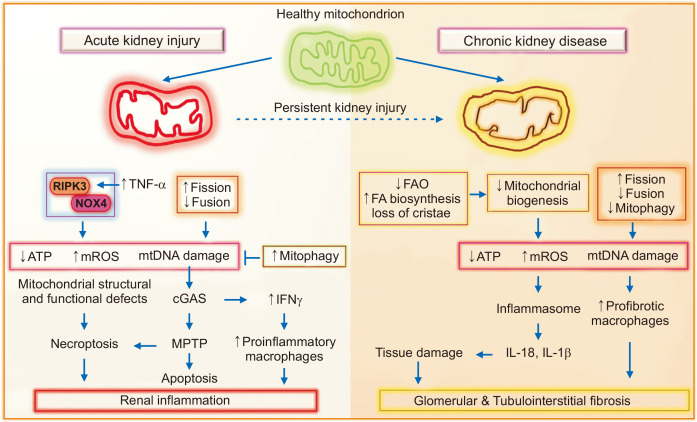
Mitochondria are energy-producing organelles that not only satisfy the high metabolic demands of the kidney but sense and respond to kidney injury-induced oxidative stress and inflammation. Kidneys are rich in mitochondria. Mitochondrial dysfunction plays a critical role in the progression of acute kidney injury and chronic kidney disease. Mitochondrial responses to specific stimuli are highly regulated and synergistically modulated by tightly interconnected processes, including mitochondrial dynamics (fission, fusion) and mitophagy. The counterbalance between these processes is essential in maintaining a healthy network of mitochondria. Recent literature suggests that alterations in mitochondrial dynamics are implicated in kidney injury and the progression of kidney diseases. A decrease in mitochondrial fusion promotes fission-induced mitochondrial fragmentation, but a reduction in mitochondrial fission produces excessive mitochondrial elongation. The removal of dysfunctional mitochondria by mitophagy is crucial for their quality control. Defective mitochondrial function disrupts cellular redox potential and can cause cell death. Mitochondrial DNA derived from damaged cells also act as damage-associated molecular patterns to recruit immune cells and the inflammatory response can further exaggerate kidney injury. This review provides a comprehensive overview of the role of mitochondrial dysfunction in acute kidney injury and chronic kidney disease. We discuss the processes that control mitochondrial stress responses to kidney injury and review recent advances in understanding the role of mitochondrial dysfunction in inflammation and tissue damage through the use of different experimental models of kidney disease. We also describe potential mitochondria-targeted therapeutic approaches.

Keywords: Acute kidney injury; Inflammation; Kidney diseases; Mitochondria; Oxidative stress.


Conflict of interest statement

Conflicts of interest

The spouse of Mary E. Choi is a cofounder and shareholder and serves on the Scientific Advisory Board of Proterris, Inc. The remaining authors have no conflicts of interest to declare.

Figures

Figure 1
Figure 1. Mitochondrial fusion, fission, and mitophagy in maintaining mitochondrial homeostasis.
Balance between the opposing processes of mitochondrial fusion and fission is essential for mitochondrial homeostasis. Mitochondrial fusion is mediated by the outer mitochondrial membrane fusion proteins mitofusin 1 and 2 (MFN1 and MFN2) and the inner mitochondrial membrane fusion protein optic atrophy 1 (OPA1). The fusion of two mitochondria generates a hyperfused mitochondrion that can produce more ATP during stress. Mitochondrial fission protein dynamin-related protein 1 (DRP1) translocates to the mitochondria and binds to mitochondrial fission 1 (FIS1) to induce mitochondrial fragmentation. The damaged mitochondria with reduced membrane potential generated by mitochondrial fragmentation are then recycled via mitophagosome formation and degradation by mitophagy.
Figure 2
Figure 2. Role of mitochondrial structural and functional defects during acute kidney injury (AKI) and chronic kidney disease (CKD).
Mitochondrial aberrations play important roles in promoting glomerular and tubulointerstitial inflammation and fibrosis during AKI and CKD. The contributors to mitochondrial dysfunction-induced renal inflammation during AKI include: 1) activation of necroptosis via tumor necrosis factor-alpha (TNFα) and receptor-interacting protein kinase-3 (RIPK3)-mediated NADPH oxidase-4 (NOX4) recruitment to the mitochondria or through the mitochondrial permeability transition pore (MPTP), 2) mitochondrial fragmentation through increased fission and decreased fusion, and 3) damaged mitochondria-derived mitochondrial DNA (mtDNA)-mediated inflammatory response through the activation of the cyclic GMP-AMP synthase (cGAS) stimulator of interferon genes (STING) pathway. The activation of mitophagy after AKI could be a renoprotective response against mitochondrial-derived reactive oxygen species (mROS) production. Reduction in mitochondrial fatty acid oxidation (FAO), loss of cristae, and impaired mitochondrial biogenesis and decreases in mitochondrial fusion and mitophagy resulting in mitochondrial fragmentation, superoxide production, and reduced adenosine triphosphate (ATP) content all contribute to an increase in tissue damage and inflammatory and fibrotic responses in CKD. FA, fatty acid; IFNγ, interferon gamma; IL, interleukin.

References

    1. Maekawa H, Inoue T, Ouchi H, et al. Mitochondrial damage causes inflammation via cGAS-STING signaling in acute kidney injury. Cell Rep. 2019;29:1261–1273.e6. doi: 10.1016/j.celrep.2019.09.050. - DOI - PubMed
    1. Chung KW, Dhillon P, Huang S, et al. Mitochondrial damage and activation of the STING pathway lead to renal inflammation and fibrosis. Cell Metab. 2019;30:784–799.e5. doi: 10.1016/j.cmet.2019.08.003. - DOI - PMC - PubMed
    1. Bhargava P, Schnellmann RG. Mitochondrial energetics in the kidney. Nat Rev Nephrol. 2017;13:629–646. doi: 10.1038/nrneph.2017.107. - DOI - PMC - PubMed
    1. Honda T, Hirakawa Y, Nangaku M. The role of oxidative stress and hypoxia in renal disease. Kidney Res Clin Pract. 2019;38:414–426. doi: 10.23876/j.krcp.19.063. - DOI - PMC - PubMed
    1. Tran M, Tam D, Bardia A, et al. PGC-1α promotes recovery after acute kidney injury during systemic inflammation in mice. J Clin Invest. 2011;121:4003–4014. doi: 10.1172/JCI58662. - DOI - PMC - PubMed

LinkOut - more resources