Review
. 2020 Oct;33(5):339-346.
doi: 10.1097/QCO.0000000000000669.

Subarachnoid neurocysticercosis: emerging concepts and treatment

Affiliations
Review

Subarachnoid neurocysticercosis: emerging concepts and treatment

Theodore E Nash et al. Curr Opin Infect Dis. 2020 Oct.

Abstract

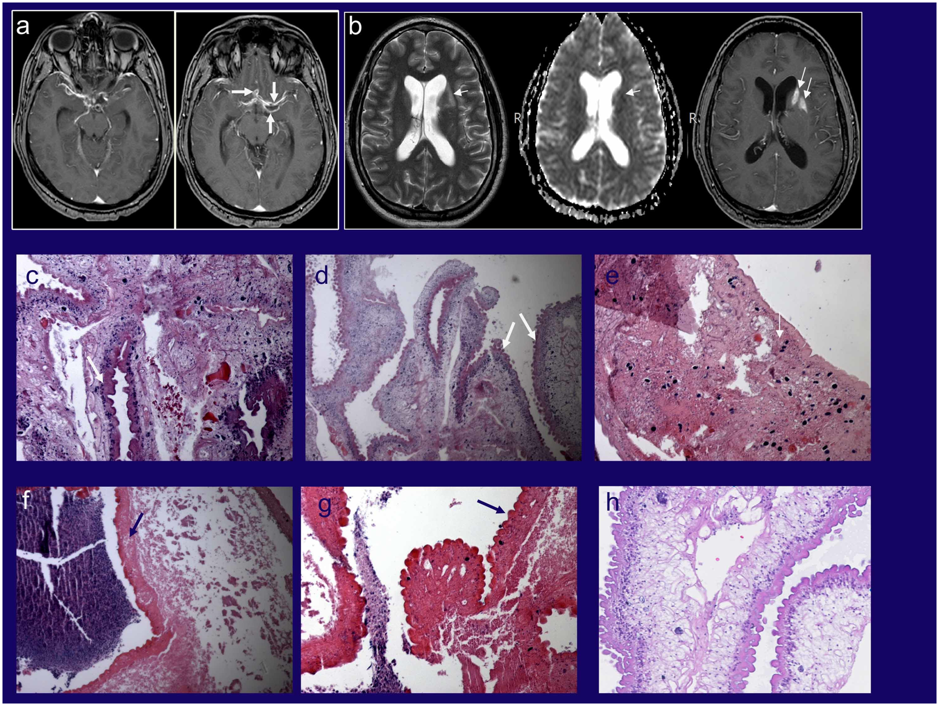
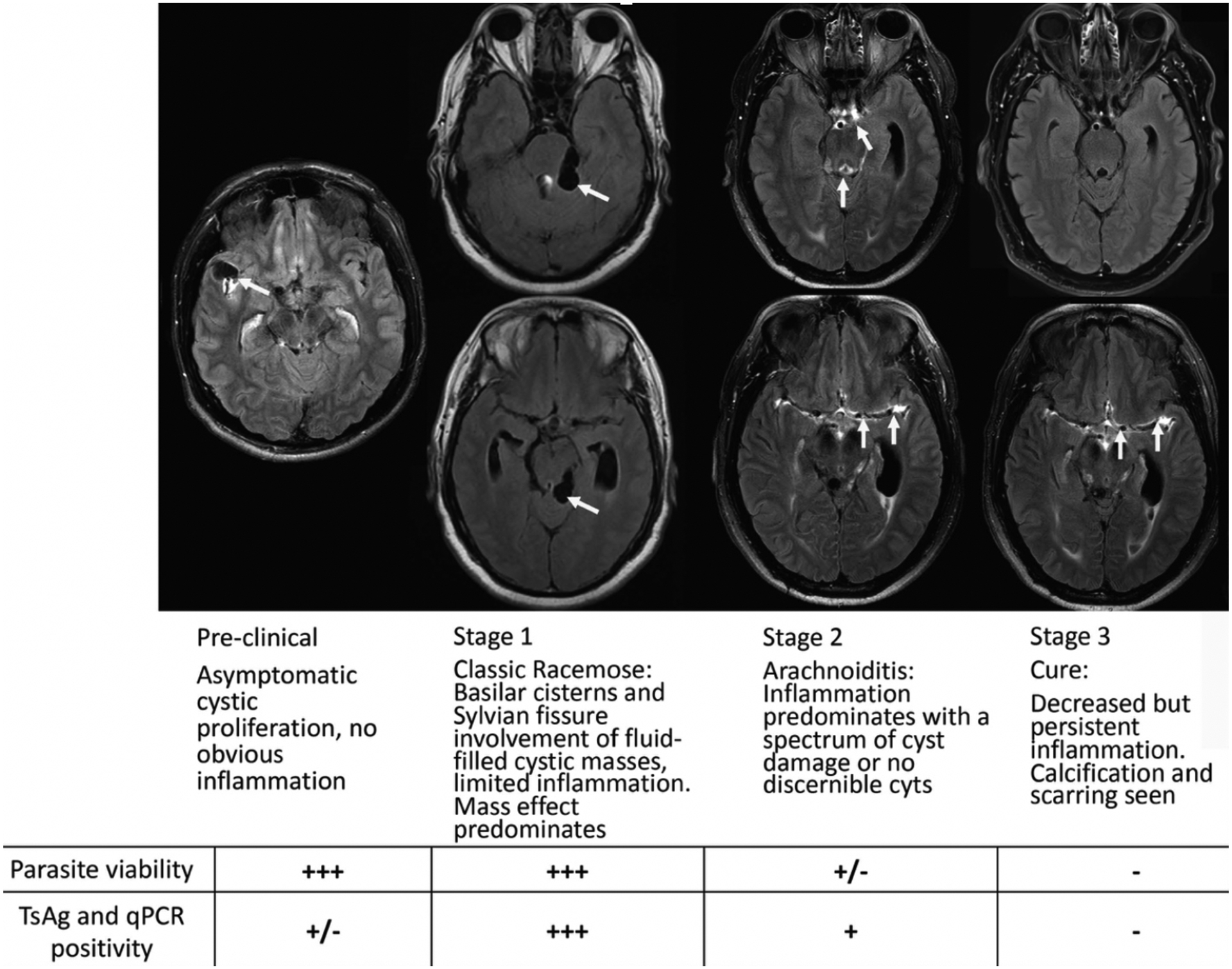
Purpose of review: Subarachnoid neurocysticercosis (SUBNCC) is caused by a morphologically unique proliferative form of Taenia solium involving the subarachnoid spaces. Prolonged therapy based upon the pathophysiology of SUBNCC and long-term follow-up have shed light on the course of disease and led to highly improved outcomes.

Recent findings: SUBNCC has a prolonged incubation period of between 10 and 25 years characterized by cyst proliferation and growth and invasion of contiguous spaces leading to mass effect (Stage 1). With induction of the host-immune responses, cysts degenerate leading to a predominately inflammatory arachnoiditis (Stage 2) causing hydrocephalus, infarcts, and other inflammatory based neurological manifestations. Inactive disease (Stage 3) may occur naturally but mostly is a result of successful treatment, which generally requires prolonged intensive anthelminthic and antiinflammatory treatments. Cerebral spinal fluid cestode antigen or cestode DNA falling to nondetectable levels predicts effective treatment. Prolonged treatment with extended follow-up has resulted in moderate disability and no mortality. Repeated short intensive 8-14-day courses of treatment are also used, but long-term outcomes and safety using this strategy are not reported.

Summary: SUBNCC gives rise to a chronic arachnoiditis. Its unique ability to proliferate and induce inflammatory responses requires long-term anthelmintic and antiinflammatory medications.


Conflict of interest statement

Conflicts of Interest: none

Figures

Figure 1
Figure 1. Radiology and histopathology of SUBNCC
MRI imaging before treatment of a patient who presented primarily with arachnoiditis and lacunar infarcts who underwent brain biopsy for diagnosis (Stage 2) (11). (A) Post contrast T1-weighted images showing abnormal enhancement along the courses of the middle and anterior cerebral arteries bilaterally with associated cystic changes (white arrows). (B) Axial T2, apparent diffusion coefficient maps and axial post contrast T1-weighted images showing subacute infarcts in the left basal ganglia and white matter (black arrows). (C-G) Various histopathology brain biopsy samples from the patient demonstrating variable morphologies of the racemose form. (C, D) Viable parasite with tegument (wide white arrows) showing disorganized growth and a variety and different types and arrangements of subtegumental tissue elements including amorphous substance, parasite cells and calcareous corpuscles better shown in (E) (narrow white arrow). (E-G) Degenerated but still recognizable cestode tissue with associated adjacent necrotic host response (hematoxylin staining amorphous material). (H) Histopathology from another patient with racemose cysts showing a different morphology that resembles the cyst wall, which lacks calcareous corpuscles that are found the neck of the cyst. The presence of viable and non-viable residual parasite in an untreated patient is expected and one cause of long-lasting subarachnoid inflammation. Source: Panels A and B from reference .
Figure 2
Figure 2. Stages of subarachnoid neurocysticercosis
Post-contrast Fluid Attenuation Inversion Recovery (FLAIR) sequence Magnetic resonance imaging (MRI) images without gadolinium (Stage 1) and with gadolinium (Stages 2 and 3) are shown with pertinent findings highlighted by white arrows. The preclinical stage demonstrates an asymptomatic patient brought to medical attention after brain MRI following trauma showing cysts in the Sylvian fissure without surrounding enhancement or edema. Stages 1–3 show the same patient over the course of a 9-year period. Stage 1 shows bulky cysts in the quadrigeminal cistern causing mass effect but without enhancement or edema (not shown but evident in post gadolinium sequences). Stage 2 shows enhancement within the aqueduct (also had significant ventricular disease) and along the middle cerebral artery. Even at presentation there was significant enhancement along the middle cerebral arteries in gadolinium injected sequences. Commonly, different regions of brain have evolved at a slower or faster rate and are in a different Stage state. Pure Stage 1 with only cyst enlargement is not common, some inflammation is usually present. Stage 3 shows resolution of previous enhancement except along the middle cerebral artery. Parasite viability and T. solium antigen (TsAg) and qPCR positivity from the cerebral spinal fluid (CSF) from each stage are also shown. This patient’s course and imaging are detailed in reference .

References

    1. Garcia HH, Nash TE, Del Brutto OH. Clinical symptoms, diagnosis, and treatment of neurocysticercosis. Lancet Neurology. 2014;13(12):1202–15. - PMC - PubMed
    1. Nash TE, Ware JM, Mahanty S. Intraventricular Neurocysticercosis: Experience and Long-Term Outcome from a Tertiary Referral Center in the United States. Am J Trop Med Hyg. 2018;98(6):1755–62. - PMC - PubMed
    1. Hinojosa-Juarez AC, Sandoval-Balanzario M, McManus DP, Monroy-Ostria A. Genetic similarity between cysticerci of Taenia solium isolated from human brain and from pigs. Infection Genetics and Evolution. 2008;8(5):653–6. - PubMed
    1. Rabiela MT, Rivas-Hernandez A, Rodriquez-Ibarra J, Castilo-Medina S, Cancino FdM. Anatomopathological Aspects of Human Brain Cysticercosis In: Flisser A, Willms k, Laclette JP, Larralde C, Ridaura C, Beltran F, editors. Cysticercosis: Present State of Knowledge and Prespectives. New York, New York: Academic Press; 1982. p. 179–200.
    1. Rabiela MT, Rivas A, Flisser A. Morphological types of Taenia solium cysticerci. Parasitol Today. 1989;5(11):357–9. - PubMed

Publication types

MeSH terms