. 2020 Aug 31;11(1):4360.
doi: 10.1038/s41467-020-18231-z.

The cellular and molecular landscape of hypothalamic patterning and differentiation from embryonic to late postnatal development

Affiliations

The cellular and molecular landscape of hypothalamic patterning and differentiation from embryonic to late postnatal development

Dong Won Kim et al. Nat Commun. .

Erratum in

Abstract

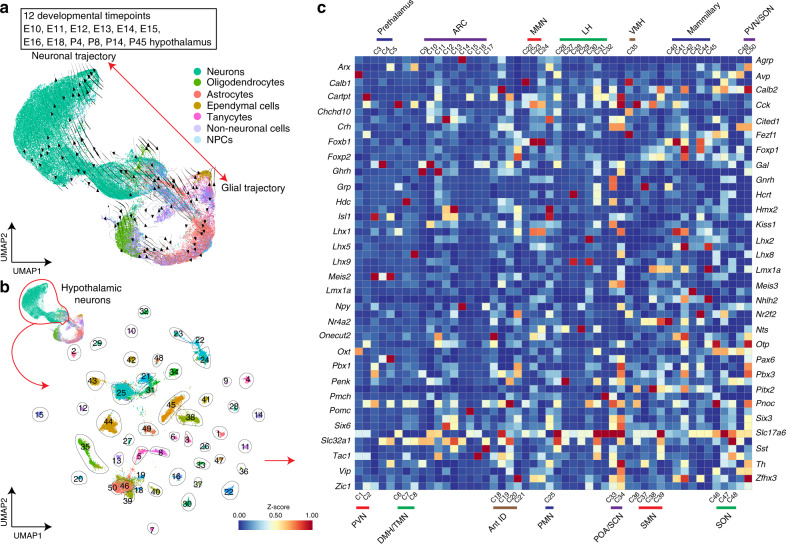
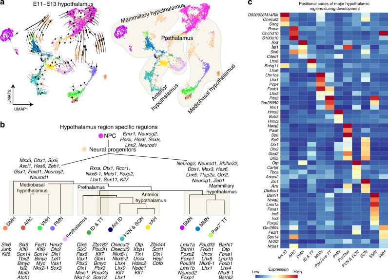
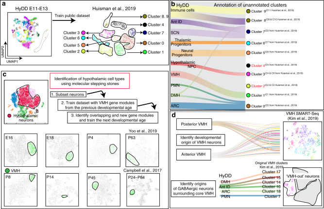
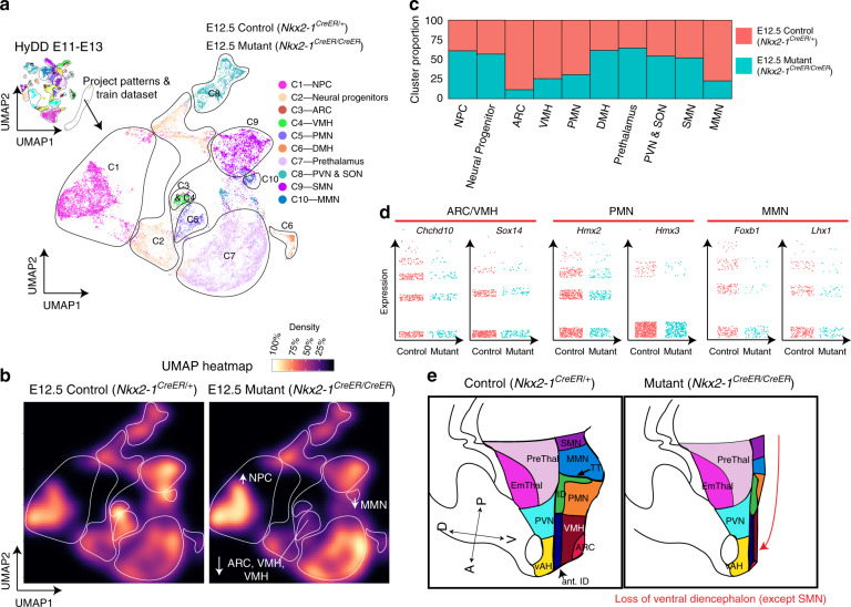
The hypothalamus is a central regulator of many innate behaviors essential for survival, but the molecular mechanisms controlling hypothalamic patterning and cell fate specification are poorly understood. To identify genes that control hypothalamic development, we have used single-cell RNA sequencing (scRNA-Seq) to profile mouse hypothalamic gene expression across 12 developmental time points between embryonic day 10 and postnatal day 45. This identified genes that delineated clear developmental trajectories for all major hypothalamic cell types, and readily distinguished major regional subdivisions of the developing hypothalamus. By using our developmental dataset, we were able to rapidly annotate previously unidentified clusters from existing scRNA-Seq datasets collected during development and to identify the developmental origins of major neuronal populations of the ventromedial hypothalamus. We further show that our approach can rapidly and comprehensively characterize mutants that have altered hypothalamic patterning, identifying Nkx2.1 as a negative regulator of prethalamic identity. These data serve as a resource for further studies of hypothalamic development, physiology, and dysfunction.


Conflict of interest statement

The authors declare no competing interests.

Figures

Fig. 1
Fig. 1. Overview of generation of the hypothalamus scRNA-seq dataset.
a UMAP plot showing scRNA-seq data obtained from the developing diencephalon (including the prethalamus and hypothalamus) between E10 and E16, E18, P4, P8, P14, and P45. RNA velocity marks neuronal or glial (oligodendrocytes, astrocytes, ependymal cells, and tanycytes) trajectories. b UMAP plot showing neuronal clusters across the entire course of hypothalamus development. c Heatmap showing subtypes of neuronal clusters based on neuropeptide and transcription factor expression.
Fig. 2
Fig. 2. Specification of hypothalamic patterning during embryonic development.
a UMAP plot showing E11–E13 developing diencephalon with RNA velocity trajectories (left), and UMAP plot showing the four main molecularly distinct regions of the developing hypothalamus and prethalamus (mediobasal hypothalamus, mammillary hypothalamus, anterior hypothalamus, and prethalamus) (right). b Dendrogram showing a developmental hierarchy of the entire diencephalon and associated regulons. c Heatmap showing a key subset of pattern-specific genes in major hypothalamic regions and prethalamus.
Fig. 3
Fig. 3. Utilizing HyDD to infer the identity and origin of individual cell types.
a The HyDD dataset was used to train a previously published scRNA-seq on E15.5 hypothalamus obtained through selective dissection of Pomc-EGFP-expressing cells. b Alluvial plot showing HyDD clusters (left) matched to clusters from Huisman et al. (right). Note that 2 clusters (clusters 2 and 4) from Huisman et al. do not match the HyDD dataset. c Using the molecular stepping stone approach to identify VMH neurons (green) across the entire developmental ages by identification of shared sets of gene modules that can demarcate the VMH across the entire hypothalamus scRNA-seq dataset. d HyDD dataset is used to identify the developmental origins of previously annotated subtypes of glutamatergic neurons of the core VMH (top), and to identify the developmental origins of GABAergic neurons surrounding the core VMH (bottom).
Fig. 4
Fig. 4. scRNA-seq-based analysis of Nkx2-1-deficient developing hypothalamus.
a UMAP plot showing clusters from combined scRNA-seq dataset of control (Nkx2-1CreER/+) and Nkx2-1 mutant line (Nkx2-1CreER/CreER), in which clusters were obtained by training the dataset using HyDD markers. b UMAP heatmap plot showing distribution of individual clusters between control (left) and Nkx2-1 mutants (right). c Bar graph showing the distribution of individual clusters between control and Nkx2-1 mutants. d Jitter plots of regional marker genes (Chchd10 and Sox14—ARC/VMH, Hmx2 and Hmx3—PMN, Foxb1 and Lhx1—MMN between control and Nkx2-1 mutants. e Schematic showing overall hypothalamic phenotype between control and Nkx2-1 mutants. Note the absence of ventral diencephalic structures (except the supramammillary nucleus), and the relative expansion of the prethalamus in Nkx2-1 mutants (right).

References

    1. Bedont JL, Newman EA, Blackshaw S. Patterning, specification, and differentiation in the developing hypothalamus. Wiley Interdiscip. Rev. Dev. Biol. 2015;4:445–468. - PMC - PubMed
    1. Shimogori T, et al. A genomic atlas of mouse hypothalamic development. Nat. Neurosci. 2010;13:767–775. - PMC - PubMed
    1. Xie Y, Dorsky RI. Development of the hypothalamus: conservation, modification and innovation. Development. 2017;144:1588–1599. - PMC - PubMed
    1. Kent MA, Peters RH. Effects of ventromedial hypothalamic lesions on hunger-motivated behavior in rats. J. Comp. Physiol. Psychol. 1973;83:92–97. - PubMed
    1. Kruk MR, et al. Discriminant analysis of the localization of aggression-inducing electrode placements in the hypothalamus of male rats. Brain Res. 1983;260:61–79. - PubMed

Publication types

Substances