[Preprint]. 2020 Aug 30:2020.08.30.273920.
doi: 10.1101/2020.08.30.273920.

Structural classification of neutralizing antibodies against the SARS-CoV-2 spike receptor-binding domain suggests vaccine and therapeutic strategies

Affiliations

Structural classification of neutralizing antibodies against the SARS-CoV-2 spike receptor-binding domain suggests vaccine and therapeutic strategies

Christopher O Barnes et al. bioRxiv. .

Update in

  • SARS-CoV-2 neutralizing antibody structures inform therapeutic strategies.
    Barnes CO, Jette CA, Abernathy ME, Dam KA, Esswein SR, Gristick HB, Malyutin AG, Sharaf NG, Huey-Tubman KE, Lee YE, Robbiani DF, Nussenzweig MC, West AP Jr, Bjorkman PJ. Barnes CO, et al. Nature. 2020 Dec;588(7839):682-687. doi: 10.1038/s41586-020-2852-1. Epub 2020 Oct 12. Nature. 2020. PMID: 33045718 Free PMC article.

Abstract

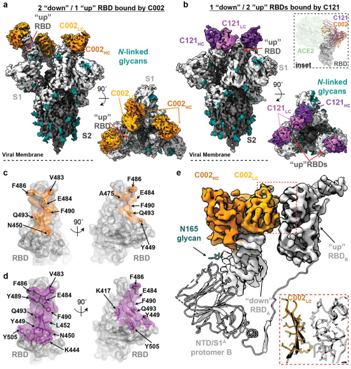
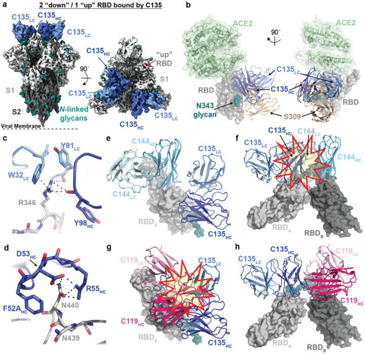
The COVID-19 pandemic presents an urgent health crisis. Human neutralizing antibodies (hNAbs) that target the host ACE2 receptor-binding domain (RBD) of the SARS-CoV-2 spike1-5 show therapeutic promise and are being evaluated clincally6-8. To determine structural correlates of SARS-CoV-2 neutralization, we solved 8 new structures of distinct COVID-19 hNAbs5 in complex with SARS-CoV-2 spike trimer or RBD. Structural comparisons allowed classification into categories: (1) VH3-53 hNAbs with short CDRH3s that block ACE2 and bind only to "up" RBDs, (2) ACE2-blocking hNAbs that bind both "up" and "down" RBDs and can contact adjacent RBDs, (3) hNAbs that bind outside the ACE2 site and recognize "up" and "down" RBDs, and (4) Previously-described antibodies that do not block ACE2 and bind only "up" RBDs9. Class 2 comprised four hNAbs whose epitopes bridged RBDs, including a VH3-53 hNAb that used a long CDRH3 with a hydrophobic tip to bridge between adjacent "down" RBDs, thereby locking spike into a closed conformation. Epitope/paratope mapping revealed few interactions with host-derived N-glycans and minor contributions of antibody somatic hypermutations to epitope contacts. Affinity measurements and mapping of naturally-occurring and in vitro-selected spike mutants in 3D provided insight into the potential for SARS-CoV-2 escape from antibodies elicited during infection or delivered therapeutically. These classifications and structural analyses provide rules for assigning current and future human RBD-targeting antibodies into classes, evaluating avidity effects, suggesting combinations for clinical use, and providing insight into immune responses against SARS-CoV-2.


Figures

Extended Data Figure 1:
Extended Data Figure 1:. X-ray structure and epitope mapping of VH3–53 hNAb C102.
a, X-ray structure of C102 Fab – RBD331–518 complex. b, C102 CDR loops mapped on the RBD surface. b, Surface representation of C102 epitope colored by C102 HC (dark green) and LC (light green) interactions. c, CDRH1, CDRH2 and d, CDRH3 interactions with RBD residues. Potential H-bond contacts are illustrated as dashed lines. f, Left: Overlay of C102-RBD crystal structure (cartoon) with C105-S trimer cryoEM density (PDB 6XCM, EMD-22127) illustrating conserved binding to RBD epitope in an “up” conformation. Right: The C102 epitope is sterically occluded when aligned to a “down” RBD conformation (red and yellow star). SARS-CoV-2 S domains are dark gray (S2 domain) and light gray (S1 domain); the C105 Fab is yellow-green. g, Alignment of selected CDRH3 sequences for VH3–53/VH3–66 SARS-CoV-2 neutralizing antibodies (IMGT definition). h, Overlay of hNAb COVA2–39 Fab (lime green and lemon, from COVA2–39-RBD structure, PDB 7JMP) and C144 Fab (blue, from C144-S structure) aligned on a RBDA of C144 epitope. COVA2–39 adopts a distinct conformation relative to the C102-like VH3–53/short CDRH3 NAb class and to C144, recognizing its RBD epitope only in an “up” RBD conformations due to steric clashes (red and yellow star) with the N343RBD-associated glycan on the adjacent RBD. i, Polyreactivity assay. IgGs were evaluated for binding to baculovirus extracts to assess non-specific binding. Polyreactive positive control IgGs were NIH45–46, NIH45–46G54W, and 45–46m2. Negative controls were bovine serum albuminn (BSA) and IgGs N6 and 3BNC117. Relative Light Unit (RLU) values are presented as the mean and standard deviation of triplicate measurements.
Extended Data Figure 2.
Extended Data Figure 2.. Overview of VH3–53/VH3–66 hNAb structures.
a, Superimposition of VH and VL domains of C102 with other VH3–53/VH3–66 NAbs (top) and RMSD calculations (bottom). b, BSA comparisons for the indicated Fab/RBD structures. BSAs were calculated using PDBePISA and a 1.4 Å probe.
Extended Data Figure 3.
Extended Data Figure 3.. Cryo-EM data processing and validation for C144-S, C002-S, and C121-S complexes.
Representative micrograph, 2D class averages, gold-standard FSC plots, and local resolution estimations for a-c, C144-S 6P, d-f, C002-S 2P, and g-I, C121-S 2P. For the C002-S dataset, two classes were resolved: State 1, C002 Fabs bound to 3 “down” RBDs, and State 2, C002 Fabs bound to 2 “down”/1 “up” RBD. For the C121-S 2P dataset, two classes were resolved: State 1, C121 Fabs bound to 2 “down”/1 “up” RBD and State 2, C121 Fabs bound to 1 “down”/2 “up” RBDs.
Extended Data Figure 4.
Extended Data Figure 4.. Cryo-EM processing, validation, and reconstruction for C119-S and C104-S complexes.
a, 3.6 Å cryo-EM reconstruction for a C119-S trimer complex. b, 3.7 Å cryo-EM reconstruction for a C104-S trimer complex. Representative micrograph, 2D class averages, gold-standard FSC plot, and local resolution estimation for c-e, C119-S2P and, d-f, C104-S. Both complexes revealed binding of Fabs to both “down” and “up” RBD conformations.
Extended Data Figure 5.
Extended Data Figure 5.. Primary and secondary epitopes of class 2 hNAbs.
a-c, Primary epitopes for C002 (panel a), C121 (panel b), and C119 (panel c) on “down” RBD. A secondary epitope is observed if a Fab is bound to an adjacent “up” RBD for these NAbs. Antibody paratopes are represented as cartoons. A similar interaction in the C104-S structure is not shown due to low local resolution on the “up” RBD. d-g, Primary epitopes for C119 (panel d), C104 (panel e), P2B-2F6 (panel f; PDB 7BWJ), and BD23 (panel g, PDB 7BYR). The existence of secondary epitopes for P2B-2F6 and BD23 cannot be determined because the P2B-2F6 epitope was determined from a crystal structure with an RBD, and the BD23-S cryo-EM structure showed only one bound Fab. h, Measurement of Cα distance between the C-termini of adjacent C121 CH1 domains (residue 222HC on each Fab). Measurements of this type were used to evaluate whether intra-spike crosslinking by an IgG binding to a single spike trimer was possible for hNAbs in Extended Data Table 1.
Extended Data Figure 6.
Extended Data Figure 6.. Mapping somatic hypermutations (SHMs) of SARS-CoV-2 NAbs.
a-f, Somatic hypermutations in HC and LC V gene segments for C002 (panel a), C121 (panel b), C119 (panel c), C144 (panel d), C102 (panel e) and C135 (panel f) are shown as spheres on the antibody VH and VL domains (ribbon representations). The primary RBD epitope is shown asa light gray surface; secondary RBD epitope for C144 is in dark gray.
Extended Data Figure 7.
Extended Data Figure 7.. Cryo-EM structure of C110-S complex and epitope mapping.
a, 3.8 Å cryo-EM reconstruction of C110-S trimer complex. b, Composite model of C110-RBD (purple and gray, respectively) overlaid with the SARS-CoV-2 NAb REGN-10987 (yellow, PDB 6XDG) and soluble ACE2 (green, PDB 6M0J). Model was generated by aligning structures on 188 RBD Cα atoms. c-f, Surface representation of RBD epitopes for c, C135 (blue), d, S309 (brown, PDB 6WSP), e, C110 (purple) and f, REGN-10987 (yellow, PDB 6XDG). Given the low resolution of the antibody-RBD interface, epitopes were assigned by selection of any RBD residue within 7 Å of any antibody Cα atom. Mutation sites found in sequence isolates (green) and in laboratory selection assays (red) are shown. Representative micrograph, 2D class averages, gold-standard FSC plot, and local resolution estimation for g-i, C135-S 2P and, j-l, C110-S 2P. Both complexes revealed binding of Fabs to both 2 “down”/1 “up” RBD conformations.
Extended Data Figure 8.
Extended Data Figure 8.. Possibilities for simultaneous engagement of C144 and C135 on spikes with different combinations of “up” and “down” RBDs.
Modeling of C144 (light blue) and C135 (dark blue) VH-VL domains on different RBD conformations. Steric clashes are shown as a red and yellow star.
Extended Data Figure 9.
Extended Data Figure 9.. SPR binding data for hNAbs.
Kinetic and equilibrium constants for binding to unaltered RBD (indicated as wt) and mutant RBDs are shown in tables beside structures of a representative NAb-RBD complex for each class. Residues that were mutated are highlighted as colored sidechains on a gray RBD surface. Antibody VH-VL domains are shown as cartoons. Kinetic and equilibrium constants for NAbs that contact adjacent RBDs on S trimer (C144, C002, C119, and C121) do not account for contacts to a secondary RBD since binding was assayed by injected monomeric RBDs over immobilized IgGs. * indicates kinetic constants determined from a two-state binding model.
Extended Data Figure 10:
Extended Data Figure 10:. Summary of hNAbs.
a, Structural depiction of a representative NAb from each class binding its RBD epitope. b, Composite model illustrating non-overlapping epitopes of NAbs from each class bound to a RBD monomer. c, Epitopes for SARS-CoV-2 NAbs. RBD residues involved in ACE2 binding are boxed in green. Diamonds represent RBD residues contacted by the indicated antibody.
Figure 1.
Figure 1.. Cryo-EM structure of the C144-S complex illustrates a distinct VH3–53 hNAb binding mode.
a, 3.2 Å cryo-EM density for C144-S trimer complex revealing C144 binding to a closed (3 RBDs “down”) spike conformation. b, Overlay of C102 Fab (from C102-RBD crystal structure; Extended Data Fig. 1) and C144 Fab (from C144-S structure) aligned on a RBD monomer. ACE2 (PDB 6M0J; light green surface) is aligned on the same RBD for reference. C144 adopts a distinct conformation relative to the C102-like VH3–53/short CDRH3 NAb class, allowing binding to the “down” RBD conformation on trimeric spike, whereas C102-like NAbs can only bind “up” RBDs. c, Quaternary epitope of C144 involving bridging between adjacent RBDs via the CDRH3 loop. d,e, Close-up view of CDRH3-mediated contacts on adjacent protomer RBD (dark gray). C144 CDRH3 residues F100D and W100E are buried in a hydrophobic pocket comprising the RBD α1 helix, residue F374RBD and the N343RBD-glycan. f, Surface representation of C144 epitope (light blue) across two adjacent RBDs. RBD epitope residues (defined as residues containing atom(s) within 4 Å of a Fab atom) are labeled in black.
Figure 2.
Figure 2.. Cryo-EM structures of class 2 C002 and C121 hNAbs show binding to “up” and “down” RBDs.
a,b, Cryo-EM densities for C002-S (panel a; 3.4 Å) and C121-S complexes (panel b; 3.7 Å) revealing binding of C002 or C121 to both “down” and “up” RBDs. Inset: Alignment of C002 and C121 Fabs on the same RBD. ACE2 is represented as a green surface for reference. c,d, Surface representations of C002 epitope (orange, panel c) and C121 epitope (purple, panel d) on the RBD surface (gray). RBD epitope residues (defined as residues containing atom(s) within 4 Å of a Fab atom) are labeled in black. e, C002 forms inter-protomer contacts via binding to an adjacent “up” RBD conformation on the surface of the trimer spike (also observed for class 2 C121-, C119-, and C104-S structures, see Extended Data Fig. 5). Red box: Close-up of adjacent “up” RBD and C002 LC interface.
Figure 3.
Figure 3.. Details of common RBD interactions among class 2 hNAbs.
Conserved interactions between the RBD and CDRs of class 2 NAbs as observed for a-d, C144 (HC: cyan, LC: sky blue), e-h, C002 (HC: dark orange, LC: light orange), and i-l, C121 (HC: purple, LC: pink). Primary and secondary epitopes on adjacent “down” RBDs are shown for C144. Secondary epitopes for C002 and C121, which require adjacent “up” RBDs, are shown in Extended Data Fig. 5. RBDs are gray; potential H-bonds and pi-pi stacking interactions (panel d, Y33LC and F486RBD; panel h, Y92LC and F486RBD; panel l, Y91LC and F486RBD) are indicated by dashed lines.
Figure 4.
Figure 4.. Cryo-EM structure of S complexed with the class 3 (non-ACE2 blocking) hNAb C135.
a, 3.5 Å cryo-EM density of C135-S complex. b, Composite model of C135-RBD (blue and gray, respectively) overlaid with the SARS-CoV-2 NAb S309 (sand, PDB 6WPS) and soluble ACE2 (green, PDB 6M0J). The model was generated by aligning on 188 RBDCα atoms. c-d, C135 CDRH (dark blue) and CDRL (light blue) interactions with residues R346RBD (panel c) and N440RBD (panel d). Potential pi-pi stacking interactions in c and H-bonds in c and d are illustrated by dashed black lines. e-f, Model of RBD interactions of NAbs C135 (class 3) and C144 (class 2) demonstrating that both Fabs can bind simultaneously to a single monomeric RBD (panel e), but would clash if bound to adjacent “down” RDBs on S trimer (panel f). Steric clashes indicated by a red and yellow star in f. g-h, Model of RBD interaction of NAbs C135 (class 3) and C119 (class 2) demonstrating that both Fabs cannot bind simultaneously to a single monomeric RBD (panel g), but do not clash if bound to adjacent “down” RDBs on S trimer (panel h). Steric clashes indicated by a red and yellow star in g.

References

    1. Baum A. et al. REGN-COV2 antibody cocktail prevents and treats SARS-CoV-2 infection in rhesus macaques and hamsters. bioRxiv 10.1101/2020.08.02.233320(2020). - DOI - PMC - PubMed
    1. Baum A. et al. Antibody cocktail to SARS-CoV-2 spike protein prevents rapid mutational escape seen with individual antibodies. Science 369, 1014–1018 (2020). - PMC - PubMed
    1. Rogers T.F. et al. Rapid isolation of potent SARS-CoV-2 neutralizing antibodies and protection in a small animal model. Science 10.1126/science.abc7520(2020). - DOI - PMC - PubMed
    1. Zost S.J. et al. Potently neutralizing and protective human antibodies against SARS-CoV-2. Nature 584, 443–449 (2020). - PMC - PubMed
    1. Robbiani D.F. et al. Convergent antibody responses to SARS-CoV-2 in convalescent individuals. Nature 584, 437–442 (2020). - PMC - PubMed

Extended Data References

    1. Barnes C.O. et al. Structures of Human Antibodies Bound to SARS-CoV-2 Spike Reveal Common Epitopes and Recurrent Features of Antibodies. Cell 182, 828–842 e16 (2020). - PMC - PubMed
    1. Wu Y. et al. A noncompeting pair of human neutralizing antibodies block COVID-19 virus binding to its receptor ACE2. Science 10.1126/science.abc2241(2020). - DOI - PMC - PubMed
    1. Yuan M. et al. Structural basis of a shared antibody response to SARS-CoV-2. Science 10.1126/science.abd2321(2020). - DOI - PMC - PubMed
    1. Wu N.C. et al. An alternative binding mode of IGHV3–53 antibodies to the SARS-CoV-2 receptor binding domain. bioRxiv 10.1101/2020.07.26.222232(2020). - DOI - PMC - PubMed
    1. Wang B. et al. Bivalent binding of a fully human IgG to the SARS-CoV-2 spike proteins reveals mechanisms of potent neutralization. bioRxiv 10.1101/2020.07.14.203414(2020). - DOI

Publication types