

Unleashing the therapeutic potential of apoptotic bodies
- PMID: 32869835
- PMCID: PMC7609033
- DOI: 10.1042/BST20200225
Unleashing the therapeutic potential of apoptotic bodies
Abstract
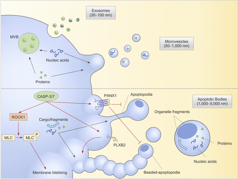
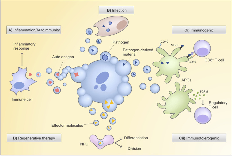
Extracellular vesicles (EVs), membrane-bound vesicles that are naturally released by cells, have emerged as new therapeutic opportunities. EVs, particularly exosomes and microvesicles, can transfer effector molecules and elicit potent responses in recipient cells, making them attractive therapeutic targets and drug delivery platforms. Furthermore, containing predictive biomarkers and often being dysregulated in various disease settings, these EVs are being exploited for diagnostic purposes. In contrast, the therapeutic application of apoptotic bodies (ApoBDs), a distinct type of EVs released by cells undergoing a form of programmed cell death called apoptosis, has been largely unexplored. Recent studies have shed light on ApoBD biogenesis and functions, promisingly implicating their therapeutic potential. In this review, we discuss many strategies to develop ApoBD-based therapies as well as highlight their advantages and challenges, thereby positioning ApoBD for potential EV-based therapy.
Keywords: apoptosis; apoptotic bodies; extracellular vesicles; therapeutics.
© 2020 The Author(s).
Conflict of interest statement
The authors declare that there are no competing interests associated with the manuscript.
Figures
Similar articles
-
Immune Cell-Derived Extracellular Vesicles - Functions and Therapeutic Applications.Trends Mol Med. 2019 May;25(5):382-394. doi: 10.1016/j.molmed.2019.02.003. Epub 2019 Mar 7. Trends Mol Med. 2019. PMID: 30853173 Review.
-
Translating extracellular vesicle packaging into therapeutic applications.Front Immunol. 2022 Aug 15;13:946422. doi: 10.3389/fimmu.2022.946422. eCollection 2022. Front Immunol. 2022. PMID: 36045692 Free PMC article. Review.
-
Circulating Extracellular Vesicles As Biomarkers and Drug Delivery Vehicles in Cardiovascular Diseases.Biomolecules. 2021 Mar 5;11(3):388. doi: 10.3390/biom11030388. Biomolecules. 2021. PMID: 33808038 Free PMC article. Review.
-
Biology and Role of Extracellular Vesicles (EVs) in the Pathogenesis of Thrombosis.Int J Mol Sci. 2019 Jun 11;20(11):2840. doi: 10.3390/ijms20112840. Int J Mol Sci. 2019. PMID: 31212641 Free PMC article. Review.
-
Extracellular vesicles as novel drug delivery systems to target cancer and other diseases: Recent advancements and future perspectives.F1000Res. 2023 Mar 23;12:329. doi: 10.12688/f1000research.132186.1. eCollection 2023. F1000Res. 2023. PMID: 37868300 Free PMC article. Review.
Cited by
-
Apoptotic Extracellular Vesicles from Supernumerary Tooth-Derived Pulp Stem Cells Transfer COL1A1 to Promote Angiogenesis via PI3K/Akt/VEGF Pathway.Int J Nanomedicine. 2024 Jul 8;19:6811-6828. doi: 10.2147/IJN.S466136. eCollection 2024. Int J Nanomedicine. 2024. PMID: 39005959 Free PMC article.
-
Apoptotic vesicles derived from human red blood cells promote bone regeneration via carbonic anhydrase 1.Cell Prolif. 2024 Feb;57(2):e13547. doi: 10.1111/cpr.13547. Epub 2023 Sep 11. Cell Prolif. 2024. PMID: 37697490 Free PMC article.
-
Deciphering Molecular and Signaling Pathways of Extracellular Vesicles-Based Therapeutics for Alzheimer's Disease.Mol Neurobiol. 2025 Jul 18. doi: 10.1007/s12035-025-05216-6. Online ahead of print. Mol Neurobiol. 2025. PMID: 40679697 Review.
-
ApoBDs: a paradigm shift from cellular debris to therapeutic vehicles.Front Endocrinol (Lausanne). 2025 Jul 17;16:1626796. doi: 10.3389/fendo.2025.1626796. eCollection 2025. Front Endocrinol (Lausanne). 2025. PMID: 40747304 Free PMC article. Review.
-
Microglial Extracellular Vesicles as Vehicles for Neurodegeneration Spreading.Biomolecules. 2021 May 21;11(6):770. doi: 10.3390/biom11060770. Biomolecules. 2021. PMID: 34063832 Free PMC article. Review.
References
Publication types
MeSH terms
Substances
LinkOut - more resources
Full Text Sources