. 2020 Sep 1;5(18):e141593.
doi: 10.1172/jci.insight.141593.

LILRB3 (ILT5) is a myeloid cell checkpoint that elicits profound immunomodulation

Affiliations

LILRB3 (ILT5) is a myeloid cell checkpoint that elicits profound immunomodulation

Muchaala Yeboah et al. JCI Insight. .

Abstract

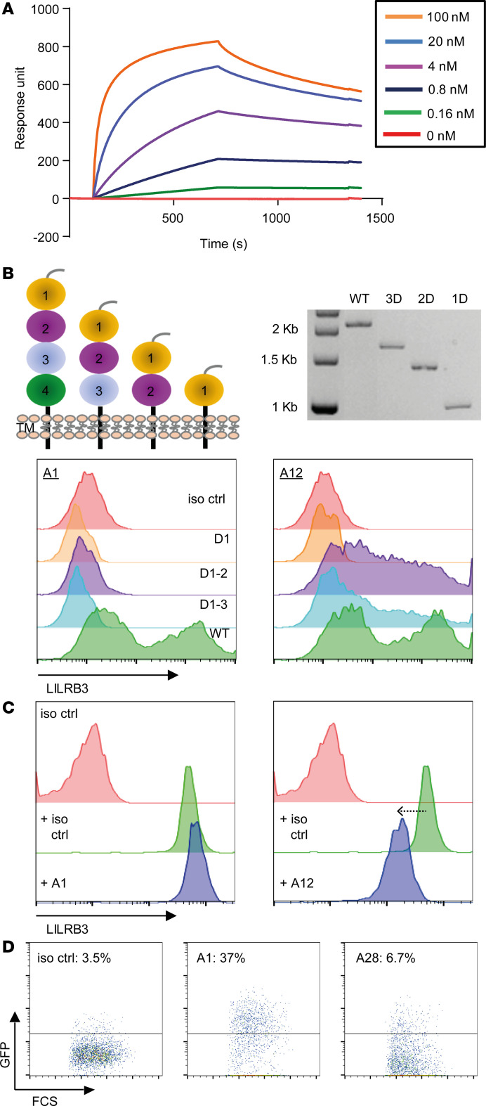
Despite advances in identifying the key immunoregulatory roles of many of the human leukocyte immunoglobulin-like receptor (LILR) family members, the function of the inhibitory molecule LILRB3 (ILT5, CD85a, LIR3) remains unclear. Studies indicate a predominant myeloid expression; however, high homology within the LILR family and a relative paucity of reagents have hindered progress toward identifying the function of this receptor. To investigate its function and potential immunomodulatory capacity, a panel of LILRB3-specific monoclonal antibodies (mAbs) was generated. LILRB3-specific mAbs bound to discrete epitopes in Ig-like domain 2 or 4. LILRB3 ligation on primary human monocytes by an agonistic mAb resulted in phenotypic and functional changes, leading to potent inhibition of immune responses in vitro, including significant reduction in T cell proliferation. Importantly, agonizing LILRB3 in humanized mice induced tolerance and permitted efficient engraftment of allogeneic cells. Our findings reveal powerful immunosuppressive functions of LILRB3 and identify it as an important myeloid checkpoint receptor.

Keywords: Immunology; Immunotherapy; Macrophages; Monocytes.


Conflict of interest statement

Conflict of interest: MSC is a retained consultant for BioInvent International and receives research funding from BioInvent International. AR receives funding from BioInvent International. TS, UM, UT, BH, AL, MM, and BF are employees of BioInvent International.

Figures

Figure 1
Figure 1. Generation of fully human mAbs against LILRB3.
(A) Schematic of antibody generation by phage-display via 3 independent “panning” techniques; (i) immobilized target (LILRB3), (ii) biotinylated target and excess nontarget (LILRB1), and (iii) LILRB3-transfected cell lines (from left to right). Biopanning was performed against generated target protein using an scFv library; “nontarget” cross-reactive scFv clones were removed by competition, and target-specific scFv clones were then eluted and converted to a soluble format, sequenced, and screened by various cell- and protein-based assays. (B and C) Screening of generated LILRB3 clones. (B) FMAT and (C) ELISA were performed and scFv clones screened against LILRB3 target– and LILRB1/LILRB2 nontarget–transfected CHO-S cells and extracellular LILRB1 protein, respectively. The relative binding to each target was calculated, with target-specific scFv clones depicted in yellow and the irrelevant isotype control shown in green. Nonbinding and cross-reactive scFv clones depicted in blue. (D) Screening of LILRB3 scFv clones by high-throughput flow cytometry. PBMCs (left plot) or LILR-transfected CHO-S (middle plot) cells were incubated with His-tagged scFv supernatants, followed by secondary anti-His staining. Where transfected CHO-S cells were used, LILRB1- and LILRB2-transfected cells were used as nontargets for LILRB3. Clones were compared against both gated CD14+ monocytes and target-transfected CHO-S cells (right plot). LILRB3-specific clones highlighted in yellow, nonspecific or nonbinding clones in red, and isotype control in green. (E) Specificity of LILRB3 clones against human LILR-transfected 2B4 cells. LILRB3 mAbs were tested against cells stably transfected with the indicated LILR family members by flow cytometry; a representative specific clone (A16; top panel) and a nonspecific cross-reactive clone (A30; bottom panel) are shown. (FG) Testing the specificity of directly fluorochrome-labeled LILRB3 clones against primary cells by flow cytometry. (F) Fresh whole peripheral blood stained with either APC-labeled LILRB3 (represented by clone A16) or an irrelevant human (h) IgG1 isotype control as well as various leukocyte surface markers, as indicated. Dot plots and histograms are representative of multiple donors indicating gating of each leukocyte subset as indicated: T cells, B cells, NK cells, monocytes, and granulocytes. (G) Graph showing relative expression of LILRB3 on each leukocyte subset. One-way ANOVA test performed (*P < 0.05; **P < 0.005); n = 5 independent donors (each color represents an individual donor). (EF) Histogram pink and blue traces indicate staining with irrelevant isotype control or LILRB3 mAb, respectively.
Figure 2
Figure 2. Characterization of LILRB3 antibodies.
(A) LILRB3 mAb affinity assessed by SPR. LILRB3-hFc recombinant protein was immobilized, and various LILRB3 mAbs flowed across the chip. Representative LILRB3 clone A16 shown. (B) LILRB3 domain epitope mapping. HEK293F cells transfected with WT LILRB3 (full-length extracellular portion), D1–3, D1–2, or D1 were stained with LILRB3 clones, followed by an anti-hIgG secondary antibody. Schematic of domain constructs and restriction digest of each DNA construct shown (top panel). Histograms showing staining of 2 representative clones differentially binding to color-coded cells expressing WT (D4), D1–3, D1–2, and D1 (bottom panel; n = 3 independent experiments). (C) Ability of generated mAbs to cross-block binding of a commercial LILRB3 mAb (clone 222821). PBMCs were stained with unconjugated LILRB3 antibody clones and subsequently stained with a directly conjugated 222821 mAb and analyzed by flow cytometry; representative clones displayed (A1 nonblocking; A12 partial blocking), as indicated. (D) LILRB3 2B4 reporter cells were incubated with 10 μg/mL LILRB3 antibodies overnight to assess receptor signaling potential as judged by GFP induction measured by flow cytometry; representative clones with percentage of GFP expression shown (n = 2 independent experiments).
Figure 3
Figure 3. LILRB3 ligation regulates T cell activation and proliferation.
CFSE-labeled PBMCs were stimulated with antibodies against human CD3 (0.02 μg/mL) and CD28 (5 μg/mL) in the presence or absence of isotype control (iso ctrl) or LILRB3 mAb (10 μg/mL) and proliferation measured through CFSE dilution after 3–5 days. (A) LILRB3 mAbs were deglycosylated (Degly) through PNGase treatment, as confirmed by SDS-PAGE; representative clone A1 shown. (B) Assessing T cell activation and proliferation following treatment. Light microscopy images following PBMC stimulation in culture. CD8+ T cell proliferation was assessed through CFSE dilution; plots and images from a donor with profound A1-induced inhibition shown, histograms (% proliferation indicated) and microscopy images shown (original magnification, ×10). (C) Assessing the effects of deglycosylated LILRB3 mAbs on T cell proliferation. CFSE dilution of CD8+ T cells, treated with the representative LILRB3 mAb, was assessed by flow cytometry. Data normalized to anti-CD3/CD28–treated samples and mean represented by solid bars. One-way ANOVA performed (**P < 0.005; ***P < 0.0005); n = 13–20 independent donors (each color represents an individual donor).
Figure 4
Figure 4. LILRB3 ligation induces tolerance in vivo.
(A) Schematic of the generation of humanized mice and subsequent treatment regimens and monitoring. (B) Expression of LILRB3 on human myeloid cells in humanized mice. Representative flow cytometry plots (gated on live single cells) showing gating strategy and the restricted expression of LILRB3 on hCD45+ peripheral blood hCD14+ myeloid cells; isotype control in pink and LILRB3 mAb staining depicted in blue. (C) The effect of agonistic LILRB3 mAb on engraftment of allogeneic cells in humanized mice. Age- and sex-matched humanized mice were injected with 200 μg LILRB3 mAb (clone A1) or an isotype-matched (hIgG1) control mAb (iso ctrl) on day 0 and 4, i.v. and i.p., respectively. On day 7, mice were injected i.p. with 1 × 10⁷ nonautologous luciferase+ human lymphoma cells. Lymphoma cell growth was monitored over time using an IVIS imager, and (D) humanized mice were sacrificed upon the development of signs of terminal tumor development. Survival data were analyzed using log-rank test (*P < 0.01); representative data from 3 independent experiments (3 individual HSPC donors) shown (n = 3 mice/group).
Figure 5
Figure 5. Human LILRB3 ligation reprograms human primary myeloid cells.
Freshly isolated human peripheral CD14+ monocytes were treated with an isotype control (iso ctrl) or a human LILRB3 mAb (clone A1) and then assessed. (A) Agonistic LILRB3 mAb (clone A1) affects monocyte morphology. Light microscopy images following overnight treatment of freshly isolated CD14+ monocytes with indicated mAbs in culture (original magnification, ×10; left panel). Images of treated monocytes were analyzed and length of monocytes quantified (right panel). A total of 200–500 individual cells were analyzed per image. Combined data from 3 independent donors shown; lines indicate median; 2-tailed paired t test performed (***P < 0.0001). (B) Transcriptomic analysis of LILRB3-treated monocytes reveals upregulation of M2-associated genes compared with controls. RNA was extracted from cells following mAb treatment (~18 hours) and subjected to RNA-Seq. Red depicts genes that were significantly upregulated, and green depicts genes that were significantly downregulated compared with isotype control–treated cells (n = 5–6 independent donors). (C) Ligation of LILRB3 on primary human CD14+ monocytes downregulated M1-associated genes. GSEA graph showing a significant enrichment for M1-polarizing genes in LILRB3-treated monocytes versus isotype control, respectively. UP; upregulated, normalized enrichment score (NES) = –1.68; family-wise error rate (FWER); P < 0.001. (D) qPCR analysis of selected genes following LILRB3 ligation on monocytes using an agonistic LILRB3 mAb (A1), a nonagonistic LILRB3 mAb (A28), or an isotype control (iso ctrl). Data were normalized to GAPDH mRNA levels and standardized to the levels of isotype control–treated monocytes. Fold difference data were log10 transformed. One-way ANOVA with Bonferroni’s multiple-comparisons test was performed (*P < 0.005). (E) GSEA showing negative correlation with IFN-γ (NES = –2.17; FWER P < 0.001), IFN-α (NES = –2.3; FWER P < 0.001), and allograft rejection (NES = –1.58; FWER P = 0.14) signaling elements and positive correlation with oxidative phosphorylation (NES = 2; FWER P < 0.001).

Similar articles

Cited by

References

    1. Borges L, Hsu ML, Fanger N, Kubin M, Cosman D. A family of human lymphoid and myeloid Ig-like receptors, some of which bind to MHC class I molecules. J Immunol. 1997;159(11):5192–5196. - PubMed
    1. Colonna M, et al. A common inhibitory receptor for major histocompatibility complex class I molecules on human lymphoid and myelomonocytic cells. J Exp Med. 1997;186(11):1809–1818. doi: 10.1084/jem.186.11.1809. - DOI - PMC - PubMed
    1. van der Touw W, Chen HM, Pan PY, Chen SH. LILRB receptor-mediated regulation of myeloid cell maturation and function. Cancer Immunol Immunother. 2017;66(8):1079–1087. doi: 10.1007/s00262-017-2023-x. - DOI - PMC - PubMed
    1. Hudson LE, Allen RL. Leukocyte Ig-like receptors - a model for MHC class I disease associations. Front Immunol. 2016;7:281. - PMC - PubMed
    1. Burshtyn DN, Morcos C. The expanding spectrum of ligands for leukocyte Ig-like receptors. J Immunol. 2016;196(3):947–955. doi: 10.4049/jimmunol.1501937. - DOI - PubMed

Publication types

MeSH terms