

The Effects of Ascorbic Acid and U-74389G on Renal Ischemia-Reperfusion Injury in a Rat Model
- PMID: 32871775
- PMCID: PMC7652495
- DOI: 10.21873/invivo.12063
The Effects of Ascorbic Acid and U-74389G on Renal Ischemia-Reperfusion Injury in a Rat Model
Abstract
Background/aim: U-74389G and ascorbic acid protect the cells from oxidation. This study aimed to depict their role in ischemia-reperfusion injury in a renal rat model.
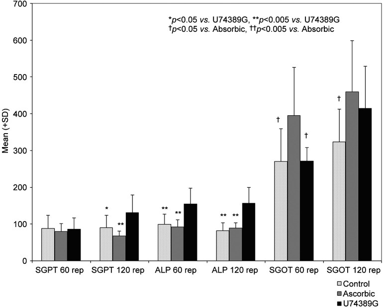
Materials and methods: Sixty Wistars rats were randomized into six groups of 10 animals each. Group A Ischemia 30 min, reperfusion 60 min; Group B Ischemia 30 min, reperfusion 120 min; Group C Ischemia 30 min, ascorbic acid administration, reperfusion 60 min; Group D Ischemia 30 min, ascorbic acid administration, reperfusion 120 min; Group E Ischemia 30 min, U-74389G administration, reperfusion 60 min; Group F Ischemia 30 min, U-74389G administration, reperfusion 120 min. We then collected tissue and blood samples.
Results: Histology and the significantly decreased malondialdehyde and tumor necrosis factor-α levels indicated that ascorbic acid was superior to U-74389G, at pre-defined time intervals.
Conclusion: Ascorbic acid and U-74389G ameliorated renal damage induced by ischemia-reperfusion injury, suggesting a therapeutic effect.
Keywords: Antioxidants; ascorbic acid; kidney; reperfusion injury.
Copyright© 2020, International Institute of Anticancer Research (Dr. George J. Delinasios), All rights reserved.
Conflict of interest statement
The Authors declare no conflicts of interest regarding this study.
Figures


Similar articles
-
Effects of U-74389G (21-Lazaroid) and Ascorbic Acid on Liver Recovery After Acute Ischemia and Reperfusion in Rats.In Vivo. 2015 Sep-Oct;29(5):585-94. In Vivo. 2015. PMID: 26359418
-
The effect of U-74389G on pancreas ischemia-reperfusion injury in a swine model.J Surg Res. 2014 Apr;187(2):450-7. doi: 10.1016/j.jss.2013.11.1082. Epub 2013 Nov 16. J Surg Res. 2014. PMID: 24332939
-
The effect of U-74389G on liver recovery after acute liver ischemia-reperfusion injury in a swine model.J Surg Res. 2009 Jan;151(1):10-4. doi: 10.1016/j.jss.2008.01.024. Epub 2008 Feb 27. J Surg Res. 2009. PMID: 18468628
-
The effect of the antioxidant drug "U-74389G" on oophoritis during ischemia reperfusion injury in rats.Antiinflamm Antiallergy Agents Med Chem. 2014;13(2):103-7. doi: 10.2174/1871523013666140804230111. Antiinflamm Antiallergy Agents Med Chem. 2014. PMID: 25091819
-
Ascorbic acid in solid organ transplantation: A literature review.Clin Nutr. 2022 Jun;41(6):1244-1255. doi: 10.1016/j.clnu.2022.04.004. Epub 2022 Apr 12. Clin Nutr. 2022. PMID: 35504167 Review.
Cited by
-
The efficiency of oxerutin on apoptosis and kidney function in rats with renal ischemia reperfusion injury.Ulus Travma Acil Cerrahi Derg. 2022 Mar;28(3):344-351. doi: 10.14744/tjtes.2021.15740. Ulus Travma Acil Cerrahi Derg. 2022. PMID: 35485553 Free PMC article.
-
The potential renoprotective effect of Raloxifene in renal ischemia-reperfusion injury in a male rat model.J Med Life. 2023 Aug;16(8):1274-1281. doi: 10.25122/jml-2023-0100. J Med Life. 2023. PMID: 38024816 Free PMC article.
References
MeSH terms
Substances
LinkOut - more resources
Full Text Sources