

Multiple schwannoma of the seminal vesicle: A case report
- PMID: 32872015
- PMCID: PMC7437769
- DOI: 10.1097/MD.0000000000021603
Multiple schwannoma of the seminal vesicle: A case report
Abstract
Rationale: Schwannomas of the seminal vesicles are extremely rare, and only cases of single seminal vesicle schwannomas have been reported. Here, we report a case of multiple schwannoma of the seminal vesicle.
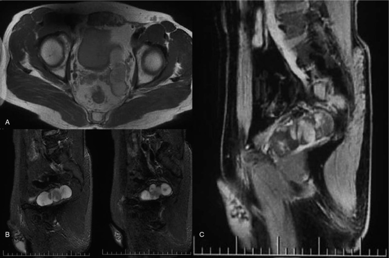
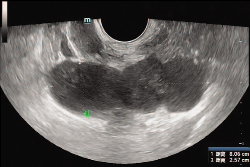
Patient concerns: We report a rare case of multiple schwannoma of the seminal vesicle that occurred in a 48-year-old man during physical examination. Multiple mixed masses in the left region of the seminal vesicle were documented with transrectal ultrasonography and magnetic resonance imaging. The patient presented no clinical symptoms, no family history of the disease and no history of genetic disease.
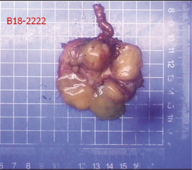
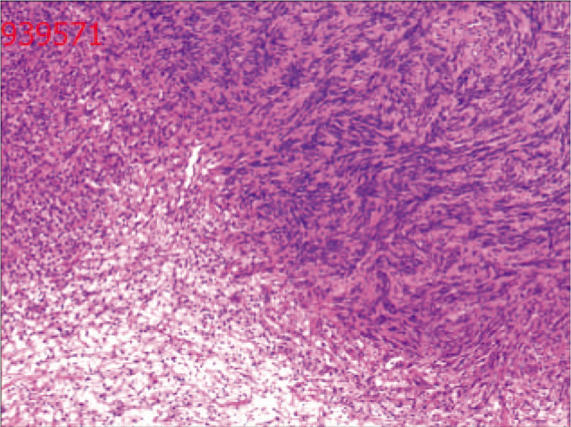
Diagnosis: Postoperative pathology revealed a diagnosis of seminal vesical schwannoma.
Interventions: The patient underwent robotic-assisted laparoscopic surgery to remove the mass.
Outcomes: The patient recovered rapidly and the length of hospitalization was 6 days after operation. At present, there is no recurrence in 10 month follow up.
Lessons: Whether benign or malignant, single or multiple, schwannomas still need to be diagnosed by pathology because of the limitations of examination methods. Surgical resection is still the preferred treatment.
Conflict of interest statement
The authors have no conflicts of interests to disclose.
Figures
References
-
- Mankin HJ, Mankin KP. Schwannoma: a rare benign tumor of soft tissues. Musculoskelet Surg 2014;98:95–9. - PubMed
-
- Iqbal N, Zins J, Klienman GW. Schwannoma of the seminal vesicle. Conn Med 2002;66:259–60. - PubMed
-
- Latchamsetty KC, Elterman L, Coogan CL. Schwannoma of a seminal vesicle. Urology 2002;60:515. - PubMed
-
- Han P, Wei Q, Yang YR. Neurilemmoma of a seminal vesicle. Chin Med J (Engl) 2007;120:1383–4. - PubMed
-
- Boeren K, De Bruecker Y, Vankan Y, et al. Schwannoma of the seminal vesicle. JBR-BTR 2011;94:96. - PubMed
Publication types
MeSH terms
LinkOut - more resources
Full Text Sources
