

Complete ureteral necrosis after injury sustained during lumbar disc surgery: A case report
- PMID: 32872054
- PMCID: PMC7437836
- DOI: 10.1097/MD.0000000000021727
Complete ureteral necrosis after injury sustained during lumbar disc surgery: A case report
Abstract
Introduction: Reports pertaining to ureteral injury sustained during lumbar disc surgery are rare; most ureteral injuries in this setting involve laceration or transection.
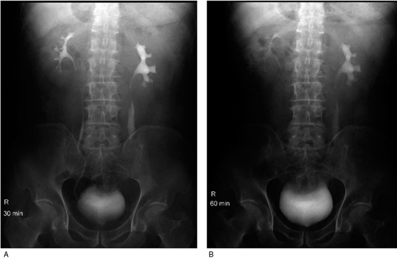
Patient concerns: We report a rare case of a 55-year-old man who presented with complete left ureteral necrosis 20 days after sustaining ureteral transection during lumbar disc surgery.
Diagnosis: The patient presented with seroperitoneum caused by left ureteral injury; post-operative histopathological examination of surgical specimen after discectomy had revealed ureter-like tissue. Exploratory laparoscopic surgery revealed necrosis of a long segment of ureter, which was not amenable to treatment with conventional methods.
Intervention: We used a spiral bladder muscle flap with vascular pedicles to repair the ureteral defect.
Outcomes: Post-operative period was uneventful and the patient showed good recovery.
Conclusion: Spiral bladder muscle flap with vascular pedicles may be used to repair extensive ureteric injury.
Conflict of interest statement
The authors report no conflicts of interest.
Figures


Similar articles
-
Insidious intraoperative ureteral injury as a complication in oblique lumbar interbody fusion surgery: a case report.BMC Res Notes. 2017 Jun 6;10(1):193. doi: 10.1186/s13104-017-2509-9. BMC Res Notes. 2017. PMID: 28587633 Free PMC article.
-
Iatrogenic Ureteral Injury as a Complication of Posterior or Lateral Lumbar Spine Surgery: A Systematic Review of the Literature.World Neurosurg. 2020 Mar;135:280-296. doi: 10.1016/j.wneu.2019.12.107. Epub 2019 Dec 27. World Neurosurg. 2020. PMID: 31887462
-
[Iatrogenic injury of the lumbar ureter and iliac vessels after lumbar discectomy: urologic treatment using kidney autotransplantation].Actas Urol Esp. 2002 Jul-Aug;26(7):504-8. doi: 10.1016/s0210-4806(02)72819-2. Actas Urol Esp. 2002. PMID: 12224434 Spanish.
-
Ureteral injury after posterior lumbar discectomy with interbody screw fixation.BMJ Case Rep. 2013 Oct 8;2013:bcr2013200383. doi: 10.1136/bcr-2013-200383. BMJ Case Rep. 2013. PMID: 24105384 Free PMC article.
-
Iatrogenic ureteral injury during retroperitoneal laparoscopy for large renal cysts: What we learned and a review of the literature.J Xray Sci Technol. 2021;29(1):185-196. doi: 10.3233/XST-200804. J Xray Sci Technol. 2021. PMID: 33459688 Review.
References
-
- He J, Xiao S, Wu Z, et al. Microendoscopic discectomy versus open discectomy for lumbar disc herniation: a meta-analysis. Eur Spine J 2016;25:1373–81. - PubMed
-
- Shriver MF, Xie JJ, Tye EY, et al. Lumbar microdiscectomy complication rates: a systematic review and meta-analysis. Neurosurg Focus 2015;39:E6. - PubMed
-
- Gangai MP. Ureteral injury incident to lumbar disc surgery. Case report. J Neurosurg 1972;36:90–2. - PubMed
-
- Hekal IA, Mohsen T, Nabeeh A. Ureteric injury after lumbosacral discectomy: a case report and review of the literature. J Trauma 2008;64:1387–91. - PubMed
-
- Mc KH, Haird HH, Justis HR. Management of ureteral injuries. J Am Med Assoc 1954;154:202–5. - PubMed
Publication types
MeSH terms
LinkOut - more resources
Full Text Sources
Medical
