. 2020 Aug 28;8(3):16.
doi: 10.3390/jdb8030016.

Col11a1a Expression Is Required for Zebrafish Development

Affiliations

Col11a1a Expression Is Required for Zebrafish Development

Makenna J Hardy et al. J Dev Biol. .

Abstract

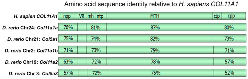
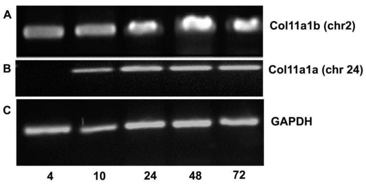
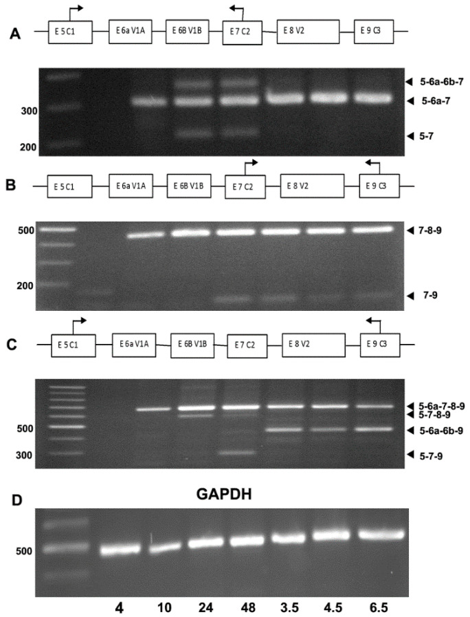
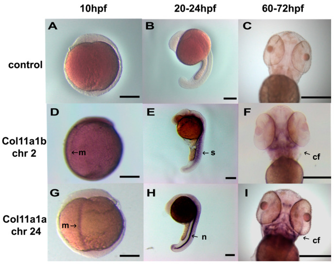
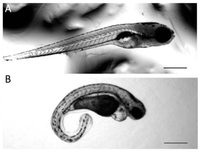
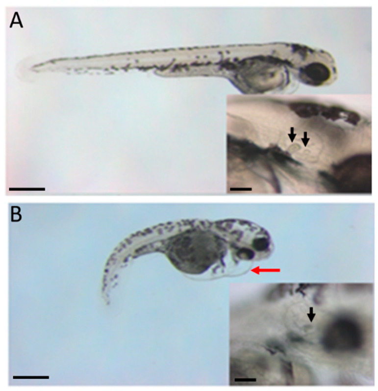
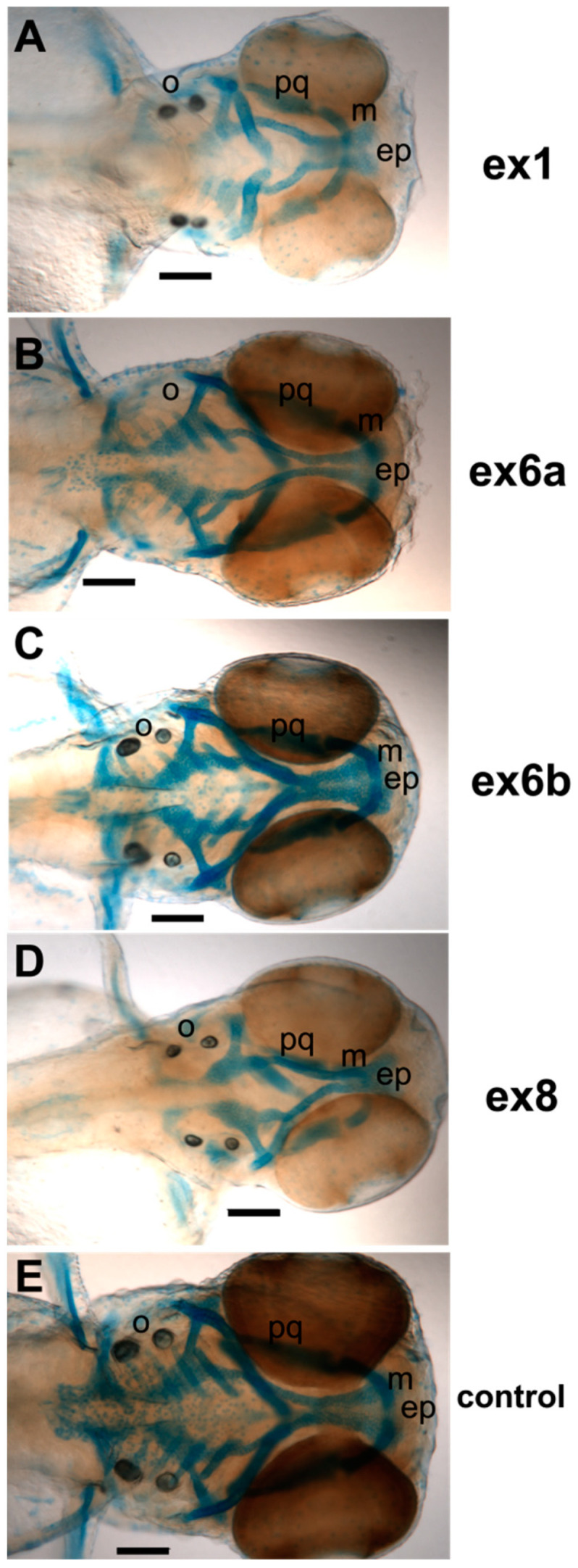
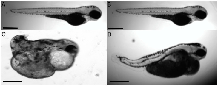
The autosomal dominant chondrodystrophies, the Stickler type 2 and Marshall syndromes, are characterized by facial abnormalities, vision deficits, hearing loss, and articular joint issues resulting from mutations in COL11A1. Zebrafish carry two copies of the Col11a1 gene, designated Col11a1a and Col11a1b. Col11a1a is located on zebrafish chromosome 24 and Col11a1b is located on zebrafish chromosome 2. Expression patterns are distinct for Col11a1a and Col11a1b and Col11a1a is most similar to COL11A1 that is responsible for human autosomal chondrodystrophies and the gene responsible for changes in the chondrodystrophic mouse model cho/cho. We investigated the function of Col11a1a in craniofacial and axial skeletal development in zebrafish using a knockdown approach. Knockdown revealed abnormalities in Meckel's cartilage, the otoliths, and overall body length. Similar phenotypes were observed using a CRISPR/Cas9 gene-editing approach, although the CRISPR/Cas9 effect was more severe compared to the transient effect of the antisense morpholino oligonucleotide treatment. The results of this study provide evidence that the zebrafish gene for Col11a1a is required for normal development and has similar functions to the mammalian COL11A1 gene. Due to its transparency, external fertilization, the Col11a1a knockdown, and knockout zebrafish model systems can, therefore, contribute to filling the gap in knowledge about early events during vertebrate skeletal development that are not as tenable in mammalian model systems and help us understand Col11a1-related early developmental events.

Keywords: Col11a1a; Marshall syndrome; Stickler type 2 syndrome; alternative splicing; collagen; fibrochondrogenesis; minor fibrillar collagen; zebrafish.


Conflict of interest statement

The authors declare no conflict of interest.

Figures

Figure 1
Figure 1
Amino acid sequence identity between Homo sapien and Danio rerio genes. Amino propeptide (npp), variable region (VR), minor helix (mh), amino telopeptide (ntp), major triple helix (MTH), carboxyl telopeptide (ctp), and carboxyl propeptide (cpp). Percentages shown indicate identity between human COL11A1 and the zebrafish gene. Homology is observed within the npp, mh and ntp, MTH, and the ctp and cpp domains, while the degree of identity is very low for the VR. Amino acid sequence identity for other minor fibrillar collagens is shown in comparison to human COL11A1. The D. rerio chromosome 2 Col11a1 locus identity corresponds to Col11a1b.
Figure 2
Figure 2
RT-PCR indicates that Col11a1a (chr24) is expressed between 10 and 72 hpf and Col11a1b (chr2) is expressed at 4 hpf through 72 hpf. (A) Using primers to amplify a 470 bp fragment, Col11a1b (chr2) mRNA was detected in embryos and larval fish. Time post-fertilization is indicated at the bottom of gels as 4, 10, 24, 48, and 72 hpf. (B) Col11a1a (chr24) was detected at 10 hpf through 72 hpf. (C) GAPDH was included as housekeeping gene control.
Figure 3
Figure 3
RT-PCR demonstrates alternative splicing patterns in the expression of Col11a1a isoforms. (A) PCR primers hybridizing to sequences within exons 5 and 7 were used to investigate the inclusion and exclusion of exons 6a and 6b overtime during development. Expression was detected as early as 10 hpf and continued throughout development to the last time point queried in this study, which was 6.5 dpf. (B) PCR primers hybridizing to sequences within exon 7 and 9 were used to investigate the inclusion and exclusion of exon 8 overtime during development. Exon 8 was included in the most predominant form of Col11a1a at all time points investigated. However, exon 8 was skipped in some forms of Col11a1a, joining exon 7 directly to exon 9, as shown by the PCR band migrating below 200 kilobases. (C) PCR primers hybridizing to sequences within exons 5 and 9 were used to investigate the complexity of splice form expression across the variable region of Col11a1a in zebrafish. The predominant splice form included exons 6a and 8, in agreement with observations shown in panels A and B. Alternative patterns of expression were observed to exclude exons 6a and 6b but include exon 8 at 24 hpf. Additionally, exclusion of exons 6a, 6b, and 8 resulted in the expression of the splice form comprising exons 5-7-9 migrating at approximately 300 kilobases at 48 hpf. (D) GAPDH was included as housekeeping gene control to confirm RNA content in samples representing distinct time points in development. The identity of the PCR product was verified by DNA sequencing.
Figure 4
Figure 4
In situ hybridization of Col11a1a (chr 24) and Col11a1b (chr 2). Wild-type embryos were treated with pSPT-18 control riboprobe (AC), Col11a1b (chr2) ex6-7-8-9 riboprobe (DF), and Col11a1a (chr24) ex6a-7-8-9 (GI). Embryos were observed at 10 hpf (A,D,G), 20–24 hpf (B,E,H), and 60–72 hpf (C,F,I). Expression was limited to the embryonic midline (m) at 10 hpf. At 20–24 hpf, expression was most pronounced in the notochord (n) for Col11a1a (chr24) and in the somites (s) for Col11a1b (chr2). At 60–72 hpf, developing craniofacial structures showed high levels of expression in addition to the notochord observed at 24 hpf seen for Col11a1a (chr24). Col11a1b (chr2) was also apparent in the craniofacial (cf) structures at 60–72 hpf in addition to the somites. Scale bars = 250 µm.
Figure 5
Figure 5
Body length and curvature changes due to Col11a1a-MOe1 AMO knockdown. (A) Control AMO injection observed at 72 hpf. (B) Col11a1a-MOe1 AMO injection observed at 72 hpf. Zebrafish embryos treated with the transcriptional start site-specific AMO for Col11a1a resulted in increased curvature and decreased body length. Additionally, heart edema was observed, as shown in B. Scale bar = 500 µm.
Figure 6
Figure 6
Cardiac, body length, and curvature changes due to Col11a1b-MOe1 AMO knockdown. (A) Control AMO injection, observed at 72 hpf. (B) Col11a1b-MOe1 AMO injection, observed at 72 hpf. Body length shortening, the curvature of the primary axis, and edema of the heart are apparent in zebrafish embryos treated with the transcriptional start site-specific AMO for Col11a1b. Otoliths are affected as indicated by arrows within the insets. Two arrows in A (inset) indicate the position of two otoliths in control zebrafish embryos. One arrow in B (inset) indicates the presence of only one otolith. Pericardial edema is indicated by the red arrow in B. Scale bar = 200 µm. Scale bars in inset represent 50 µm.
Figure 7
Figure 7
Alcian blue staining of craniofacial cartilage in 72 hpf zebrafish morphants of Col11a1a. A range of severity was observed among embryos of Col11a1a morphants. (A) Antisense morpholino oligonucleotide targeting the translational start site of Col11a1a-MOe1 morphants demonstrate reduced Alcian blue staining, disorganized cartilage, and shortened jaw. (B) Col11a1a-MOe6a morphants demonstrate reduced Alcian blue staining, shortened Meckel’s cartilage, and an absence of otoliths. (C) Col11a1a-MOe6b show relatively little effect and are similar to the control zebrafish. (D) Col11a1a-MOe8 show reduced Alcian blue staining. (E) Standard AMO control. Abbreviations palatoquadrate (pq); Meckel’s cartilage (m); ethmoid plate (ep); otolith (o). Scale bars = 200 µm.
Figure 8
Figure 8
CRISPR/Cas9-mediated homozygous and heterozygous knockout of Col11a1a shows a similar but more severe outcome compared to AMO knockdown. (A) Wild-type 72 hpf embryo compared to (C) homozygous Col11a1a −/− knockout embryo at 72 hpf showing the severe effect of the complete absence of Col11a1a in early embryogenesis. Homozygous offspring were raised to adulthood and bred to wild type to generate heterozygous Col11a1 −/+ offspring. (B) Wild-type 72 hpf embryo. (D) Heterozygous Col11a1a −/+ embryo at 72 hpf. Scale bar = 200 µm.

Similar articles

Cited by

References

    1. Goldring M.B., Tsuchimochi K., Ijiri K. The control of chondrogenesis. J. Cell. Biochem. 2006;97:33–44. doi: 10.1002/jcb.20652. - DOI - PubMed
    1. Hall B.K., Miyake T. Divide, accumulate, differentiate: Cell condensation in skeletal development revisited. Int. J. Dev. Biol. 1995;39:881–893. - PubMed
    1. Glenister T.W. An embryological view of cartilage. J. Anat. 1976;122:323–330. - PMC - PubMed
    1. Zylińska B., Silmanowicz P., Sobczyńska-Rak A., Jarosz Ł., Szponder T. Treatment of articular cartilage defects: Focus on tissue engineering. In Vivo. 2018;32:1289–1300. doi: 10.21873/invivo.11379. - DOI - PMC - PubMed
    1. Riley B.B., Phillips B.T. Ringing in the new ear: Resolution of cell interactions in otic development. Dev. Biol. 2003;261:289–312. doi: 10.1016/S0012-1606(03)00245-8. - DOI - PubMed

LinkOut - more resources