Review
. 2020 Aug 28;12(9):821.
doi: 10.3390/pharmaceutics12090821.

Silver Nanomaterials for Wound Dressing Applications

Affiliations
Review

Silver Nanomaterials for Wound Dressing Applications

Priya Dharshini Krishnan et al. Pharmaceutics. .

Abstract

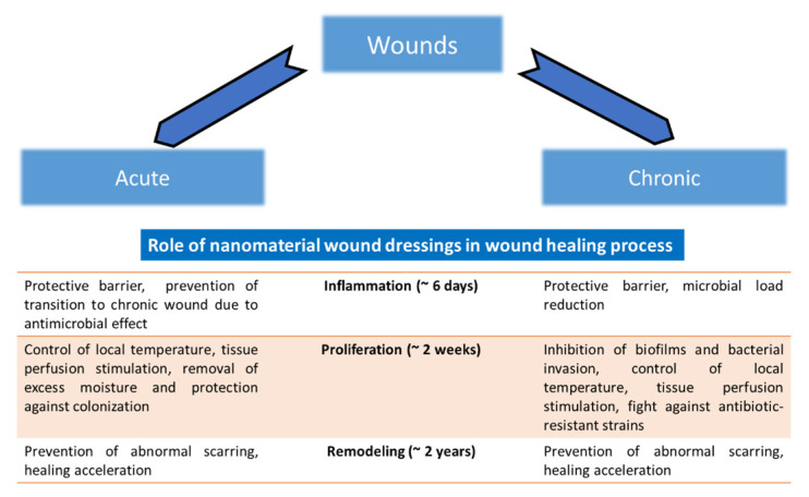
Silver nanoparticles (AgNPs) have recently become very attractive for the scientific community due to their broad spectrum of applications in the biomedical field. The main advantages of AgNPs include a simple method of synthesis, a simple way to change their morphology and high surface area to volume ratio. Much research has been carried out over the years to evaluate their possible effectivity against microbial organisms. The most important factors which influence the effectivity of AgNPs against microorganisms are the method of their preparation and the type of application. When incorporated into fabric wound dressings and other textiles, AgNPs have shown significant antibacterial activity against both Gram-positive and Gram-negative bacteria and inhibited biofilm formation. In this review, the different routes of synthesizing AgNPs with controlled size and geometry including chemical, green, irradiation and thermal synthesis, as well as the different types of application of AgNPs for wound dressings such as membrane immobilization, topical application, preparation of nanofibers and hydrogels, and the mechanism behind their antimicrobial activity, have been discussed elaborately.

Keywords: antibacterial effect; nanosilver; synthesis route; therapeutic activity.


Conflict of interest statement

The authors declare no conflict of interest.

Figures

Figure 1
Figure 1
Nanomaterials with potential use for wound treatment. Nanomaterials cover several groups of materials including nanoparticles, nanocomposites and different coatings and scaffolding materials. Nanoparticles can be divided into two groups depending on their chemical basis, which comprises inorganic (metal—Ag, Au, Zn, Cu etc.; non-metal—for example, Se) and organic (polymeric and non-polymeric) nanoparticles. From the perspective of nanocomposites, four groups can be recognized: porous materials, colloids, copolymers and gels. For the wound coating and scaffolds, hydrogels, nanofibers, films and membranes can be used. Adapted from [25] under a CC BY 4.0 license.
Figure 2
Figure 2
Effect of silver nanoparticles (AgNPs) on the single bacterial cell. (A) AgNPs penetrate through the cell membrane and cause its disruption; intracellular metabolites are released via the disrupted membrane. (B) AgNPs release silver ions (Ag+) to generate reactive oxygen species (ROS) and induce oxidative stress. (C) ROS are, for example, responsible for lipid peroxidation, which significantly changes the distribution of intracellular and extracellular metabolites and causes a change in membrane permeability, because lipid strains are repulsed. Interaction of AgNPs and Ag+ with proteins can affect (D) DNA enzymatic replication machinery, (E) DNA transcription, (F) translation of DNA to polypeptide/protein chain on ribosomes (red-brown ovals), (G) proton efflux pumps, (H) electron transport chain (represented by blue, yellow and green ovals) and creation of energetic sources of cells (ATP). Adapted from [31], Copyright Springer Nature, 2013.
Figure 3
Figure 3
Two basic methods of screening of the antibacterial effect: (A) Effect of AgNPs in concentrations of 5 (1), 2.5 (2), 1.25 (3) and 0.625 mg/mL (4) in the agar plate disk diffusion test on S. aureus cells after 24 h of incubation. Growth inhibition zones are marked by red circles. (B) The growth curve of S. aureus without (control) or with 0.1 mg/mL AgNPs showed optical density of 620 nm (OD620), dependent on the time of growth.
Figure 4
Figure 4
Wound treatment with silver nanomaterials. Nanomaterials can be applied directly to the wound (solution or powder nanoparticles) or nanomaterial-containing cloth and fabrics can be used. Hydrogels and water-soluble polymers are being used as non-compact materials (they are easily rubbed on the skin); on the contrary, compact materials comprising fibers, membranes and films are firm and non-soluble.
Figure 5
Figure 5
Summary of antimicrobial wound dressings with nanomaterials. The role of wound dressings using nanomaterials in various phases of wound healing (which comprise inflammation, proliferation and remodeling of the wound) is shown (the approximate duration of each phase is indicated in parentheses). Mostly, the effect is achieved due to the physical protection of the wound, acceleration of healing and broad antimicrobial action. Adapted from [25] under a CC BY 4.0 license.
Figure 6
Figure 6
Flowchart of choice of articles for review, “Silver Nanoparticles for Wound Dressing Applications“. In the first phase, articles were searched according to the following keywords: “silver nanoparticles in wounds”; “silver nanoparticles and therapeutic application”; “chemical synthesis of AgNPs”; “green synthesis of AgNPs”; “irradiation synthesis of AgNPs”; “thermal synthesis of AgNPs”. The following databases were used: Web of Science (WoS) core collection; PubMed and Embase, searching since 2000. Completely, more than 11,000 articles were found (5809 WoS, 3018 PubMed and 2679 Embase). Condensate extraction of articles from this phase was used for writing introduction and parts about the synthesis of particles. In the second phase, we were interested only in articles with the keywords “AgNPs for wound dressing applications”. More than 600 articles were found (444 WoS, 83 PubMed and 140 Embase). Some of these articles were used for writing the main section concerning different means of preparing AgNPs wound dressings. It is seen that using AgNPs, especially in wound dressings, comprises the effort of many work teams from around the world; thus, it is impossible to describe all techniques, materials and types of synthesis of AgNPs. We tried to choose several representative examples for all types of synthesis and the most frequent using polymeric materials to give the reader a complex view of the problematic nature of using AgNPs in wound dressing. The articles which were preferred were articles from 2007, which describe mainly the use of AgNPs. The articles were selected also upon impact factor (average impact factor is 6.31) and quartile of the journal (Q1 and Q2). Finally, 129 articles were chosen to write this review.
Figure 7
Figure 7
The process of preparation of AgNPs. (A) A plant/fungus is (B) homogenized, extracted, centrifugated and filtrated; subsequently (C), silver ions in the form of 0.1 M AgNO3 are added in a 1:1 (v/v) ratio. (D) AgNPs are formed within 24 h. (E) AgNPs are precipitated with methanol in a 1:1 (v/v) ratio for 1 h. (F) The solution of AgNPs is centrifuged for 15 min, 4000 g. (G)The supernatant is discarded and AgNPs are dried by lyophilization for 24 h.
Figure 8
Figure 8
A model example of green synthesis of AgNPs. As mentioned in Figure 7, the green synthesis of AgNPs is based on using extracts from plants or fungi, which contain a giant spectrum of different oxidative and reductive agents. These molecules reduce silver ions (Ag+) to elementary silver (Ag0). Compounds which influence the reduction and capping of originating AgNPs are, for example, proteins, alkaloids or polysaccharides. Different types of nanoparticles can be prepared depending on reaction conditions (metal concentration, extract concentration, length of synthesis, stabilization agents, etc.).

References

    1. Augustine R., Zahid A.A., Hasan A., Wang M., Webster T.J. CTGF loaded electrospun dual porous core-shell membrane for diabetic wound healing. Int. J. Nanomed. 2019;14:8573–8588. doi: 10.2147/IJN.S224047. - DOI - PMC - PubMed
    1. Garcia-Villen F., Faccendini A., Aguzzi C., Cerezo P., Bonferoni M.C., Rossi S., Grisoli P., Ruggeri M., Ferrari F., Sandri G., et al. Montmorillonite-Norfloxacin nanocomposite intended for healing of infected wounds. Int. J. Nanomed. 2019;14:5051–5060. doi: 10.2147/IJN.S208713. - DOI - PMC - PubMed
    1. Pang S.C., Gao Y., Wang F.B., Wang Y.Y., Cao M.X., Zhang W.J., Liang Y., Song M.Y., Jiang G.B. Toxicity of silver nanoparticles on wound healing: A case study of zebrafish fin regeneration model. Sci. Total Environ. 2020;717:10. doi: 10.1016/j.scitotenv.2020.137178. - DOI - PubMed
    1. Azeredo J., Azevedo N.F., Briandet R., Cerca N., Coenye T., Costa A.R., Desvaux M., Di Bonaventura G., Hébraud M., Jaglic Z., et al. Critical review on biofilm methods. Crit. Rev. Microbiol. 2017;43:313–351. doi: 10.1080/1040841X.2016.1208146. - DOI - PubMed
    1. Percival S.L., Salisbury A.M., Chen R. Silver, biofilms and wounds: Resistance revisited. Crit. Rev. Microbiol. 2019;45:223–237. doi: 10.1080/1040841X.2019.1573803. - DOI - PubMed

LinkOut - more resources