Review
. 2020 Aug 28;9(9):1983.
doi: 10.3390/cells9091983.

Transient Receptor Potential Canonical (TRPC) Channels: Then and Now

Affiliations
Review

Transient Receptor Potential Canonical (TRPC) Channels: Then and Now

Xingjuan Chen et al. Cells. .

Abstract

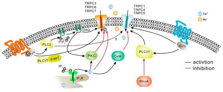
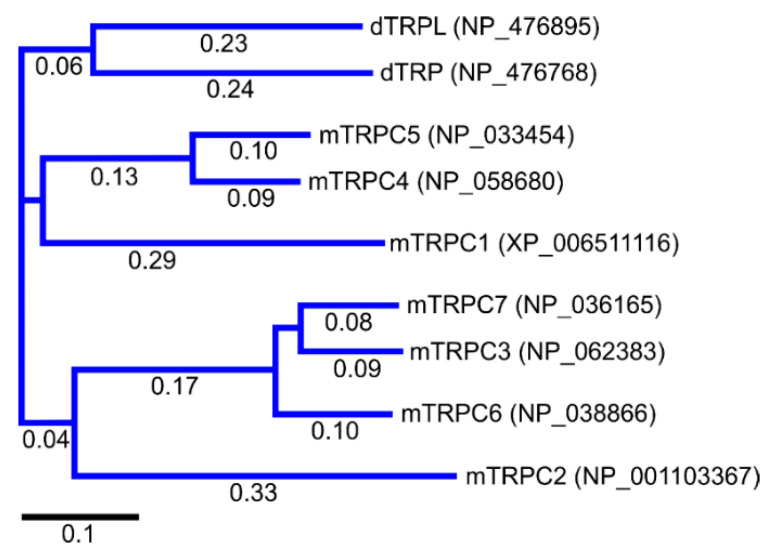
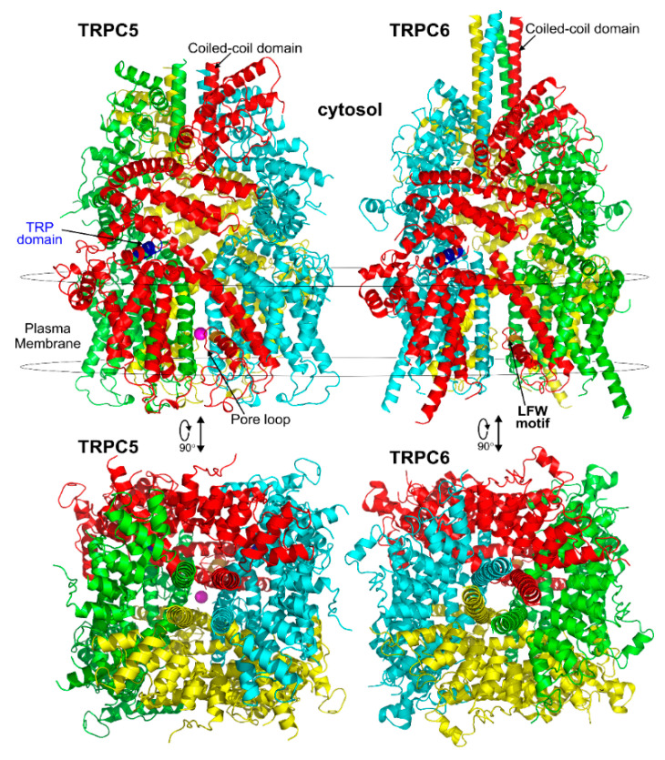
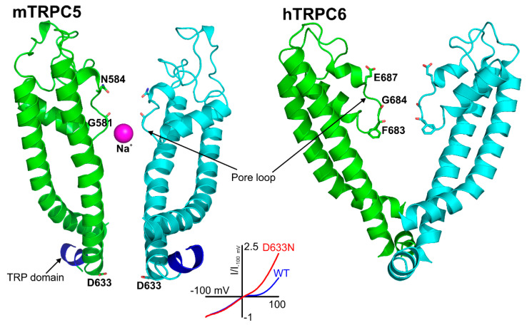
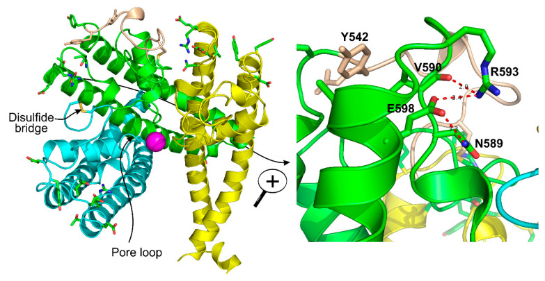
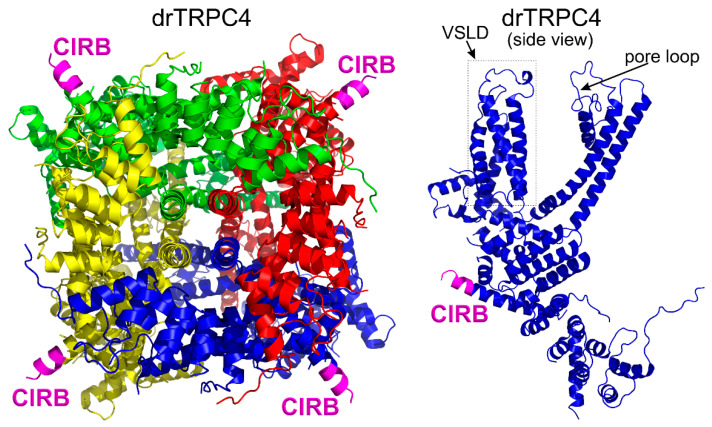
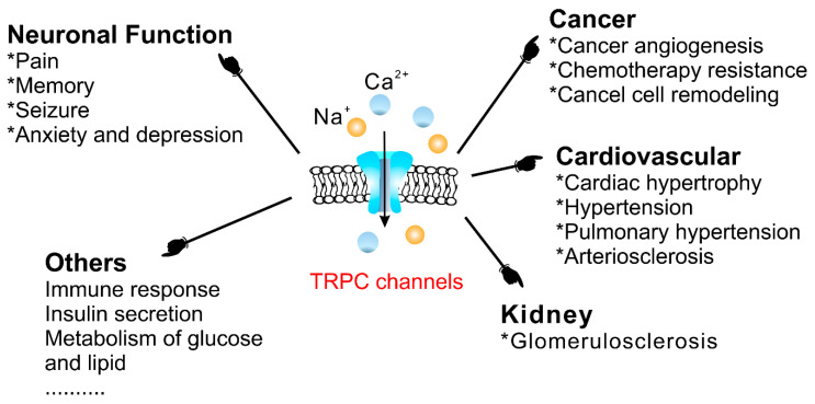
Twenty-five years ago, the first mammalian Transient Receptor Potential Canonical (TRPC) channel was cloned, opening the vast horizon of the TRPC field. Today, we know that there are seven TRPC channels (TRPC1-7). TRPCs exhibit the highest protein sequence similarity to the Drosophila melanogaster TRP channels. Similar to Drosophila TRPs, TRPCs are localized to the plasma membrane and are activated in a G-protein-coupled receptor-phospholipase C-dependent manner. TRPCs may also be stimulated in a store-operated manner, via receptor tyrosine kinases, or by lysophospholipids, hypoosmotic solutions, and mechanical stimuli. Activated TRPCs allow the influx of Ca2+ and monovalent alkali cations into the cytosol of cells, leading to cell depolarization and rising intracellular Ca2+ concentration. TRPCs are involved in the continually growing number of cell functions. Furthermore, mutations in the TRPC6 gene are associated with hereditary diseases, such as focal segmental glomerulosclerosis. The most important recent breakthrough in TRPC research was the solving of cryo-EM structures of TRPC3, TRPC4, TRPC5, and TRPC6. These structural data shed light on the molecular mechanisms underlying TRPCs' functional properties and propelled the development of new modulators of the channels. This review provides a historical overview of the major advances in the TRPC field focusing on the role of gene knockouts and pharmacological tools.

Keywords: TRPC; TRPC knockouts; TRPC modulators; calcium influx; cation channels.


Conflict of interest statement

The authors declare no conflict of interest. The funders had no role in the collection, analyses, or interpretation of data; in the writing of the manuscript, or in the decision to publish the results.

Figures

Figure 6
Figure 6
The mechanisms involved in regulating TRPC activity. Gαq/11-coupled-receptor activation leads to PLC-mediated hydrolysis of phosphatidylinositol 4,5-bisphosphate (PIP2) and production of inositol 1,4,5-trisphosphate (IP3) and diacylglycerols (DAG). IP3 activates IP3 receptors (IP3R) on the endoplasmic reticulum resulting in stored Ca2+ release and Ca2+ store depletion. DAG can directly activate TRPC3, TRPC6, and TRPC7 channels [40,123]. Additionally, DAG in conjunction with Ca2+ can activate PKC, which may in turn phosphorylate TRPC channels. Phosphorylation by PKC inhibits TRPC3/TRPC6 activity [120]. Ca2+ may decrease TRPC channel activity directly or via calmodulin (CaM). TRPC4 channel activity can be elicited not only downstream of Gαq/11, but also via Gαi/o protein interaction. Besides DAG, TRPC3 and TRPC6 channels have been reported to be activated in PLCγ, IP3 or β-arrestin-1 dependent manner.
Figure 1
Figure 1
The phylogenetic tree of Drosophila melanogaster TRP channels and mouse TRPC channels. The multiple sequence alignment was performed using the MUSCLE algorithm. The length of branches is shown under the lines, indicating the number of substitutions per site. The scale bar is also included under the plot for convenience. Based on the phylogenic tree, mouse TRPCs can be subdivided into four groups: TRPC1, TRPC2, TRPC4/5 and TRPC3/6/7. MegAlign Pro 17 of Lasergene 17 software was used to align and construct the tree. The TRP protein accession numbers are shown on the right from the name of each protein.
Figure 2
Figure 2
Cryo-EM structures of TRPC5 and TRPC6 channels. Each subunit in these TRPC structures was color-coded as red, green, yellow, and cyan for better identification. The TRP domain conserved in all TRPC channels is colored in blue in the red subunits of the shown structures. The LFW motif, which is located in the pore helix and colored in bright orange in the figure, is critical for the function of all TRPC channels because this protein segment is important for positioning the pore loop of the channel. Substituting AAA for LFW residues in a TRPC subunit renders it as a dominant negative. Dominant negative subunits are able to quench the activity of the functional subunits in heteromeric TRPC channels, which is a useful strategy to study TRPC channel roles in various organ systems. The mouse TRPC5 and human TRPC6 atomic coordinates were from PDB ID#: 5AEI and PDB ID#: 5YX9, respectively. A Na+ cation in the selectivity filter of TRPC5 channels is shown as a magenta sphere.
Figure 3
Figure 3
Structural architecture of the pore region of TRPC5 and TRPC6 channels. Only two subunits are shown for clarity (mouse TRPC5-pdb: 5aei and human TRPC6-pdb: 5yx9). The residues involved in controlling TRPC5 and TRPC6 cation selectivity are indicated within the pore loop of the channels. The role of the E687 residue in controlling TRPC6 Ca2+ permeability was identified by the Klaus Groschner group in 2011 [104,105], whereas the importance of the N584 residue for determining the TRPC5 channel’s Ca2+ selectivity was identified in a screen by Chen et al. in 2017 [56]. TRPC5 is inhibited by intracellular Mg2+ in a voltage-dependent manner, with a S6 transmembrane helix residue, D633, being responsible for that signature property of TRPC5 [79]. The inset shows the current–voltage relationships of wild type and the D633N mutant of TRPC5. The D633N mutant exhibits a reduced Mg2+ sensitivity, whereas the D636N mutant has a current–voltage relationship similar to that of the wild type TRPC5 [79]. The solved structure of TRPC5 confirmed that the D633 residue is located within the cation conduction pathway, whereas neighboring D636 residue faces away [103]. The mouse TRPC5 and human TRPC6 atomic coordinates were from PDB ID#: 5AEI and PDB ID#: 5YX9, respectively.
Figure 4
Figure 4
The structures of three out of four TRPC5 subunits are shown in the left panel. The position of the antigen for the E3 antibody which inhibits TRPC5 activity is shown in wheat color. This E3 antibody was developed by the Beech group [108]. The disulfide bridge is shown in yellow within the cyan subunit of TRPC5. Na+ in the cation conduction pathway of TRPC5 is depicted as a magenta sphere. The right panel shows a magnified view of the green subunit. The R593 residue which Chen et al. [56] named as “molecular fulcrum” is labeled, and its interactions with neighboring residues are shown using the red dotted lines. The Y542 residue that is involved in regulating Gd3+ sensitivity of TRPC5 is shown in wheat color. The mouse TRPC5 atomic coordinates were from PDB ID#: 5AEI.
Figure 5
Figure 5
The atomic structure of apo Danio rerio TRPC4 (drTRPC4). VSLD stands for the voltage sensor-like domain: (Left) a view at the drTRPC4 protein from the cytosol; and (Right) a side view at drTRPC4. CIRB stands for the Ca2+-calmodulin-IP3 receptor binding domain. The CIRB domains are colored in magenta. The drTRPC4 atomic coordinate were from pdb ID: 6g1k [101].
Figure 7
Figure 7
The physiological and pathophysiological roles of TRPC channels.

References

    1. Minke B., Wu C., Pak W.L. Induction of photoreceptor voltage noise in the dark in Drosophila mutant. Nature. 1975;258:84–87. doi: 10.1038/258084a0. - DOI - PubMed
    1. Cosens D.J., Manning A. Abnormal electroretinogram from a Drosophila mutant. Nature. 1969;224:285–287. doi: 10.1038/224285a0. - DOI - PubMed
    1. Wong F., Hokanson K.M., Chang L.T. Molecular basis of an inherited retinal defect in Drosophila. Invest. Ophthalmol. Vis. Sci. 1985;26:243–246. - PubMed
    1. Montell C., Rubin G.M. Molecular characterization of the Drosophila trp locus: A putative integral membrane protein required for phototransduction. Neuron. 1989;2:1313–1323. doi: 10.1016/0896-6273(89)90069-X. - DOI - PubMed
    1. Wong F., Schaefer E.L., Roop B.C., LaMendola J.N., Johnson-Seaton D., Shao D. Proper function of the Drosophila trp gene product during pupal development is important for normal visual transduction in the adult. Neuron. 1989;3:81–94. doi: 10.1016/0896-6273(89)90117-7. - DOI - PubMed

Publication types

Substances

LinkOut - more resources