Review
. 2020 Aug 28;25(17):3945.
doi: 10.3390/molecules25173945.

Unravelling the Photoprotective Mechanisms of Nature-Inspired Ultraviolet Filters Using Ultrafast Spectroscopy

Affiliations
Review

Unravelling the Photoprotective Mechanisms of Nature-Inspired Ultraviolet Filters Using Ultrafast Spectroscopy

Temitope T Abiola et al. Molecules. .

Abstract

There are several drawbacks with the current commercially available ultraviolet (UV) filters used in sunscreen formulations, namely deleterious human and ecotoxic effects. As a result of the drawbacks, a current research interest is in identifying and designing new UV filters. One approach that has been explored in recent years is to use nature as inspiration, which is the focus of this review. Both plants and microorganisms have adapted to synthesize their own photoprotective molecules to guard their DNA from potentially harmful UV radiation. The relaxation mechanism of a molecule after it has been photoexcited can be unravelled by several techniques, the ones of most interest for this review being ultrafast spectroscopy and computational methods. Within the literature, both techniques have been implemented on plant-, and microbial-inspired UV filters to better understand their photoprotective roles in nature. This review aims to explore these findings for both families of nature-inspired UV filters in the hope of guiding the future design of sunscreens.

Keywords: nature-inspired; photochemistry; photophysics; photoprotection; sunscreens; ultrafast spectroscopy.


Conflict of interest statement

The authors declare no conflict of interest.

Figures

Figure 1
Figure 1
Chemical structures of ultraviolet (UV) filters found in nature including (a) the plant-based sinapic acid (SA) and sinapoyl malate (SM), with the malate group added to the sinapic acid shown in blue; and (b) the core components of mycosporines and mycosporine-like amino acids synthesized by microorganisms, with the chromophores shown in red. See main text for definitions of R/R1/R2. Stereocenters have not been included in the structures.
Figure 2
Figure 2
Scheme showing a typical transient electronic absorption spectroscopy (TEAS) experimental setup.
Figure 3
Figure 3
Transient absorption spectra (TAS) displayed as false colour maps of (1) SA and (2) SM in (a) dioxane, (b) acetonitrile and (c) methanol, respectively. Reproduced and adapted with permission from [104], licensed under CC-BY. © 2015 American Chemical Society.
Figure 4
Figure 4
Relaxation scheme of SM and SA proposed by Baker et al. [104] Reproduced and adapted with permission from [104], licensed under CC-BY. © 2015 American Chemical Society.
Figure 5
Figure 5
UV/visible spectra showing the photoisomerisation of (a) SA, (b) SA and (c) SM2−, with the group added to SA shown in blue, at varying duration of pulsed irradiation. Corresponding structures are shown under UV/visible spectra. Reproduced and adapted with permission from [106]. © 2017 American Chemical Society.
Figure 6
Figure 6
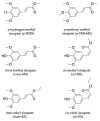
Chemical structures of sinapate ester derivatives including p-hydrogen methyl sinapate (p-HMS), p-methoxy methyl sinapate (p-OMEMS), trans-methyl sinapate (trans-MS) and cis-methyl sinapate (cis-MS) studied by Zhao et al. [111,113]. Also shown are trans-ethyl sinapate (trans-ES) and cis-ethyl sinapate (cis-ES) studied by Horbury et al. [114].
Figure 7
Figure 7
TAS displayed as false colour maps of (a) cis-ES and (b) trans-ES in cyclohexane photoexcited at 319 nm. Plot to show the TAS taken at ∆t = 2 ns of (c) cis-ES and (d) trans-ES overlaid with ∆UV/visible spectra. Reproduced and adapted with permission from [114], licensed under CC BY 3.0. Published by the Royal Society of Chemistry.
Figure 8
Figure 8
(a) Steady-state emission spectra of trans-MS (black solid line) and cis-MS (red solid line) at room temperature. The corresponding emission spectra at 77 K are shown as dotted lines. (b) Schematic of the proposed relaxation scheme of MS by Zhao et al. [113]. Reproduced and adapted with permission from [113]. © 2019 American Chemical Society.
Figure 9
Figure 9
(a) Structure of diethyl sinapate (DES) with the group added to the monoester precursor of ES shown in blue. (b) Long-term photostability of DES. UV/visible spectra of DES in C12-C15 alkyl benzoate, at varying durations of irradiation at 335 nm and replicating solar intensity. (c) Normalized TAS displayed as a false colour map of DES in C12-15 alkyl benzoate (AB) deposited on a synthetic skin mimic (termed DES VC/AB), photoexcited at 335 nm. The timescale is plotted linearly from −0.5 to 10 ps, then as a log scale from 10 to 100 ps. Reproduced and adapted with permission from [115], licensed under CC BY 4.0. Published by Springer Nature.
Figure 10
Figure 10
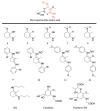
Structures of mycosporine motifs studied by Losantos et al. [89] and Woolley et al. [85], and gadusol/gadusolate studied by Losantos et al. [87].
Figure 11
Figure 11
Computed minimum energy path (MEP) of molecule 1 by Losantos et al. [89] where f is the oscillator strength. Reproduced and adapted with permission from [89]. © 2017 Wiley-VCH Verlag GmbH & Co. KGaA, Weinheim.
Figure 12
Figure 12
Critical points along the MEP for (a) gadusol and (b) gadusolate computed by Losantos et al. [87] where the energies are reported in kcal mol−1 relative to the electronic ground-state minimum. Reproduced and adapted with permission from [87], licensed under CC BY-NC-ND 4.0. © 2015 The Authors. Published by Wiley-VCH Verlag GmbH & Co. KGaA.
Figure 13
Figure 13
Structure of mycosporine-glycine at different pHs determined by Matsuyama et al. [129].
Figure 14
Figure 14
Structures of mycosporine-like amino acid (MAA) motifs studied by Losantos et al. [84,89] and Woolley et al. [85], and the natural MAAs studied by Sampedro [86], Koizumi et al. [88] and Hatakeyama et al. [90].
Figure 15
Figure 15
Computed MEP of molecule 6 by Losantos et al. [89], where f is the oscillator strength. Reproduced and adapted with permission from [89]. © 2017 Wiley-VCH Verlag GmbH & Co. KGaA, Weinheim.
Figure 16
Figure 16
Critical points along the MEP for molecule 11 computed by Losantos et al. [84], where the energies are reported in kcal mol-1 with respect to the electronic ground-state minimum. Where two numbers are present, the first number corresponds to the S1 energy, and the second number corresponds to the S0 energy at the specified geometry. Reproduced from [84], with permission from the PCCP Owner Societies.
Figure 17
Figure 17
TAS displayed as false colour maps for molecules 9–12, respectively, in methanol photoexcited at (a) 306 nm, (b) 330 nm, (c) 341 nm and (d) 353 nm, studied by Losantos et al. [84]. Reproduced and adapted from [84], with permission from the PCCP Owner Societies.
Figure 18
Figure 18
(a) Structure of porphyra-334 elucidated by Klisch et al. [134], and (b) structure of shinorine at different pHs determined by Matsuyama et al. [129].

References

    1. Lacis A.A., Hansen J. A parameterization for the absorption of solar radiation in the Earth’s atmosphere. J. Atmos. Sci. 1974;31:118–133. doi: 10.1175/1520-0469(1974)031<0118:APFTAO>2.0.CO;2. - DOI
    1. Frederick J.E., Snell H.E., Haywood E.K. Solar ultraviolet radiation at the Earth’s surface. Photochem. Photobiol. 1989;50:443–450. doi: 10.1111/j.1751-1097.1989.tb05548.x. - DOI
    1. Matsumi Y., Kawasaki M. Photolysis of atmospheric ozone in the ultraviolet region. Chem. Rev. 2003;103:4767–4782. doi: 10.1021/cr0205255. - DOI - PubMed
    1. Young A.R., Claveau J., Rossi A.B. Ultraviolet radiation and the skin: Photobiology and sunscreen photoprotection. J. Am. Acad. Dermatol. 2017;76:S100–S109. doi: 10.1016/j.jaad.2016.09.038. - DOI - PubMed
    1. Holick M.F. Sunlight and vitamin D for bone health and prevention of autoimmune diseases, cancers, and cardiovascular disease. Am. J. Clin. Nutr. 2004;80:1678S–1688S. doi: 10.1093/ajcn/80.6.1678S. - DOI - PubMed

Substances