Review
. 2020 Aug 28;13(9):221.
doi: 10.3390/ph13090221.

Bullous Central Serous Chorioretinopathy: A Rare and Atypical Form of Central Serous Chorioretinopathy. A Systematic Review

Affiliations
Review

Bullous Central Serous Chorioretinopathy: A Rare and Atypical Form of Central Serous Chorioretinopathy. A Systematic Review

Francesco Sartini et al. Pharmaceuticals (Basel). .

Abstract

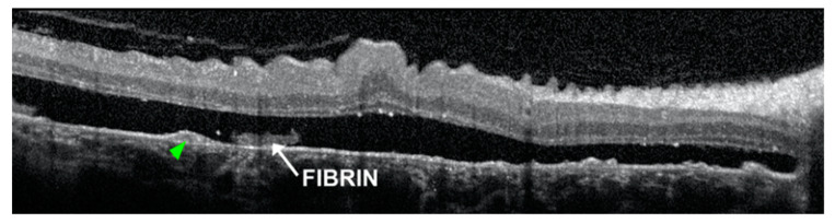
Bullous central serous chorioretinopathy (bCSCR) is a rare variant of the central serous chorioretinopathy, complicated by an exudative retinal detachment with shifting fluid. This systematic review aims to present the epidemiology, the pathogenesis, the clinical presentation, the imaging, the differential diagnosis, and the latest treatments of this disease. A total of 60 studies were identified following a literature search adhering to PRISMA guidelines. After full-text evaluation, 34 studies about bCSCR were included. bCSCR usually affects middle-aged men, and the principal risk factor is corticosteroid medications. Pathogenesis is related to an increased choroidal vessel and choriocapillaris permeability, with subsequent subretinal fluid accumulation, rich in fibrin, which may provoke the exudative retinal detachment. Clinical presentation and imaging are fundamental to distinguish bCSCR from other pathologies, avoiding unappropriated treatment. Corticosteroid withdraws (if assumed) and laser photocoagulation of leakage sites seen at angiography may speed up retinal reattachment. Verteporfin photodynamic therapy, transpupillary thermal therapy, oral eplerenone and scleral thinning surgery are other therapeutic options. An early diagnosis might prevent disease progression due to harmful medications as well as unnecessary surgery.

Keywords: argon laser photocoagulation; bullous central serous chorioretinopathy; chronic central serous chorioretinopathy; exudative retinal detachment; mineralocorticoid receptor antagonists; pachychoroid; scleral thinning surgery; transpupillary thermal therapy.


Conflict of interest statement

The authors declare no conflict of interest.

Figures

Figure 1
Figure 1
Fundus photographs reveal an exudative inferior retinal detachment macula-off (A) with a large retinal pigment epithelial tear in the temporal quadrant (B). Reproduced with permission from [14].
Figure 2
Figure 2
Optical coherence tomography (OCT) detects a pigment epithelial detachment (PED) with internal hyperreflectivity (green arrowheads) beneath the subretinal fluid, containing fibrin. Reproduced with permission from [6].
Figure 3
Figure 3
Fundus examination reveals turbid subretinal fluid at sites of neurosensory detachment (green arrows) (A); Fundus autofluorescence shows multiple areas of hypo-autofluorescence at the posterior pole (B). Reproduced with permission from [6].
Figure 4
Figure 4
Fluorescein angiography (FA) reveals multiple areas of leakage at the posterior pole in the early phase (A); with consequent pooling of the subretinal space and the dependent exudative retinal detachment (B). Indocyanine green angiography (ICGA) shows hyperpermeable and dilated choroidal vessels (C); in late phase pigment epithelial detachment (PED) it appears still hypofluorescent (D). FA shows focal area of hyperfluorescence (blowouts) within PED and an RPE tear with crescent shape (E). FA delineates a concentric RPE tear, with a retracted RPE at the center (F). Reproduced and merged with permission from [6,14].
Figure 5
Figure 5
PRISMA flowchart.

References

    1. Kaye R., Chandra S., Sheth J., Boon C.J.F., Sivaprasad S., Lotery A. Central serous chorioretinopathy: An update on risk factors, pathophysiology and imaging modalities. Prog. Retin. Eye Res. 2020 doi: 10.1016/j.preteyeres.2020.100865. - DOI - PubMed
    1. Gass J.D. Bullous retinal detachment. An unusual manifestation of idiopathic central serous choroidopathy. Am. J. Ophthalmol. 1973;75:810–821. doi: 10.1016/0002-9394(73)90887-8. - DOI - PubMed
    1. Sahu D.K., Namperumalsamy P., Hilton G.F., de Sousa N.F. Bullous variant of idiopathic central serous chorioretinopathy. Br. J. Ophthalmol. 2000;84:485–492. doi: 10.1136/bjo.84.5.485. - DOI - PMC - PubMed
    1. Otsuka S., Ohba N., Nakao K. A long-term follow-up study of severe variant of central serous chorioretinopathy. Retina. 2002;22:25–32. doi: 10.1097/00006982-200202000-00005. - DOI - PubMed
    1. Fawzi A.A., Holland G.N., Kreiger A.E., Heckenlively J.R., Arroyo J.G., Cunningham E.T., Jr. Central serous chorioretinopathy after solid organ transplantation. Ophthalmology. 2006;113:805–813. doi: 10.1016/j.ophtha.2006.01.031. - DOI - PubMed

LinkOut - more resources