

Multiple Cephalic Malformations in a Calf
- PMID: 32872674
- PMCID: PMC7552318
- DOI: 10.3390/ani10091532
Multiple Cephalic Malformations in a Calf
Abstract
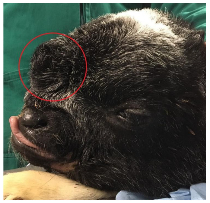
Congenital malformations of the central nervous system (CNS) can affect the CNS alone or the CNS and craniofacial structures. Here, we report an unusual and complex congenital cephalic malformation observed in a 3-day-old male crossbreed calf. Clinical examination disclosed a dome-shaped cranial vault, a flat face with a short snout, a median cleft lip, and increased intraorbital distance. The frontal region of the head was remarkable for a fluctuant, sac-like protrusion covered with haired skin. Neurologic findings suggested a multifocal intracranial lesion affecting the prosencephalon and the central vestibular system. While pathological and histopathological findings posited for a presumptive diagnosis of either hydranencephaly or holoprosencephaly associated with multiple congenital facial abnormalities, not all the findings could be definitely attributed to either of the two encephalic malformations alone. To our knowledge, a similar combination of severe congenital abnormalities affecting both the CNS and the craniofacial structures has not been reported in calves to date.
Keywords: bovine; holoprosencephaly; hydranencephaly; malformation; median cleft face syndrome.
Conflict of interest statement
The authors declare no conflict of interest.
Figures

References
-
- De Lahunta A., Noden D.M. Embryology of Domestic Animals: Developmental Mechanisms and Malformations. 1st ed. Volume 5–6. Williams & Wilkins; Baltimore, MD, USA: 1985.
-
- Hyttel P., Sinowatz F., Vejlsted M. Essentials of Domestic Animal Embryology. 1st ed. Volume 19. Saunders/Elsevier; Edinburgh, UK: 2010. pp. 338–344, 354–357.
-
- Leipold H.W., Hiraga T., Dennis S.M. Congenital Defects of the Bovine Central Nervous System. Vet. Clin. N. Am. Food Anim. Pract. 1993;9:77–91. - PubMed
-
- De Lahunta A., Glass E., Kent M. Veterinary Neuroanatomy and Clinical Neurology. 4th ed. Volume 3. Elsevier/Saunders; St. Louis, MO, USA: 2015. pp. 45–49, 64–68.
-
- Summers B.A., Cummings J.F., de Lahunta A. Veterinary Neuropathology. 1st ed. Volume 2. Mosby; St. Louis, MO, USA: 1995. pp. 71–73.
