

Feasibility and efficacy of lung ultrasound to investigate pulmonary complications in patients who developed postoperative Hypoxaemia-a prospective study
- PMID: 32873237
- PMCID: PMC7461251
- DOI: 10.1186/s12871-020-01123-6
Feasibility and efficacy of lung ultrasound to investigate pulmonary complications in patients who developed postoperative Hypoxaemia-a prospective study
Erratum in
-
Correction to: Feasibility and efficacy of lung ultrasound to investigate pulmonary complications in patients who developed postoperative Hypoxaemia-a prospective study.BMC Anesthesiol. 2020 Nov 9;20(1):281. doi: 10.1186/s12871-020-01196-3. BMC Anesthesiol. 2020. PMID: 33167910 Free PMC article.
Abstract
Background: Postoperative pulmonary complications (PPCs) and hypoxaemia are associated with morbidity and mortality. We aimed to evaluate the feasibility and efficacy of lung ultrasound (LUS) to diagnose PPCs in patients suffering from hypoxaemia after general anaesthesia and compare the results to those of thoracic computed tomography (CT).
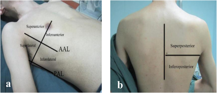
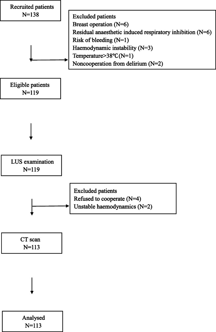
Methods: Adult patients who received general anaesthesia and suffered from hypoxaemia in the postanaesthesia care unit (PACU) were analysed. Hypoxaemia was defined as an oxygen saturation measured by pulse oximetry (SPO2) less than 92% for more than 30 s under ambient air conditions. LUS was performed by two trained anaesthesiologists once hypoxaemia occurred. After LUS examination, each patient was transported to the radiology department for thoracic CT scan within 1 h before returning to the ward.
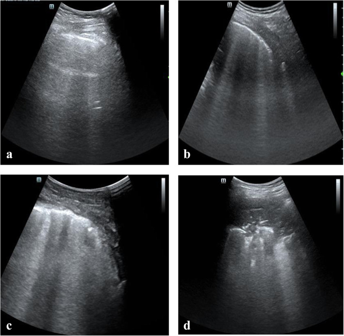
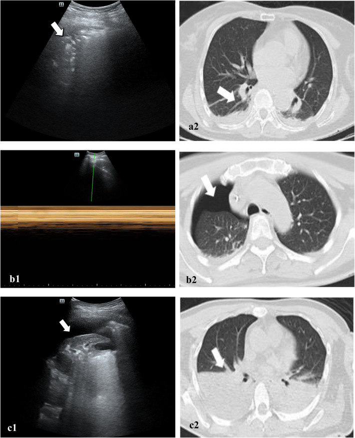
Results: From January 2019 to May 2019, 113 patients (61 men) undergoing abdominal surgery (45 patients, 39.8%), video-assisted thoracic surgery (31 patients, 27.4%), major orthopaedic surgery (17 patients, 15.0%), neurosurgery (10 patients, 8.8%) or other surgery (10 patients, 8.8%) were included. CT diagnosed 327 of 1356 lung zones as atelectasis, while LUS revealed atelectasis in 311 of the CT-confirmed zones. Pneumothorax was detected by CT scan in 75 quadrants, 72 of which were detected by LUS. Pleural effusion was diagnosed in 144 zones on CT scan, and LUS detected 131 of these zones. LUS was reliable in diagnosing atelectasis (sensitivity 98.0%, specificity 96.7% and diagnostic accuracy 97.2%), pneumothorax (sensitivity 90.0%, specificity 98.9% and diagnostic accuracy 96.7%) and pleural effusion (sensitivity 92.9%, specificity 96.0% and diagnostic accuracy 95.1%).
Conclusions: Lung ultrasound is feasible, efficient and accurate in diagnosing different aetiologies of postoperative hypoxia in healthy-weight patients in the PACU.
Trial registration: Current Controlled Trials NCT03802175 , 2018/12/05, www.ClinicalTrials.gov.
Keywords: Atelectasis; Lung ultrasound; Pleural effusion; Pneumothorax; Thoracic computed tomography.
Conflict of interest statement
The authors declare that they have no competing interests.
Figures
References
-
- Belcher AW, Khanna AK, Leung S, et al. Long-acting patient-controlled opioids are not associated with more postoperative hypoxemia than short-acting patient-controlled opioids after noncardiac surgery: a cohort analysis. Anesth Analg. 2016;123:1471–1479. - PubMed
-
- Govinda R, Kasuya Y, Bala E, et al. Early postoperative subcutaneous tissue oxygen predicts surgical site infection. Anesth Analg. 2010;111:946–952. - PubMed
Publication types
MeSH terms
Associated data
LinkOut - more resources
Full Text Sources
Medical
