Case Reports
. 2020 Aug 26;2020(8):rjaa191.
doi: 10.1093/jscr/rjaa191. eCollection 2020 Aug.

Brunner's glands hamartoma with pylorus obstruction: a case report and review of literature

Affiliations
Case Reports

Brunner's glands hamartoma with pylorus obstruction: a case report and review of literature

Mohammed A Bakir et al. J Surg Case Rep. .

Abstract

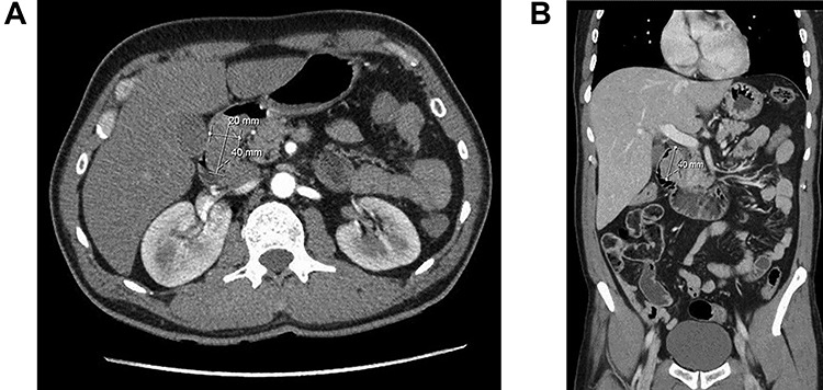
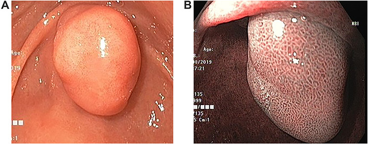
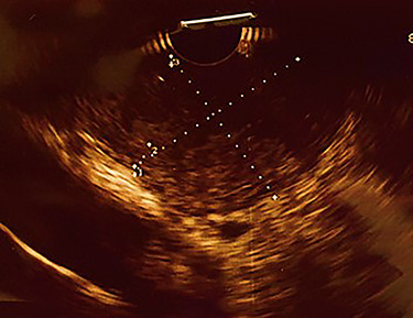
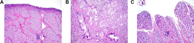
Proliferative lesions of the Brunner's glands are uncommonly encountered lesions of the small intestine, originating from the deeply seated mucosal and submucosal Brunner's glands, mainly in the duodenum. The vast majorities of these lesions are benign and include Brunner's glands hyperplasia (adenomas/nodules) and hamartomas. The etiology and pathogenesis of these lesions are not fully understood, and the diagnosis can sometimes be challenging. We report a case of Brunner's gland hamartoma in a 57-year-old man who presented with chronic dyspepsia, hematemesis and weight loss. Endoscopic and radiological investigations show a submucosal polypoid lesion at the first part of the duodenum. Routine endoscopic biopsies demonstrated normal duodenal mucosa. The lesion considered endoscopically unresectable and was surgically resected. Frozen section examination and intraoperative consultation showed unremarkable duodenal mucosa and histologically bland Brunner's glands.

Keywords: Brunner's gland hamartoma; adenoma; duodenal polyps; frozen section; hyperplasia.


Figures

Figure 1
Figure 1
Computed tomography of duodenal Brunner’s gland hamartoma. (a) Axial plane. (b) Coronal plane.
Figure 2
Figure 2
Endoscopic images. (a) Large duodenal submucosal lesion protruding through the pylorus to gastric antrum. (b) Narrow Band Imaging with normal tubulovillous mucosal patterns with regular vessels.
Figure 3
Figure 3
Endoscopic ultrasound, showing heterogeneous hypoechoic subepithelial lesion.
Figure 4
Figure 4
Histopathology of Brunner’s gland hamartoma. (a) The submucosal lesion consisted of lobules of proliferating bland Brunner’s glands, separated by fibromuscular septa; scattered lymphoid follicles were evident (Hematoxylin and Eosin × 100). (b) Irregularly-shaped ductal structures were present, mainly at the deep aspect of the lesion (Hematoxylin and Eosin × 400). (c) Focal papillary architecture was evident on the surface of the lesion (hematoxylin and eosin × 200).

References

    1. Henken EM, Forouhar F. Hamartoma of Brunner's gland causing partial obstruction of the ileum. J Can Assoc Radiol 1983;34:73–4. - PubMed
    1. Fuller JW, Maj MC, Cruse CW, Williams JW. Hyperplasia of Brunner's glands of the duodenum. Am Surg 1997;43:246–50. - PubMed
    1. Mochizuki T, Fujikuni N, Nakadoi K, Nakahara M, Tanabe K, Yonehara S, et al. Adenocarcinoma of the duodenum arising from Brunner's gland resected by partial duodenectomy: a case report. Surg Case Rep 2019;5:179. - PMC - PubMed
    1. Destek S, Gul VO. Brunner's gland hyperplasias and hamartomas in association with helicobacter pylori. Can J Gastroenterol Hepatol 2019;2019:6340565. - PMC - PubMed
    1. Yi L, Cheng Z, Qiu H, Yang J, Wang T, Liu K. A giant Brunner's gland hamartoma being treated as a pedunculated polyp: a case report. BMC Gastroenterol 2019;19:151. - PMC - PubMed

Publication types